File:Nihms275352f2.jpg

Original file (466 × 720 pixels, file size: 62 KB, MIME type: image/jpeg)
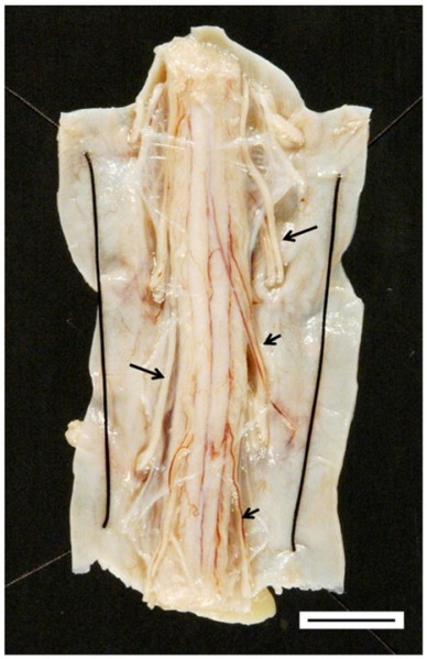
Summary[edit | edit source]
The thoracic spinal cord seen in FA. Images shows degeneration of the transverse diameter of the spinal cord. The dorsal surface of the spinal cord are thick white anterior roots (long arrows) that stand out in contrast to thin dorsal roots (short arrows).
Koeppen, A. Friedreich’s ataxia: pathology, pathogenesis, and molecular genetics. J Neuro. Sci. 2011;303;1-12.
Licensing[edit | edit source]
This work has been released into the public domain by its author. This applies worldwide. In some countries this may not be legally possible; if so: the author grants anyone the right to use this work for any purpose, without any conditions, unless such conditions are required by law.
File history
Click on a date/time to view the file as it appeared at that time.
| Date/Time | Thumbnail | Dimensions | User | Comment | |
|---|---|---|---|---|---|
| current | 21:55, 6 May 2018 | | 466 × 720 (62 KB) | Ashley Button (talk | contribs) | The thoracic spinal cord seen in FA. Images shows degeneration of the transverse diameter of the spinal cord. The dorsal surface of the spinal cord are thick white anterior roots (long arrows) that stand out in contrast to thin dorsal roots (short arro... |
You cannot overwrite this file.
File usage
The following page uses this file:

