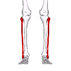
Fibula
Original Editor - Leana Louw
Top Contributors - Leana Louw and Kim Jackson
Description[edit | edit source]
There are two bones in the lower leg - the tibia and fibula. The fibula plays a key role in muscle attachment and contributes to the stability of the ankle joint.
Structure[edit | edit source]
The fibula is the thinner and posteriolaterally situated of the two lower leg bones. These two bones are connected by the tibiofibular syndesmosis, which includes the interosseous membrane.[1]
Proximal: The proximal part of the fibula features an enlarged pointed head and a small neck.
Shaft: The shaft of the fibula is twisted and triangular in cross-section. It has anterior, interosseous, and posterior borders, as well as medial, posterior, and lateral surfaces. This area is the primary site for muscle attachments.
Distal: At the distal end, the fibula extends to form the lateral malleolus, an important structure in the ankle joint, positioned inferiolaterally.
Function[edit | edit source]
The fibula's role is to act as an attachment for muscles, as well as providing stability of the ankle joint. Although it is less involved in weight-bearing compared to the tibia, it still supports a small portion of body weight, especially when the knee is flexed.[1]
Articulations[edit | edit source]
Proximal: The fibular head articulates with the lateral tibial condyle at the fibular facet, forming the proximal tibiofibular joint, a syndesmotic joint.[1]
Distal: The lateral malleolus articulates with the fibular notch of the tibia to form the distal tibiofibular joint and the talus to form the superior part of the ankle joint
Muscle attachments[edit | edit source]
The fibula provides proximal attachment for the following muscles:[1]
- Extensor digitorum longus: Superior 3/4 of medial border
- Extensor hallucis longus: Middle of anterior surface
- Fibularis tertius: Inferior 1/3 of anterior surface
- Fibularis longus: Fibular head and superior 2/3 of lateral surface
- Fibularis brevis: Inferior 2/3 of lateral surface
- Soleus: Fibular head (posterior) and superior 1/4 of posterior surface
- Flexor hallucis longus: Inferior 2/3 of posterior surface
- Flexor Digitorum Longus: Via tendon
- Tibialis posterior: Posterior surface
*Take note that above only describes fibular attachments, and that all of these muscles also has other areas of attachments not mentioned in this page.
Injuries[edit | edit source]
Fractures:
The most common area for fibula fractures are 2-6cm proximal to the distal end of the fibula. It is also often linked to ankle fractures.[1]
Surface Anatomy[edit | edit source]
- Fibula head: Very superficial and palpable at the posteriolateral aspect of the knee, level with the tibial tuberosity.
- Fibula neck: Situated distal to the lateral part of the fibula head.
- Fibula shaft: The distal 1/4 is palpable.
- Lateral malleolus: A superficial prominence on the lateral side of the ankle, distinct from the medial malleolus of the tibia.


