Martin Gruber Anastomosis
Original Editor - Nehal Shah
Top Contributors - Nehal Shah and Ewa Jaraczewska
Introduction[edit | edit source]
Anomalous means deviated from normal / routine. Anomalous innervations are routinely a normal occurrence. But if these are not recognised, they may be mistaken for a technical pitfall or an actual pathology, which may not be present. [1] Nerve communication between the median and ulnar nerves in the forearm is known as Martin-Gruber anastomosis. This implies that nerve fascicles from the Median nerve transfer to the Ulnar nerve. [2]
The prevalence of Martin Gruber Anastomosis (MGA) is between 3.3 and 40% of the population [1]with a prevalence of 11.6 % in the Indian population. [3] Median to Ulnar nerve communication was first described by Martin in 1763 [4], who proposed the possibility of such a communication. This finding was finally confirmed after 100 years by Gruber in 1870. [5]
Innervation Pattern of Hand[edit | edit source]
| Median Nerve | Ulnar Nerve |
|---|---|
| Abductor Pollicis Brevis | Abductor Digiti Minimi |
| Opponens Pollicis Brevis | Flexor Digiti Minimi |
| Flexor Pollicis Brevis | Opponens Digiti Minimi |
| 1st and 2nd Lumbrical | 3rd and 4th Lumbrical |
| Dorsal and Palmar Interossei | |
| Deep Head of Flexor Pollices Brevis | |
| Adductor Pollicis |
Studies suggest that this standard pattern is seen in 33% of cases. [6]
Common Patterns of Anamolous Innervations of Median and Ulnar nerves[edit | edit source]
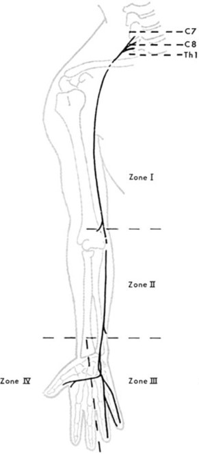
The complexity of the anatomy of the upper extremity, especially the brachial plexus and strategical anatomical zones such as the cubital tunnel, Guyon canal, and carpal tunnel, results in anomalous branching of nerves. They may form anastomosis. [2] To understand them better, Mennerfelt et al. divided the Ulnar nerve into four zones: [7]
- ANOMALOUS PATTERN OF INNERVATION IN ZONE I - Usually, the Flexor Carpi Ulnaris (FCU) muscle innervates at the level of the medial epicondyle of the Humerus. But it can innervate 4-7 cm proximal to FCU (Sunderland & Hughes, 1946)
- ANOMALOUS PATTERN OF INNERVATION IN ZONE II -
- An isolated branch of the Ulnar nerve arises in the middle of the forearm and runs down to supply the Ulnar aspect of 4th finger (Turner 1874)
- Dorsal Ulnar Cutaneous nerve, which usually arises about 5 cm above the wrist
- Variation in the level of innervation of FCU and Flexor Digitorum Profundus IV & V (Sunderland and Hoges 1946)
- Martin Gruber Anastomosis - Anastomosis between the Median and Ulnar nerves where communicating branches are from the Median to the Ulnar nerve. This is the main anomalous innervation in Zone II
- ANOMALOUS PATTERN OF INNERVATION IN ZONE III - Unusual distribution of Dorsal Ulnar Cutaneous nerve enclosing Pisiform bone (Kaplan 1963)
- ANOMALOUS PATTERN OF INNERVATION IN ZONE IV -
- Double innervation of First Dorsal Interosseus - both by Median and Ulnar nerves (Murphey, Kirklin and Finlayson (1946))
- Double innervation of Lumbricals - Double innervation of any of the Lumbricals by Median and Ulnar nerve (Brooke 1887 and Riche 1897)
- Thenar eminence supplied entirely or partly by Ulnar nerve
- Anastomosis between deep ulnar branches and motor branches of the median nerve in the radial part of the palm of the hand.
- Hypothenar muscles are innervated entirely or partly by the median nerve.
Martin Gruber Anastomosis[edit | edit source]
MGA is an anomalous innervation formed from the cross-over of median-to-ulnar motor nerve fibres, which usually occurs in the forearm. [1] It can occur bilaterally and unilaterally. It occurs bilaterally in 10-40% of detected cases. [8] In MGA, a communicating branch arises from the trunk of the Median nerve or any branches in the forearm, mainly the Anterior Interosseous nerve. This communicating branch from the Median nerve can innervate any intrinsic hand muscles supplied by the Ulnar nerve like ADM, FDI, Deep head of FPB, Adductor Pollicis or any combination of these muscles.[9]
Types of MGA[edit | edit source]
To date, various MGA classifications have been proposed by various authors. These depend upon the anatomical site of branching of the communicating branch with the most recent classification suggested by Cavalheiro et al. described below [2]
- Type I - Nerve fascicles originate from the Anterior Interosseus Nerve (AIN) distal to the elbow and join the Ulnar nerve between the proximal and middle one-third of the forearm
- Type II - Nerve fascicles originate from AIN, bifurcate into two and communicate with the Ulnar nerve at two different points in the forearm
- Type III - Nerve fascicles originate from the Median nerve, proximal to the emergence of AIN and head towards joining the Ulnar nerve. These fascicles can originate either proximal or distal to the elbow joint.
- Type IV -The communication occurs between the nerve fascicles originating from the Median and Ulnar nerve branches. They head towards FDP muscle mass.
- Type V - Nerve communications occur inside the muscle mass of FDP. This is also called intramuscular MGA
- Type VI - Nerve fascicles arise from the branch of the Median nerve to the Flexor Digitorum Superficialis muscle and head towards communication with the Ulnar nerve
Various authors have tried to classify MGA, and their findings are listed in the table below, starting from 1893 through today:[2]
| Year | Authors | Classification proposed; anastomosis between the median and ulnar nerves |
|---|---|---|
| 1893 | Thomson | Class I: anterior interosseous nerve and ulnar nerve |
| Class II: median nerve and ulnar nerve | ||
| Class III: muscle branch to deep flexor muscle of the fingers | ||
| 1931 | Hirasawa | Oblique anastomosis: anterior interosseous nerve and ulnar nerve |
| median nerve and ulnar nerve | ||
| Loop anastomosis: muscle branch to deep flexor muscle of the fingers | ||
| Combined anastomosis: combinations between others | ||
| 1976 | Kimura et al. | Type I: median nerve and ulnar nerve innervating the hypothenar muscle |
| Type II: median nerve and ulnar nerve innervating the deep flexor muscle of the fingers | ||
| Type III: median nerve and ulnar nerve innervating the thenar muscle | ||
| 1981 | Srinivasan and Rhodes | Types I, II, VI: anterior interosseous nerve and ulnar nerve or other |
| Type III: median nerve and ulnar nerve | ||
| Types IV, V: combinations of others | ||
| 1992 | Uchida and Sugioka | Type I: median nerve and ulnar nerve innervating the hypothenar muscle |
| Type II: median nerve and ulnar nerve innervating the thenar muscle | ||
| Type III: median nerve and ulnar nerve innervating the deep flexor muscle of the fingers | ||
| 1993 | Nakashima | Type Ia: anterior interosseous nerve and ulnar nerve |
| Type Ib: median nerve and ulnar nerve | ||
| Type III: combination of types Ia, Ib and II | ||
| 1995 | Oh et al. | Type I: median nerve and ulnar nerve innervating the hypothenar muscle |
| Type II: median nerve and ulnar nerve innervating the deep flexor muscle of the fingers | ||
| Type III: median nerve and ulnar nerve innervating the thenar muscle | ||
| 1999 | Shu et al. | Type I: anterior interosseous nerve and ulnar nerve |
| Type II: median nerve and ulnar nerve | ||
| Type III: muscle branch to deep flexor muscle of the fingers | ||
| Type IV: anterior interosseous nerve and ulnar nerve, muscle branch to deep flexor muscle of the fingers originating from the connection | ||
| Type V: two anastomotic branches | ||
| 2002 | Rodriguez-Niedenfuhr et al. | Pattern I: one anastomotic ramus |
| Pattern II: two anastomotic rami | ||
| Type A: anastomotic ramus originating from a branch of the median nerve to the nerve of the superficial flexor muscle of the forearm | ||
| Type B: anastomotic ramus originating from the median nerve | ||
| Type C: anastomotic ramus originating from the anterior interosseous nerve | ||
| 2005 | Lee et al. | Type I: anterior interosseous nerve and ulnar nerve |
| Type II: median nerve and ulnar nerve | ||
| Type III: muscle branch to deep flexor muscle of the fingers | ||
| Type IV: two anastomotic rami from the ulnar nerve or anterior interosseous nerve and ulnar nerve | ||
| 2015 | Cavalheiro et al | Type I: anterior interosseous nerve and ulnar nerve |
| Type II: anterior interosseous nerve and ulnar nerve (double anastomosis) | ||
| Type III: median nerve and ulnar nerve | ||
| Type IV: loop between anterior interosseous nerve and ulnar nerve with branches to deep flexor muscle of the fingers | ||
| Type V: intramuscular anastomosis | ||
| Type VI: branch from the median nerve to the superficial flexor muscle and ulnar nerve | ||
Clinical Implications of MGA[edit | edit source]
It is important to understand anatomic variations to explain paradoxical motor and sensory loss in patients. [10] When assessing Median and Ulnar nerve lesions, consider the specifics of the MGA. MGA can lead to misdiagnosis, especially in conditions like Carpal Tunnel Syndrome, Cubital Tunnel Syndrome, and Peripheral neuropathy involving the Median / Ulnar nerve. [11] MGA may cause alteration in clinical symptoms, especially in patients with carpal tunnel syndrome, where symptoms can be exacerbated or attenuated, causing motor and sensory alterations in a pattern that is different from the usual pattern. Many times, it is seen that even with a complete rupture of the Median or Ulnar nerve, some of the muscles routinely expected to become paralyzed do not present with motor deficits. This could lead to a wrong conception of the nerve being normal even when there is a cause of injury. It is also necessary to differentiate between partial and complete rupture of the nerve. For example, in lesions proximal to the communication, motor and sensory innervation remains normal, and in the complete lesion of the Median nerve, some muscles innervated by the Median nerve may not be paralysed, which can be misleading. [3]
Apart from clinical examination, Electrophysiological studies and Neuromuscular Ultrasound testing with a thorough knowledge of anomalous innervations is necessary.
References[edit | edit source]
- ↑ 1.0 1.1 1.2 Aziz Saba EK. Electrophysiological study of Martin—Gruber anastomosis in a sample of Egyptians. Egyptian Rheumatology and Rehabilitation. 2017 Oct;44:153-8.
- ↑ 2.0 2.1 2.2 2.3 Cavalheiro CS, Filho MR, Pedro G, Caetano MF, Vieira LA, Caetano EB. Clinical repercussions of Martin-Gruber anastomosis: anatomical study. Rev Bras Ortop. 2016 Feb 23;51(2):214-23.
- ↑ 3.0 3.1 Kaur N, Singla RK, Kullar JS. Martin-Gruber Anastomosis- A Cadaveric Study in North Indian Population. J Clin Diagn Res. 2016 Feb;10(2):AC09-11.
- ↑ Martin R. Tal om nervers allmänna egenskaper i människans kropp.
- ↑ Gruber W. Ueber die verbindung des nervus medianus mit dem nervus ulnaris am unterarme des menshen und der saugethiere. Arch Anat Phisiol. 1870;37:501.
- ↑ Rowntree T. Anomalous innervation of the hand muscles. The Journal of Bone and Joint Surgery. British volume. 1949 Nov;31(4):505-10.
- ↑ Mannerfelt L. Studies on the hand in ulnar nerve paralysis: a clinical-experimental investigation in normal and anomalous innervation. Acta Orthopaedica Scandinavica. 1966 Mar 1;37(sup87):3-176.
- ↑ Taams KO. Martin-Gruber connections in South Africa: an anatomical study. Journal of Hand Surgery. 1997 Jun;22(3):328-30.
- ↑ Prates LC, de Carvalho VC, Prates JC, Langone F, Esquisatto MA. The Martin-Gruber anastomosis in Brazilians: an anatomical study. Braz. J. morphol. Sci. 2003; 20(3):177-180.
- ↑ Hefny M, Sallam A, Abdellatif M, Okasha S, Orabi M. Electrophysiological Evaluation and Clinical Implication of Martin-Gruber Anastomosis in Healthy Subjects. J Hand Surg Asian Pac Vol. 2020 Mar;25(1):87-94.
- ↑ Tagil SM, Bozkurt MC, Ozçakar L, Ersoy M, Tekdemir I, Elhan A. Superficial palmar communications between the ulnar and median nerves in Turkish cadavers. Clin Anat. 2007 Oct;20(7):795-8.


