Sternoclavicular Joint Disorders: Difference between revisions
No edit summary |
No edit summary |
||
| Line 1: | Line 1: | ||
< | == Clinically Relevant Anatomy<br> == | ||
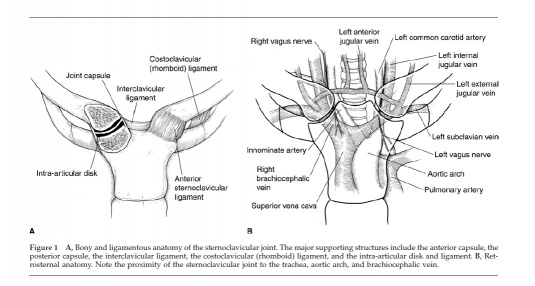
The Sternoclavicular (SC) joint is the only bony joint that connects the axial and appendicular skeletons. The SC joint is a plane synovial joint formed by the articulation of the sternum and the clavicle. Due to the joint’s articulation between the medial clavicle and the manubrium of the sternum and first costal cartilage, the joint has little bony stability. Between the medial clavicle and the manubrium is a dense fibrocartilaginous disc that separates the joints into two distinct synovial compartments. The intra-articular ligament provides joint stability and prevents medial displacement of the clavicle. This ligament originates from the junction of the first rib and sternum and passes through the SC joint and attaches to the clavicle on the superior and posterior side. The anterior and posterior sternoclavicular ligaments restrain anterior and posterior translation of the medial clavicle. The anterior and posterior sternoclavicular ligaments originates on the anterior and posterior ends of the clavicle, respectively, and inserts onto the anterior and posterior surfaces of the manubrium, respectively. The SC joint is supported superiorly by the interclavicular ligament that connects the superomedial portions of each clavicle. The blood supply to the SC joint is from the articular branches of the internal thoracic and suprascapular arteries. The SC joint is innervated by the branches of the medial suprascapular nerve. The brachiocephalic trunk, common carotid artery, and the internal jugular vein all lie directly posterior to the SC joint<ref name="Higginbotham">Higginbotham TO, Kuhn JE. Atraumatic disorders of the sternoclavicular joint. Journal of the American Academy of Orthopaedic Surgeons. 2005;13:138-145.</ref>.<br> | |||
[[Image:Scjointanatomy.jpg|535x286px|Figure adapted from: Higginbotham TO, Kuhn JE. Atraumatic disorders of the sternoclavicular joint. Journal of the American Academy of Orthopaedic Surgeons. 2005;13:138-145.]] | |||
Figure adapted from: Higginbotham TO, Kuhn JE. Atraumatic disorders of the sternoclavicular joint. Journal of the American Academy of Orthopaedic Surgeons. 2005;13:138-14 | |||
Revision as of 12:29, 17 June 2013
Clinically Relevant Anatomy
[edit | edit source]
The Sternoclavicular (SC) joint is the only bony joint that connects the axial and appendicular skeletons. The SC joint is a plane synovial joint formed by the articulation of the sternum and the clavicle. Due to the joint’s articulation between the medial clavicle and the manubrium of the sternum and first costal cartilage, the joint has little bony stability. Between the medial clavicle and the manubrium is a dense fibrocartilaginous disc that separates the joints into two distinct synovial compartments. The intra-articular ligament provides joint stability and prevents medial displacement of the clavicle. This ligament originates from the junction of the first rib and sternum and passes through the SC joint and attaches to the clavicle on the superior and posterior side. The anterior and posterior sternoclavicular ligaments restrain anterior and posterior translation of the medial clavicle. The anterior and posterior sternoclavicular ligaments originates on the anterior and posterior ends of the clavicle, respectively, and inserts onto the anterior and posterior surfaces of the manubrium, respectively. The SC joint is supported superiorly by the interclavicular ligament that connects the superomedial portions of each clavicle. The blood supply to the SC joint is from the articular branches of the internal thoracic and suprascapular arteries. The SC joint is innervated by the branches of the medial suprascapular nerve. The brachiocephalic trunk, common carotid artery, and the internal jugular vein all lie directly posterior to the SC joint[1].
Figure adapted from: Higginbotham TO, Kuhn JE. Atraumatic disorders of the sternoclavicular joint. Journal of the American Academy of Orthopaedic Surgeons. 2005;13:138-14
- ↑ Higginbotham TO, Kuhn JE. Atraumatic disorders of the sternoclavicular joint. Journal of the American Academy of Orthopaedic Surgeons. 2005;13:138-145.


