Calcaneal Fractures
Original Editor - Elien Vanderlinden as part of the Vrije Universiteit Brussel's Evidence-based Practice project
Lead Editors - Your name will be added here if you are a lead editor on this page. Read more.
Definition / Description[edit | edit source]

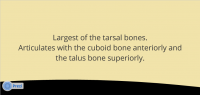
A calcaneus fracture is a heel bone fracture. The calcaneus, also called the heel bone, is the largest of the tarsal bones. It is situated at the lower and back part of the foot, forming the heel.
Together with the talus, the calcaneus forms the subtalar joint. This joint allows inversion and eversion of the foot. The midtarsal joint is comprised of two joints: The talocalcaneonavicalar and the calcaneocuboid joint.
The calcaneus has four important functions:
1. Acts as a foundation and support for the body’s weight
2. Supports the lateral column of the foot and acts as the main articulation for inversion / eversion
3. Acts as a lever arm for the gastrocnemius muscle complex
4. Makes normal walking possible
Epidemiology[edit | edit source]
Calcaneal fractures are uncommon, but serious injuries and they frequently occur in young adult men.
The annual incidence of fracture was 11.5 per 100.000. It occurred 2.4 times more frequently in males than females. In males, the incidence was 16.5/100000/year, with a peak incidence between 20-29 years of age (21.6/100000/year). In females, the overall incidence was 6.26/100000/year, with a gradual increase in incidence towards the post-menopausal years.[1]
These fractures account for 2-3% of all fractures of the body and 60% of all tarsal fractures. [2]
Mechanism of Injury / Pathological Process[edit | edit source]
Calcaneal fractures are mostly the result of a traumatic incident and high impact situation. The greater part of fractures (71,5%)[3] are sustained in falls from a height. The small amount of 18.8% of fractures occurred in the workplace. Calcaneal fractures can also occur with less severe accidents like an ankle sprain or a stress fracture.
Mostly, the injuries occur in isolation. Most seen concomitant injuries were lower limb (13.2%) or spinal injuries (6.3%).
These calcaneal fractures can be intra-articular or extra-articular. Involving one or more of the three subtalar articulating facets, 75% of all calcaneal fractures are intra-articular. These fractures have a poorer prognosis. Fractures of the calcaneal body, anterior process, sustentaculum tali, and superior tuberosity are known as extra-articular fractures and don’t involve the joint.
Characteristics / Clinical Presentation[edit | edit source]

There are certain characteristics of a calcaneal fracture:
• Sudden pain in the heel, most importantly pressure pain.
• Swelling in the heel area
• Bruising of the heel and ankle
• Generalized pain in the heel area that usually develops slowly (over several days to weeks): typically for stress fractures
• Edema
• A hematoma or pattern of ecchymosis extending distally to the sole of the foot.
• Deformity of the heel or plantar arch: Secondary to the displacement of the lateral calcaneal border outward, there is a possible widening or broadening of the heel.
• Inability or difficulty to bear weight on affected side[4]
• Limited or absent inversion / eversion of the foot
• Decreased Böhler or “tuber-joint” angle
• CT scan: Diverse views, both axial and coronal views can classify the degree of injury to the posterior facet and lateral calcaneal wall.
• X-rays or Radiographs:
o Axial x-ray: Determines primary fracture line and displays the body, tuberosity, middle and posterior facets
o Lateral x-ray: Determines Böhler angle[5]
o Oblique / Broden’s view: Determines the degree of displacement of the primary fracture line
• Heel tenderness
• Difficulty walking:
o Inability to walk
o Inability to move the foot
Differential Diagnosis[edit | edit source]
• Heel pain
• Baxter's nerve entrapment: An entrapment of the recurrent branch of the posterior tibial nerve
• Calcaneal spurs
• Plantar fasciitis: Plantar fascial pain is specific to the bottom of the heel. An MRI can be used to differentiate a calcaneal fracture from plantar fascitis.
• Retrocalcaneal bursitis: This is the formation and inflammation of a bursa at the back of the heel between the heel bone and achilles tendon. Also called Albert's Disease.
• Rheumatoid Arthritis
• Septic Arthritis
• Tarsal Tunnel Syndrome: The pain of this syndrome doesn’t decrease with rest. Other symptoms are numbness or tingling of the toes.
• Ankle instability[6]
Management / Interventions[edit | edit source]
Treatment of calcaneal fractures depends on the type of fracture and the extent of the injury.
Operative care[7][edit | edit source]
For the majority of patients, surgery is the correct form of treatment. The goal of surgery is to restore the correct size and structure of the heel. Intra-articular fractures are often treated operatively. This is possible by performing an open reduction and internal fixation of the fracture. These procedures are performed through an incision on the outside of the heel. The calcaneus is put together and held in place with a metal plate and multiple screws. This procedure decreases the possibility of developing arthritis and maximizes the potential for inversion and eversion of the foot.
Extra-articular fractures are generally treated conservatively.
| [8] | [9] |
Non-operative care[edit | edit source]
R.I.C.E.:
- Rest: The affected foot must rest and the patient is not allowed to use the foot. This is to allow the fracture to heal.
- Ice: Several times a day the patient has an ice treatment to reduce inflammation, swelling and pain.
- Compression: Bandage / Compression stocking
- Elevation: The initial management is to reduce the swelling with rest in bed with the foot slightly above heart level.
Immobilisation:
Partial or complete immobilisation is used if the fracture has not displaced the bone. Usually a cast is used to keep the fractured bone from moving. In the cast, the ankle is in neutral position and sometimes in slight eversion.
To avoid weight bearing, crutches may be needed.
Postoperative management[edit | edit source]
| [10] |
Physical Therapy:
After the surgery, active range of motion exercises may be practiced with small amounts of movement for all joints of the foot and ankle. These exercises are used to maintain and regain the ankle joint movement. When needed for the involved lower extremity, the patient may continue with elevation, icing and compression. During the therapy, the patient will progress to gradual weight bearing. Patients may find this very difficult and painful. The physiotherapist conducts joint mobilisation to all hypomobile joints.
During the treatment, progressive resisted strengthening of the gastrocnemius muscles is done by weighted exercises, toe-walking, ascending and descending stairs and plyometric exercises. When the fracture is healed, the physiotherapist will progress the weight bearing in more stressful situations. This therapy consists of gait instruction and balance practice on different surfaces.
Resources[edit | edit source]
http://ezinearticles.com/?Rehabilitation-After-Calcaneal-Fractures&id=4082480
http://orthopedics.about.com/od/footanklefractures/a/calcaneus.htm
http://xnet.kp.org/socal_rehabspecialists/ptr_library/09FootRegion/31Foot-CalcanealFracture.pdf
http://www.healthstatus.com/articles/Calcaneal_Fractures.html
Presentations[edit | edit source]
 |
Calcaneal Fractures
This presentation, created by Alice Thompson, provides an interactive insight into presentation, causes and types of calcaneal fractures as well as the evidence base for treatment options. |
Alice Thompson. Calcaneal Fractures: An interactive insight into the presentation, causes and types of calcaneal fractures. Also looking at the evidence base for treatment options. [Accessed 14/1/12 at http://prezi.com/htzzh_lneqpu/calcaneal-fractures/]
References[edit | edit source]
- ↑ Mitchell MJ, McKinley JC, Robinson CM. The epidemiology of calcaneal fractures. Royal Infirmary of Edinburgh, 2009 Dec;19(4):197-200. (Level Of Evidence: B)
- ↑ http://xnet.kp.org/socal_rehabspecialists/ptr_library/09FootRegion/31Foot-CalcanealFracture.pdf
- ↑ Mitchell MJ, McKinley JC, Robinson CM. The epidemiology of calcaneal fractures. Royal Infirmary of Edinburgh, 2009 Dec;19(4):197-200. (Level Of Evidence: B)
- ↑ B. Kienast B, Gille J, Queitsch C, Kaiser MM, Thietje R,Juergens C and Schulz AP. Early Weight Bearing of Calcaneal Fractures Treated by Intraoperative 3D-Fluoroscopy and Locked-Screw Plate Fixation. The Open Orthopaedics Journal, 2009, 3, 69-74 69. (Level Of Evidence: A2) http://www.ncbi.nlm.nih.gov/pmc/articles/PMC2738828/pdf/TOORTHJ-3-69.pdf
- ↑ Grala P, Machyñska-Buæko Z, Kierzynka G. Surgical treatment of articular calcaneal fractures. Ortopedia Traumatologia Rehabilitacja Medsportpress, 2007; 1(6); Vol. 9, 89-97. (Level Of Evidence: A2)
- ↑ http://www.eorif.com/AnkleFoot/CalcaneousFx.html#Anchor-Associated-44867
- ↑ Grala P, Machyñska-Buæko Z, Kierzynka G. Surgical treatment of articular calcaneal fractures. Ortopedia Traumatologia Rehabilitacja Medsportpress, 2007; 1(6); Vol. 9, 89-97. (Level Of Evidence: A2)
- ↑ Shajil K. CALCANEUM FRACTURE ORIF VIDEO . Available from: http://www.youtube.com/watch?v=gngTOOFmgJM [last accessed 09/02/13]
- ↑ Legal Animations. Open Reduction Internal Fixation Calcaneus Fractures . Available from: http://www.youtube.com/watch?v=UTo0c_6YTK4 [last accessed 09/02/13]
- ↑ firefighterretired. Calcaneus Fracture Physical Therapy (Nov 17, 2011). Available from: http://www.youtube.com/watch?v=ss5pF-LBn_Y [last accessed 09/02/13]

