Lunate Instability: Difference between revisions
Jamie Keller (talk | contribs) No edit summary |
Jamie Keller (talk | contribs) No edit summary |
||
| Line 52: | Line 52: | ||
A patient presenting with lunate instability will most likely have a history of trauma to the wrist that has resulted in some type of lesion to the stabilizing ligaments of the carpal bones. Additionally, a patient with a history of certain degenerative inflammatory diseases such as chondrocalcinosis, gout, aseptic bone necrosis of the scaphoid or lunate bones, or Madelung deformity (a form of misalignment) can also lead to carpal instability. <ref name="Redeker, Vogt">Redeker J, Vogt PM. Carpal instability. Chirurg. 2011 Jan; 82(1):85-93.</ref> The patient will have complaints of pain in the involved wrist, specifically around the scaphoid and lunate bones. The pain can be chronic if there is no history of trauma, or acute if the injury is the result of trauma. The pain is typically exacerbated by overuse activities, such as typing on a computer or doing push-ups, or gripping. The patient may deny “clicking” in the wrist, but report a feeling of “instability” with some activities. | A patient presenting with lunate instability will most likely have a history of trauma to the wrist that has resulted in some type of lesion to the stabilizing ligaments of the carpal bones. Additionally, a patient with a history of certain degenerative inflammatory diseases such as chondrocalcinosis, gout, aseptic bone necrosis of the scaphoid or lunate bones, or Madelung deformity (a form of misalignment) can also lead to carpal instability. <ref name="Redeker, Vogt">Redeker J, Vogt PM. Carpal instability. Chirurg. 2011 Jan; 82(1):85-93.</ref> The patient will have complaints of pain in the involved wrist, specifically around the scaphoid and lunate bones. The pain can be chronic if there is no history of trauma, or acute if the injury is the result of trauma. The pain is typically exacerbated by overuse activities, such as typing on a computer or doing push-ups, or gripping. The patient may deny “clicking” in the wrist, but report a feeling of “instability” with some activities. | ||
<h2> Diagnostic Procedures </h2> | |||
<p><i>Physical Exam:</i> | |||
</p><p>The wrist will most likely not be edematous or tender when palpated if there has been no history of trauma. Full range of motion may be preserved with a chronic type of injury. A tender dorsal scapholunate interval may be evident but it may appear normal in radiographs and the scaphoid shift test will either cause a “clunking” sound or it will be painful to the patient.<span class="fck_mw_ref" _fck_mw_customtag="true" _fck_mw_tagname="ref" name="Dyer">Dyer GS. Predynamic scapholunate instability. J Hand Surg Am. 2010 Nov;35(11):1879-1880.)</span> | |||
</p><p><br /> | |||
The wrist will most likely not be edematous or tender when palpated if there has been no history of trauma. Full range of motion may be preserved with a chronic type of injury. A tender dorsal scapholunate interval may be evident but it may appear normal in radiographs and the scaphoid shift test will either cause a “clunking” sound or it will be painful to the patient.<ref name="Dyer">Dyer GS. Predynamic scapholunate instability. J Hand Surg Am. 2010 Nov;35(11):1879-1880.)</ | </p><p><i>Palpation of Scapholunate Articulation:</i> | ||
</p><p>Palpate Lister’s tubercle on the distal radius. Move slightly distal to the tubercle and you will find the scapholunate joint. | |||
<br> | </p><p><img src="/images/5/5b/Lister%27s_Tubercle.jpg" _fck_mw_filename="Lister's Tubercle.jpg" _fck_mw_location="center" alt="" class="fck_mw_center" /> | ||
</p><p><br /> | |||
</p><p> <a href="http://www.aafp.org/afp/2004/0415/p1941.html">http://www.aafp.org/afp/2004/0415/p1941.html</a> | |||
</p><p><br /> | |||
Palpate Lister’s tubercle on the distal radius. Move slightly distal to the tubercle and you will find the scapholunate joint. | </p><p><i>Scaphoid Shift Test (Watson Test)</i> | ||
</p><p>-How to Perform: <br /> 1.) Examiner palpates and provides compression force over scaphoid (thumb on volar surface of hand—use anatomical snuff box for reference—and first finger on palmar surface) with one hand, while grasping the hand at the level of the metacarpal with the other.<br /> 2.) Examiner passively moves patient’s wrist into ulnar deviation and slight extension.<br /> 3.) Examiner passively and slowly moves patient’s wrist into radial deviation and slight flexion.<br /> 4.) Examiner releases compression on the scaphoid.<br />-Interpreting Findings: <br /> The test is positive if a “thunk” is produced OR the patient’s symptoms are reproduced when compression of the scaphoid is released.<br />-Diagnostic Accuracy<span class="fck_mw_ref" _fck_mw_customtag="true" _fck_mw_tagname="ref" name="LaStayo, Howell">LaStayo P, Howell J. Clinical provocative tests used in evaluating wrist pain: a descriptive study. J Hand Ther. 1995;8(1):10--17.</span>: | |||
</p><p> Sensitivity = .69<br /> Specificity = .66<br /> +LR = 2.0<br /> | |||
</p><p><br /> | |||
<br> | </p><p><i>Diagnostic Exam:</i> | ||
</p><p>Scapholunate Interval—This measurement is the distance between the proximal medial corner of the lunate and the proximal lateral corner of the scaphoid. A separation of greater than 2 mm may be indicative of scapholunate instability and it is referred to as the “Terry Thomas sign.” This can be measured through radiographs and should be compared with the patient’s uninvolved side. A “clenched-fist” view in a radiograph may help accentuate the space between the two bones. | |||
| </p><p><img src="/images/a/a5/Terry_Thomas-Cartoon.jpg" _fck_mw_filename="Terry Thomas-Cartoon.jpg" _fck_mw_location="center" alt="" class="fck_mw_center" /> | ||
</p><p> Dorsal View | |||
<br> | </p><p> <a href="http://www.wikiradiography.com/page/The+Terry-Tomas+Sign">http://www.wikiradiography.com/page/The+Terry-Tomas+Sign</a> | ||
</p><p><img src="/images/6/62/Terry_Thomas-Radiograph.jpg" _fck_mw_filename="Terry Thomas-Radiograph.jpg" _fck_mw_location="center" alt="" class="fck_mw_center" /> | |||
</p><p> Dorsal View-Radiograph | |||
</p><p> <a href="http://radiopaedia.org/articles/scapholunate-dissociation">http://radiopaedia.org/articles/scapholunate-dissociation</a> | |||
-How to Perform: <br> 1.) Examiner palpates and provides compression force over scaphoid (thumb on volar surface of hand—use anatomical snuff box for reference—and first finger on palmar surface) with one hand, while grasping the hand at the level of the metacarpal with the other.<br> 2.) Examiner passively moves patient’s wrist into ulnar deviation and slight extension.<br> 3.) Examiner passively and slowly moves patient’s wrist into radial deviation and slight flexion.<br> 4.) Examiner releases compression on the scaphoid.<br>-Interpreting Findings: <br> The test is positive if a “thunk” is produced OR the patient’s symptoms are reproduced when compression of the scaphoid is released.<br>-Diagnostic Accuracy< | </p> | ||
Sensitivity = .69<br> Specificity = .66<br> +LR = 2.0<br> | |||
< | |||
Scapholunate Interval—This measurement is the distance between the proximal medial corner of the lunate and the proximal lateral corner of the scaphoid. A separation of greater than 2 mm may be indicative of scapholunate instability and it is referred to as the “Terry Thomas sign.” This can be measured through radiographs and should be compared with the patient’s uninvolved side. A “clenched-fist” view in a radiograph may help accentuate the space between the two bones. | |||
Dorsal View | |||
| |||
Dorsal View-Radiograph | |||
| |||
== Outcome Measures == | == Outcome Measures == | ||
Revision as of 05:34, 20 March 2011
Be the first to edit this page and have your name permanently included as the original editor, see the editing pages tutorial for help.
|
Original Editor - Your name will be added here if you created the original content for this page. Lead Editors - Your name will be added here if you are a lead editor on this page. Read more. |
Clinically Relevant Anatomy
[edit | edit source]
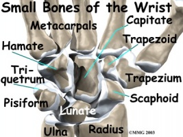
The lunate is one of the eight carpal bones located in the wrist. These eight bones are arranged into two rows with four bones in each. The proximal row, laterally to medially, consists of the scaphoid, lunate, triquetrum, and pisiform. The distal row, going in the same direction, consists of the trapezium, trapezoid, capitate and hamate. The lunate articulates laterally with the scaphoid, medially with the triquetrum, and inferiorly with the capitate and hamate.
Palmar View
http://handsonhealingpt.com/article.php?aid=384
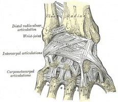
There are several different ligaments that attach to the dorsal and palmar surfaces of the carpal bones and hold them together and provide stability in the wrist. Most of the ligaments are named with respect to the two bones they connect.
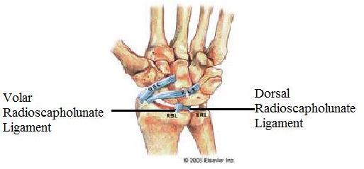
Radioscapholunate Ligaments
Dorsal View with Ligaments
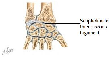
Scapholunate Interosseous Ligaments
Netter FH. Atlas of human anatomy: 4th edition. 2006: 452-456.
Mechanism of Injury / Pathological Process
[edit | edit source]
Scapholunate instability (the most common instability in the wrist) occurs when a person experiences falls on an outstretched hand (FOOSH) with the wrist positioned in extension, ulnar deviation, and intercarpal supination.[1][2] Scapholunate instability is considered to be present if at least two of the following three ligaments are injured: volar radioscapholunate ligament, scapholunate interosseous ligament, and dorsal scapholunate ligament. [3] If not treated, dynamic scapholunate instability can progress to rotatory subluxation of scaphoid, scapholunate dissociation, dorsal intercalated segment instability, and then scapholunate advanced collapse.[2]
Clinical Presentation[edit | edit source]
A patient presenting with lunate instability will most likely have a history of trauma to the wrist that has resulted in some type of lesion to the stabilizing ligaments of the carpal bones. Additionally, a patient with a history of certain degenerative inflammatory diseases such as chondrocalcinosis, gout, aseptic bone necrosis of the scaphoid or lunate bones, or Madelung deformity (a form of misalignment) can also lead to carpal instability. [4] The patient will have complaints of pain in the involved wrist, specifically around the scaphoid and lunate bones. The pain can be chronic if there is no history of trauma, or acute if the injury is the result of trauma. The pain is typically exacerbated by overuse activities, such as typing on a computer or doing push-ups, or gripping. The patient may deny “clicking” in the wrist, but report a feeling of “instability” with some activities.
Diagnostic Procedures
Physical Exam:
The wrist will most likely not be edematous or tender when palpated if there has been no history of trauma. Full range of motion may be preserved with a chronic type of injury. A tender dorsal scapholunate interval may be evident but it may appear normal in radiographs and the scaphoid shift test will either cause a “clunking” sound or it will be painful to the patient.Dyer GS. Predynamic scapholunate instability. J Hand Surg Am. 2010 Nov;35(11):1879-1880.)
Palpation of Scapholunate Articulation:
Palpate Lister’s tubercle on the distal radius. Move slightly distal to the tubercle and you will find the scapholunate joint.
<img src="/images/5/5b/Lister%27s_Tubercle.jpg" _fck_mw_filename="Lister's Tubercle.jpg" _fck_mw_location="center" alt="" class="fck_mw_center" />
<a href="http://www.aafp.org/afp/2004/0415/p1941.html">http://www.aafp.org/afp/2004/0415/p1941.html</a>
Scaphoid Shift Test (Watson Test)
-How to Perform:
1.) Examiner palpates and provides compression force over scaphoid (thumb on volar surface of hand—use anatomical snuff box for reference—and first finger on palmar surface) with one hand, while grasping the hand at the level of the metacarpal with the other.
2.) Examiner passively moves patient’s wrist into ulnar deviation and slight extension.
3.) Examiner passively and slowly moves patient’s wrist into radial deviation and slight flexion.
4.) Examiner releases compression on the scaphoid.
-Interpreting Findings:
The test is positive if a “thunk” is produced OR the patient’s symptoms are reproduced when compression of the scaphoid is released.
-Diagnostic AccuracyLaStayo P, Howell J. Clinical provocative tests used in evaluating wrist pain: a descriptive study. J Hand Ther. 1995;8(1):10--17.:
Sensitivity = .69
Specificity = .66
+LR = 2.0
Diagnostic Exam:
Scapholunate Interval—This measurement is the distance between the proximal medial corner of the lunate and the proximal lateral corner of the scaphoid. A separation of greater than 2 mm may be indicative of scapholunate instability and it is referred to as the “Terry Thomas sign.” This can be measured through radiographs and should be compared with the patient’s uninvolved side. A “clenched-fist” view in a radiograph may help accentuate the space between the two bones.
<img src="/images/a/a5/Terry_Thomas-Cartoon.jpg" _fck_mw_filename="Terry Thomas-Cartoon.jpg" _fck_mw_location="center" alt="" class="fck_mw_center" />
Dorsal View
<a href="http://www.wikiradiography.com/page/The+Terry-Tomas+Sign">http://www.wikiradiography.com/page/The+Terry-Tomas+Sign</a>
<img src="/images/6/62/Terry_Thomas-Radiograph.jpg" _fck_mw_filename="Terry Thomas-Radiograph.jpg" _fck_mw_location="center" alt="" class="fck_mw_center" />
Dorsal View-Radiograph
<a href="http://radiopaedia.org/articles/scapholunate-dissociation">http://radiopaedia.org/articles/scapholunate-dissociation</a>
Outcome Measures[edit | edit source]
Potential self report measures for individuals presenting with wrist pain are listed below.
-Disability of Arm, Shoulder, and Hand questionnaire (DASH)
-Quick DASH –This outcome measure is a shortened version of the DASH and is used to determine the patient’s physical function and symptoms.
-Brigham and Women’s Hospital Carpal Tunnel Questionnaire (CTQ) – This outcome measure is intended to assess functional status along with severity of symptoms in patients with carpal tunnel syndrome.
-Patient-Rated Wrist Evaluation Score (PRWE) – This outcome measure attempts to quantify wrist pain and disability, according to the patient, to assess the potential outcome for patients with distal radius fractures.
-Gartland and Werley Score – This is one of the most widely used outcome measures used in the clinic to evaluate wrist and hand function.
Management / Interventions
[edit | edit source]
add text here relating to management approaches to the condition
Differential Diagnosis
[edit | edit source]
As with all other cases, other potential diagnoses must be ruled out before a final diagnosis of lunate instability can be determined. These other potential diagnoses for the wrist can be categorized depending on the location of the patient’s pain/symptoms.
Radial Wrist Pain:
First Carpometacarpal Osteoarthritis
DeQuervain’s (Stenosing tenosynovitis of the tendons on the lateral border of the anatomical snuffbox)
Non-Specific Wrist Pain:
Mechanical Wrist Pain – Individuals who have symptoms suspicious of scapholunate instability with unremarkable findings on further work up may have an injury such as wrist strain or sprain, joint dysfunction, or repetitive strain injuries. Tests that differentiate between these diagnoses often do not have known reliability or diagnostic accuracy. Thus, if a specific diagnosis cannot be made with certainty, and red flags are ruled less likely, the most accurate diagnosis may be referred to as “mechanical wrist pain,” which is largely a diagnosis of exclusion. Intervention would consist of addressing the identified impairments such as joint hypomobility, decreased muscle length, or decreased strength.
Key Evidence[edit | edit source]
Outcome measures for wrist and hand – which one to choose?
Scapholunate stabilization with dynamic extensor carpi radialis longus tendon transfer
Controlled active mobilization after dorsal capsulodesis to correct capitolunate dissociation
Resources
[edit | edit source]
Links to relevant documents are cited throughout this page.
Case Studies[edit | edit source]
Scapholunate instability following dorsal wrist ganglion excision: a case report
References[edit | edit source]
References will automatically be added here, see adding references tutorial.
- ↑ Mayfield JK, Johnson RP, Kilcoyne RK. Carpal dislocations: pathomechanics and progressive perilunar instability. J Hand Surg Am. 1980;5 (3): 226-41
- ↑ 2.0 2.1 Wheeless C. Scapholunate Instability. Duke Orthopaedics presents Wheeless' Textbook of Orthopaedics Web site. http://www.wheelessonline.com/ortho/scapholunate_instability. Updated 2011. Accessed 03/02, 2011
- ↑ Scapholunate Instability. UW MSK Resident Projects Web site. http://uwmsk.org/residentprojects/scapholunate.html. Updated 2005. Accessed 03/02, 2011.
- ↑ Redeker J, Vogt PM. Carpal instability. Chirurg. 2011 Jan; 82(1):85-93.


