Metacarpal Fractures
Original Editor - --Marie Avau , Debby Decock, Farrie Bakalli, Margaux Jacobs
1. Search strategy
[edit | edit source]
To get a first global view on our topic, we inserted words like metacarpal fracture, anatomy of the hand, fractures of the metacarpals, … in Pedro and PubMed. Then, to complete every part of our subject, we continued with terms like: diagnosis of metacarpal fracture, treatment, epidemiology and characteristics and physical therapy after metacarpal fractures. With the articles and links we found with this keywords, we completed a big part of our task. After this, some of the items were still incomplete or empty, so we did a more thorough search for medical and physical therapy, examination, outcome measures and recent related research. We putted words like treatment of metacarpal fractures, outcome measures of hand fractures and physical therapy metacarpal fractures in the available databases (Pedro, PubMed, research gate, springer link, Medscape, …)
2. Definition/ Description
[edit | edit source]
A metacarpal fracture is a break in one of the five metacarpal bones of either hand. Metacarpal fractures are categorized as being fractures of the head, neck, shaft, and base (from distal at the metacarpal phalangeal joint to proximal at the wrist). [1,2 Blomberg et al, level 5,8]
Thereby we also have the Boxer fracture, this is another name for a fracture of the fourth or fifth metacarpal. This is one of the most common metacarpal fractures, in contrast with the fractures of the thumb (Bennett’s and Rolando’s fracture). [2] (Blomberg et al, level 5)
3. Clinically Relevant Anatomy
[edit | edit source]
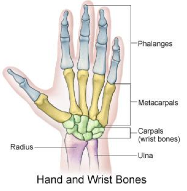
The hand is composed of 19 bones (5 metacarpals and 14 phalanges), more than 30 tendinous insertions and numerous complex structures. The metacarpals are long, thin bones which are located between the carpal bones in the wrist and the phalanges in the digits.[4,17]
Each are comprised of a base, shaft, and head. The proximal bases of the metacarpals articulate with the carpal bones, and the distal heads of the metacarpals articulate with the proximal phalanges and form the knuckles. The 1st metacarpal (of the thumb) is the thickest and shortest of these bones. The 3rd metacarpal is distinguished by a styloid process on the lateral side of its base. Soft tissues generally involved with fractures include cartilage, joint capsule, ligaments, fascia, and the dorsal hood fibers. With severe polytrauma cases, the tendons and nerves adjacent to the fracture can also be injured. [48]
Muscles in the metacarpal region: [3,14]
There are three palmar and four dorsal interossei muscles that arise from metacarpal shafts. We can also find the insertion of extensor carpi radialis longus and brevis at the base of the second and third metacarpal. This muscles assist with wrist extension and radial flexion.
In the wrist we can thereby also find the extensor carpi ulnaris muscle, which inserts on the base of metacarpal five. His function is to extend and fixe the wrist when the digits are being flexed and assists with ulnar flexion of wrist.
The abductor pollicis longus is situated in the area of the thumb. It inserts on the trapezium and base of metacarpal I and is responsible for abduction of the thumb in the frontal plane and extends the thumb at the carpometacarpal joint. At this site we can also find the opponens pollicis, which inserts on metacarpal I and functions as a flexor of metacarpal I to oppose the thumb to the fingertips.
The final muscle at this site is the opponens digiti minimi, it inserts on the medial surface of metacarpal V, flexes metacarpal V at carpometacarpal joint.


