Search results
Page title matches
File:Knee-joint-meniscus.jpg (324 × 345 (29 KB)) - 21:17, 30 September 2013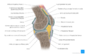
File:Knee joint - Kenhub.png Knee joint(1,400 × 896 (728 KB)) - 14:10, 16 March 2022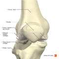
File:Knee joint anterior aspect Primal.png (660 × 660 (279 KB)) - 21:36, 7 December 2020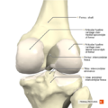
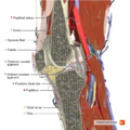
File:Knee joint posterior aspect Primal.png (660 × 660 (289 KB)) - 21:37, 7 December 2020- ...systematic approach to diagnosing common musculoskeletal conditions of the knee.] SMRJ 2020; 4(2). </ref> ...rmth, joint pain and joint deformity.<ref name="Stephen">Stephen J, et al. Joint tenderness overview. DSHI systems 2010(5)</ref>16 KB (2,455 words) - 18:42, 24 May 2022
- [[File:Total knee arthroplasty.jpg|right|frameless|312x312px]] ...ery are removed. New prosthetic pieces are then replaced within knee joint joint. The components consist of the femoral condyle component, the tibial platea10 KB (1,456 words) - 08:40, 5 April 2024
File:Sagittal section of the knee joint Primal.png (660 × 660 (607 KB)) - 21:46, 7 December 2020- ...ritical mechanism that play an important role in terminal extension of the knee. * There is an observable rotation of the knee during flexion and extension.6 KB (944 words) - 17:36, 17 January 2023
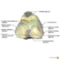
File:Ligaments of the knee joint superior aspect Primal.png (990 × 990 (409 KB)) - 21:37, 7 December 2020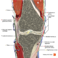
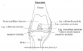
File:Coronal section of the knee joint 1 Primal.png (660 × 660 (658 KB)) - 21:44, 7 December 2020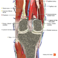
File:Coronal section of the knee joint 2 Primal.png (660 × 660 (629 KB)) - 21:45, 7 December 2020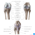
File:Overview of the knee joint (anterior and posterior views) - Kenhub.png Overview of the knee joint (anterior and posterior views)(1,400 × 1,400 (1.04 MB)) - 04:53, 30 March 2022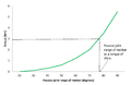
File:Torque displacement curve for the ankle joint with the knee extended.png (1,159 × 753 (16 KB)) - 01:15, 23 June 2019
File:Figure 3. Torque and displacement curve for the ankle joint with the knee extended.pdf Figure 3. Torque and displacement curve for the ankle joint with the knee extended(0 × 0 (94 KB)) - 00:46, 21 June 2019


_-_Kenhub.png)

Page text matches
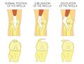
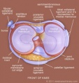
File:Patella Dislocation and Subluxation - Shutterstock Image - ID 637362388.jpg ...a or kneecap. Anatomy of human knee joint, front view of straight and bent knee. Aksanaku. Shutterstock.com(1,300 × 1,000 (382 KB)) - 12:06, 14 June 2022- The Knee joint is a complex joint and the the largest in the body [[Category:Joints]] [[Category:Knee - Anatomy]]5 members (0 subcategories, 0 files) - 17:28, 28 August 2019
- ...body forward. This category contains pages relating to the anatomy of the knee. [[Category:Anatomy]] [[Category:Knee]]38 members (4 subcategories, 0 files) - 01:05, 29 August 2019

File:Figure 5.0.jpg Manual therapy at knee joint(276 × 183 (5 KB)) - 00:47, 8 May 2018- #REDIRECT [[Joint Line Tenderness of the Knee]]47 bytes (7 words) - 03:54, 16 September 2014
File:Heleen fig1.jpg anatomical structure of the knee joint, fig 1(800 × 484 (28 KB)) - 16:48, 30 December 2010- ...body forward. This category contains all the articles that relate to the Knee.<br>166 members (7 subcategories, 0 files) - 17:11, 18 August 2019
- ...mitations not allowing the clinician to analyse the ROM and track the knee joint during eg [[Walking - Muscles Used|walking]] or maximum squat. Motion captu Knee flexion2 KB (230 words) - 10:13, 16 April 2022
- ...a tap test or ballottement test''' is used to examine the knee swelling or knee effusion. This test is also know as '''dancing knee sign.'''980 bytes (148 words) - 19:38, 15 August 2020
- ...hysical therapy to provide active and passive mobilization of the affected joint.[1] They often requires an extensive rehabilitation. [4] In most of the cas586 bytes (95 words) - 16:58, 20 May 2011
File:Condyles of femur LD.B.0190.004.L.jpg Condyles of femur. Interior of right knee joint, anterior view. Image #190-4(640 × 458 (110 KB)) - 18:48, 13 February 2020
File:Guideline for the management of knee and hip osteoarthritis .png management of hip and knee osteoarthritis. The objective of this guideline is to present the best ava other than joint replacement for the hip and knee.(1,920 × 1,080 (344 KB)) - 01:38, 26 February 2019File:Figure 3. Torque and displacement curve for the ankle joint with the knee extended.pdf Figure 3. Torque and displacement curve for the ankle joint with the knee extended(0 × 0 (94 KB)) - 00:46, 21 June 2019
File:Condyles of femur with cruciate ligaments LD.B.0190.005.L.jpg Interior of right knee joint, anterior cruciate ligament . Basset image ID Image #190-5(640 × 458 (111 KB)) - 18:51, 13 February 2020File:Knee joint - Kenhub.png Knee joint(1,400 × 896 (728 KB)) - 14:10, 16 March 2022File:Overview of the knee joint (anterior and posterior views) - Kenhub.png Overview of the knee joint (anterior and posterior views)(1,400 × 1,400 (1.04 MB)) - 04:53, 30 March 2022- ...ined from a french word. The test is usually used to check for [[Knee|knee joint]] effusion. ...sed 21 January 2020)</ref>. Then the test is repeated on the contralateral joint for comparison.2 KB (262 words) - 18:37, 3 February 2020
- ...swelling to the joint the morning after the injury. She does not feel her knee is improving and is now off work. ...for more than 30 minutes and up and down the stairs is also difficult. The joint is not locking / clicking or giving way.1 KB (220 words) - 00:02, 12 March 2018
- [[File:Sagittal section of the knee joint Primal.png|thumb|Knee Joint ]] ...]] (Hoffa’s disease) is one of the causes of [[Anterior Knee Pain|anterior knee pain]].<ref name=":0">Kumar D, Alvand A, Beacon JP. Impingement of infrapat3 KB (385 words) - 02:48, 9 January 2022
- ...anni CW, Kuo R, Tejwani N, et al: Isolated gastrocnemius tightness. J Bone Joint Surg Am 2002;84(6):962-970.</ref>. ...degrees. The test is considered positive when DF at the AJ is greater with knee flexed than extended.3 KB (368 words) - 12:24, 7 April 2023


