Structure and Function of the Cervical Spine: Difference between revisions
No edit summary |
No edit summary |
||
| Line 8: | Line 8: | ||
*'''Inferior cervical group:''' made up by C3 to C7. | *'''Inferior cervical group:''' made up by C3 to C7. | ||
{{#ev:youtube|RNUpMNd_u1U|250}} <div class="row"><div class="col-md-6 col-md-offset-3"><div class="text-right"><ref>Randale Sechrest. Cervical Spine Anatomy (eOrthopod). Available from: http://www.youtube.com/watch?v=RNUpMNd_u1U[last accessed 04/06/18]</ref></div></div></div> | |||
===== '''Superior cervical Group:''' ===== | ===== '''Superior cervical Group:''' ===== | ||
Revision as of 18:49, 4 June 2018
Introduction:[edit | edit source]
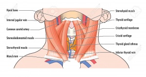
Cervical spine spine starts at base of skull superiorly,ends above thoracic spine inferiorly and it could be called (neck).Neck joins head with trunk and limbs and it works as a major conduit for structures between them. Flexibility of neck movement allows and maximise necessary positions to head functions and its sensory organs. There are many important structures in neck area like nerves, muscles,arteries,veins, vertebrates, lymphatics,glands, oesophagus and trachea. Due to these important structures and lack of bony protection in neck area,it is considered as a region of vulnerability. The main arterial blood supply for head and neck is carotid arteries and principal venous drainage is jugular veins.Carotid and Jugular are commonly injured in penetrating wound of neck. Brachial plexues of nerves originates in neck and travels inferiorly to upper limb. In anterior aspect of neck,there is thyroid cartilage (largest cartilage of thyroid and trachea)[1].
Bony Structure of Neck:[edit | edit source]
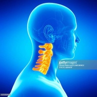
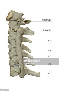
It consists of 7 cervical vertebrates from C1 to C7, hyoid bone,manubrium of sternum and clavicles[1]. Cervical spine has a lordotic curve ( C shaped curve )[2]. According to peculiarities of cervical vertebrates,it could be divided in to two groups:[3][4]
- Superior cervical group: made up by C1 and C2.
- Inferior cervical group: made up by C3 to C7.
Superior cervical Group:[edit | edit source]
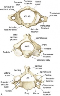
C1 called atlas and C2 called axis and they have some differences than other vertebrae, Atlas C1 is ring like shape , it has no body and no spinous process,
C2 looks like other cervical vertebrae but most distinctive feature is odontoid process or tooth which placed vertically on superior surface of vertebral body[6] with two articular facets( anterior and posterior) which articulate with atlas bone and atlas transverse ligament. C2 has smaller and triangular vertebral foramen[7].
Inferior Cervical Group:[edit | edit source]
It is made up of 5 vertebrea from C3 TO C7 with similar characteristics like smaller vertebral body with two rafes at lateral sides directed Superiorly called spinous processes,two pedicles directed backwards and transverse process located anteriorly.C7 may be considered typical or atypical but has two distinct features. The first is that unlike the rest of the cervical vertebrae, is that the vertebral artery does not traverse the transverse foramen. The second is that it contains a long spinous process, also known as “vertebra prominens.”[8] .
For more details about cervical vertebrae anatomy,please follow this link : https://www.physio-pedia.com/index.php?title=Cervical_Vertebrae&oldid=174634.[edit | edit source]
Cervical spine joints:[edit | edit source]
Joints between vertebrae are made for spine mobility, Movements of superior cervical spine joint and inferior cervical spine joints functionally completing each others allowing movements like rotation,flexion,extension and inclination of head.[6]
Superior cervical spine joints:[edit | edit source]
- Atanto-occipital joint :is aligned to permit movement of nodding[9] (Flexion and extension) and turning(Lateral flexionand rotation)peculiar at this level. For more details please follow this link, Index.php?title=Atlanto-occipital joint&oldid=184161.
- Atlanto- Axial joint : It compromise 3 synovial joints one central Atlanto- Odontoid joint and 2 Lateral Atlanto- Axial joints.Allows about 50% of cervical rotation,flextion about 10 degrees and limited extention. For more details please follow this link: Index.php?title=Atlanto-axial joint&oldid=174590.
Inferior Cervical spine joints: joints between adjacent cervical vertebrae[edit | edit source]
Intervertebral (inter-body)joints: below C2 adjacent cervical vertebrae are linked by intervertebral disc at inter-body joint which is termed symphysis .Discs allow and restrain movement .These joints are saddle articuation.It is reinforced by anterior longitudinal ligament anteriorly and Posterior longitudinal ligament Posteriorly,[10]
Apophyseal Joints: it is formed by articulation of inferior facets of vertebrae and superior facet of adjacent vertebrae.Direction and range of movement of these joints depend on orientation of articular facets.These joints allow flexion, extension, rotation and lateral flexion.Degenerative changes at these joints are very common due to weight-bearing functions[10]. For more details please follow this link :: https://www.physio-pedia.com/index.php?title=Facet_Joints&oldid=182072
The Uncovertebral Joints (Joints of Luschka) : Opinions are divided whether these structures are joints or pseudoarthrosis. The clinical significance of these structures is the high tendency of developing degenerative changes that may impinge vertebral artery,cervical nerve roots or anterior part of spinal cord.They allow for flexion and extension and limit lateral flexion in the cervical spine.They prevent posterior linear translation movements of the vertebral bodies,Important in providing stability and guiding the motion of the cervical spine[11].For more details please follow this link:Index.php?title=Uncovertebral Joints&oldid=173523
Ligaments of Cervical Spine:[edit | edit source]
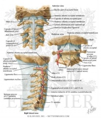
-Craniovertebral Ligaments: Stability of this region depends on integrity of ligaments of upper cervical spine and this has an important consideration in examining and treating cervical region.Ligaments from anterior to posterior:
- Anterior atlanto-occipital membrane: it connects between foramen-magnum above and atlas below, it continues with anterior longitudinal ligament.
- The Apical ligament: it is short and attaches anterior part of foramen- magnum.
- The Alar ligaments: they are symmetrically placed and inserted onto occipital -condyles. Rotation to right are limited to by left alar ligament and vice-verca. Damage of Alar ligaments by trauma or inflammatory disease can result in increase axial rotation between occiput and atlas and atlas and axis [10] for more details please follow link ; : https://www.physio-pedia.com/index.php?title=Alar_ligaments&oldid=184151.
- Membrane of Tectoria: Connecting posterior surface of body of axis to the basiocciput. It is prolongation of posterior longitudinal ligament and it is found in internal surface of vertebral canal For more details please follow link; Index.php?title=Tectorial membrane&oldid=171265.
- Transverse ligament of Atlas.
- Accessory Atanto- Axial Ligaments .
- Posteri Atlanto- Occipital Membrane.
- Lateral Atlanto- Occipital Ligamments.
Lower Cervical Ligaments:[edit | edit source]
- Anterior longitudinal Ligament : it is strong band lies anterior to vertebral body .It is relaxed in flexion and taut in extension.
- Posterior longitudinal Ligament: it is posterior to vertebral bodies and lies in vertebral canal.It stretches in neck flexion and relaxes in neck extension.
- The Ligamenta Flava : It is yellow elastic tissue , it connects laminae of adjacent vertebrae. It allows flexion to occur and prevent hyper- flexion by breaking movement at end of range.
- Ligamentum Nuchae: it is a fibro-elastic membrane which extends from occiput to spine of all cervical vertebrae. It helps in stability of head and neck especially in head flextion /acceleration injuries. It limits flextion and provides an attachment to Trapezius and Splenius capitis. For more details please follow link; Index.php?title=Ligamentum nuchae&oldid=172982.
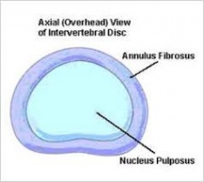
Inter-vertebral Discs: It make about 25% of cervical spine height ,no disc between occiput and C1 or between C1 and C2[12].Disc consists of both nucleus pulposus and annulus fibrosis.[edit | edit source]
Cervical Nerve Roots:[edit | edit source]
Although there are 7 cervical vertebraes,there are 8 nerve roots as there is a root between occiput and C1. The roots are named for the vertebrae below[12] .
References:[edit | edit source]
- ↑ 1.0 1.1 Lang, J., 1993. Clinical anatomy of the cervical spine. Stuttgart; New York: Thieme.
- ↑ Cervical Spine Anatomy. University of Maryland Clinical Center. http://www.umm.edu/programs/spine/health/guides/cervical-spine-anatomy. accessed Apirl 2018.
- ↑ Spine, S.C., 2015. Anatomy of the Cervical Spine. Cervical Spine: Minimally Invasive and Open Surgery, p.1.
- ↑ Kim, D.H., Vaccaro, A.R., Dickman, C.A., Cho, D., Lee, S. and Kim, I., 2013. Surgical Anatomy and Techniques to the Spine E-Book. Elsevier Health Sciences.
- ↑ Randale Sechrest. Cervical Spine Anatomy (eOrthopod). Available from: http://www.youtube.com/watch?v=RNUpMNd_u1U[last accessed 04/06/18]
- ↑ 6.0 6.1 Spine, S.C., 2015. Anatomy of the Cervical Spine. Cervical Spine: Minimally Invasive and Open Surgery, p.1.
- ↑ Ross, J.S. and Moore, K.R., 2015. Diagnostic Imaging: Spine E-Book. Elsevier Health Sciences.
- ↑ Panjabi MM , Duranceau J , Goel V , Oxland T , Takata K Spine [01 Aug 1991, 16(8):861-869]
- ↑ Jeffreys, E., 2013. Disorders of the cervical spine. Butterworth-Heinemann.
- ↑ 10.0 10.1 10.2 Middleditch, A. and Oliver, J., 2005. Functional anatomy of the spine. Elsevier Health Sciences.
- ↑ Hartman J. Anatomy and clinical significance of the uncinate process and uncovertebral joint: A comprehensive review. Clin Anat. 2014 Jan 22. doi: 10.1002/ca.22317.
- ↑ 12.0 12.1 Magee, D.J., 2014. Orthopedic physical assessment-E-Book. Elsevier Health Sciences.
