Supraspinatus Tear: Difference between revisions
No edit summary |
No edit summary |
||
| Line 3: | Line 3: | ||
== <br><u>1. Search Strategy</u> == | == <br><u>1. Search Strategy</u> == | ||
Databases used: Pubmed, Web of Sience, google scholar VUBIS catologus, the book ‘Oefentherapie voor schouderaandoeningen’ van A. Cools. | Databases used: Pubmed, Web of Sience, google scholar VUBIS catologus, the book ‘Oefentherapie voor schouderaandoeningen’ van A. Cools. | ||
Supraspinatus tear – rotator cuff tear – supraspinatus rupture – rotator cuff rupture – supraspinatus tendon tear – supraspinatus strain – supraspinatus injuries – supraspinatus avulsion – shoulder disabilities | Supraspinatus tear – rotator cuff tear – supraspinatus rupture – rotator cuff rupture – supraspinatus tendon tear – supraspinatus strain – supraspinatus injuries – supraspinatus avulsion – shoulder disabilities | ||
Most successful keyword was supraspinatus tear or in combination with rotator cuff tear. <br><br> | Most successful keyword was supraspinatus tear or in combination with rotator cuff tear. <br><br> | ||
<br> | <br> | ||
== 2. Definition/Description == | == 2. Definition/Description == | ||
<br>A supraspinatus tear is a tear or rupture of the tendonof the supraspinatus muscle. The supraspinatus is part of the rotator cuff of the shoulder. The rotator cuff consists of M. Supraspinatus, M. Infraspinatus, M. Subscapularis and M. Teres minor (<R> http://www.physio-pedia.com/Rotator_Cuff). Most of the time it is accompanied with another rotator cuff muscle tear (<R> http://www.physio-pedia.com/Rotator_Cuff_Tears ) . This tear can occur in 2 ways. Due to a trauma or due repeated micro-trauma. The supraspinatus tear can be partial or full thickness tear<sup>1</sup> . A partial tear means that the soft tissue (the muscle fibers) will not be completely disrupted. A complete tear on the other hand means that all the muscle fibers are disrupted. It is common that disrupted tendons begin by fraying and when the damage progresses the partial tear evolves into a complete tear<sup>1</sup>. Most of the time the tear occurs in the tendon or as an avulsion from the greater tuberosity<sup>2 </sup>. The supraspinatus muscle is responsible for the abduction of the upper limb<sup>1</sup>.<br> | <br>A supraspinatus tear is a tear or rupture of the tendonof the supraspinatus muscle. The supraspinatus is part of the rotator cuff of the shoulder. The rotator cuff consists of M. Supraspinatus, M. Infraspinatus, M. Subscapularis and M. Teres minor (<R> http://www.physio-pedia.com/Rotator_Cuff). Most of the time it is accompanied with another rotator cuff muscle tear (<R> http://www.physio-pedia.com/Rotator_Cuff_Tears ) . This tear can occur in 2 ways. Due to a trauma or due repeated micro-trauma. The supraspinatus tear can be partial or full thickness tear<sup>1</sup> . A partial tear means that the soft tissue (the muscle fibers) will not be completely disrupted. A complete tear on the other hand means that all the muscle fibers are disrupted. It is common that disrupted tendons begin by fraying and when the damage progresses the partial tear evolves into a complete tear<sup>1</sup>. Most of the time the tear occurs in the tendon or as an avulsion from the greater tuberosity<sup>2 </sup>. The supraspinatus muscle is responsible for the abduction of the upper limb<sup>1</sup>.<br> | ||
<br> | <br> | ||
== 3. Clinical Relevant Anatomy == | == 3. Clinical Relevant Anatomy == | ||
| Line 21: | Line 21: | ||
<br>The shoulder joint is made up of three bones: the humerus (the upper arm), the scapula (shoulder blade) and the clavicle (collarbone). The head of the humerus fits into a fordable socket in the scapula so it is a ball-and-socket joint<sup>4</sup> . The supraspinatus muscle is located on the back of the shoulder. This muscle arises from the supraspinous fossa of the scapula and inserts on the greater tubercle of the humerus. The supraspinatus muscle is innervated by the nervus supraspinatus (C5-C6)<sup>5</sup> .<br> <br>The supraspinatus muscle is part of the rotator cuff. The rotator cuff is a group of muscles<sup>4</sup>. It consists of the supraspinatus muscle, the infraspinatus muscle, the teres minor and the subscapularis. These muscles with their tendons stabilize, control and move the shoulder <sup>6</sup>. It also prevents from luxation of the shoulder. | <br>The shoulder joint is made up of three bones: the humerus (the upper arm), the scapula (shoulder blade) and the clavicle (collarbone). The head of the humerus fits into a fordable socket in the scapula so it is a ball-and-socket joint<sup>4</sup> . The supraspinatus muscle is located on the back of the shoulder. This muscle arises from the supraspinous fossa of the scapula and inserts on the greater tubercle of the humerus. The supraspinatus muscle is innervated by the nervus supraspinatus (C5-C6)<sup>5</sup> .<br> <br>The supraspinatus muscle is part of the rotator cuff. The rotator cuff is a group of muscles<sup>4</sup>. It consists of the supraspinatus muscle, the infraspinatus muscle, the teres minor and the subscapularis. These muscles with their tendons stabilize, control and move the shoulder <sup>6</sup>. It also prevents from luxation of the shoulder. | ||
The rotator cuff covers the head of the humerus and keeps it into place. The muscles help to lift and rotate the arm. <br>Position of the arm to be able to palpate the tendon to apply deep transvers friction or Ultrasound to the full width of the tendon: the shoulder should be rotated internally (forearm behind the back) so the supraspinatus tendon appears more in the frontal plane of the shoulder. | The rotator cuff covers the head of the humerus and keeps it into place. The muscles help to lift and rotate the arm. <br>Position of the arm to be able to palpate the tendon to apply deep transvers friction or Ultrasound to the full width of the tendon: the shoulder should be rotated internally (forearm behind the back) so the supraspinatus tendon appears more in the frontal plane of the shoulder. | ||
<u>Joints: </u><u></u><br>-Glenohumeral<br>-Acromioclavicular<br>-Sternoclavicular<br>-Scapulathoracal<br><br> | <u>Joints: </u><u></u><br>-Glenohumeral<br>-Acromioclavicular<br>-Sternoclavicular<br>-Scapulathoracal<br><br> | ||
<br> | <br> | ||
== 4. Epidemiology/Etiology == | == 4. Epidemiology/Etiology == | ||
The etiology is multifactorial consisting of age-related degeneration , microtrauma and macrotrauma. The incidence increases with the age. The half of individuals have a supraspinatus tear in their 80s. Smoking, hypercholesterolemia, and genetics affect the development of tearing. Substantial full-thickness tears progress and enlarge with time. Pain, or worsening pain, usually signals tear progression in asymptomatic and symptomatic tears and should warrant further investigation if the tear is treated conservatively. Smaller symptomatic full-thickness tears ( < 1 cm ) and partial – thickniss tears have a slower rate of progression and can be treated with a nonoperative treatment. In both cases increasing pain should alert physicians to obtain further imaging as it can signal tear progression. Larger symptomatic full-thickness tears ( > 1cm -1,50 cm )have a high rate of progression and should be considered for earlier surgical repair in younger patients if the tear is reparable and the muscle degeneration is limited. This is important to avoid irreversible changes to the cuff. A nonoperative treatment can be used in older patients ( older than 70 years ) who have a chronic tear , in patients with irreparable tears with irreversible changes , in patients of any age with small (<1 cm) full-thickness tears or in patients without a full-thickness tear. An operative treatment can be used in acute tears (>1 cm-1.5 cm) or young patients with full-thickness tears who have a significant risk for the development of irreparable rotator cuff changes.<sup>8</sup> | The etiology is multifactorial consisting of age-related degeneration , microtrauma and macrotrauma. The incidence increases with the age. The half of individuals have a supraspinatus tear in their 80s. Smoking, hypercholesterolemia, and genetics affect the development of tearing. Substantial full-thickness tears progress and enlarge with time. Pain, or worsening pain, usually signals tear progression in asymptomatic and symptomatic tears and should warrant further investigation if the tear is treated conservatively. Smaller symptomatic full-thickness tears ( < 1 cm ) and partial – thickniss tears have a slower rate of progression and can be treated with a nonoperative treatment. In both cases increasing pain should alert physicians to obtain further imaging as it can signal tear progression. Larger symptomatic full-thickness tears ( > 1cm -1,50 cm )have a high rate of progression and should be considered for earlier surgical repair in younger patients if the tear is reparable and the muscle degeneration is limited. This is important to avoid irreversible changes to the cuff. A nonoperative treatment can be used in older patients ( older than 70 years ) who have a chronic tear , in patients with irreparable tears with irreversible changes , in patients of any age with small (<1 cm) full-thickness tears or in patients without a full-thickness tear. An operative treatment can be used in acute tears (>1 cm-1.5 cm) or young patients with full-thickness tears who have a significant risk for the development of irreparable rotator cuff changes.<sup>8</sup> | ||
<sup></sup><br>Rotator cuff tears were associated with older patients, males, a history of trauma and affected the dominant arm. Patients have also a reduced forward elevation , external rotation and abduction. We can conclude that the risk factors for a tear consist of a history of trauma, dominant arm and age. <sup>9</sup> | <sup></sup><br>Rotator cuff tears were associated with older patients, males, a history of trauma and affected the dominant arm. Patients have also a reduced forward elevation , external rotation and abduction. We can conclude that the risk factors for a tear consist of a history of trauma, dominant arm and age. <sup>9</sup> | ||
<sup></sup><br>Injury and degeneration are the two main causes of rotator cuff tears. | <sup></sup><br>Injury and degeneration are the two main causes of rotator cuff tears. | ||
<br>An acute tear can be get when you fall down on your outstretched arm or when you lift something too heavy. This type of tear can occur with other shoulder injuries, such as a broken collarbone or dislocated shoulder. | <br>An acute tear can be get when you fall down on your outstretched arm or when you lift something too heavy. This type of tear can occur with other shoulder injuries, such as a broken collarbone or dislocated shoulder. | ||
<br>Most of the cases are the result of a wearing down of the tendon that occurs slowly over time. That is called a degenerative tear. This degeneration increases with the age and are more common in the dominant arm. When you have a degenerative tear in one shoulder , you have a greater risk for a tear in the opposite shoulder, even if you have no pain in the opposite shoulder. | <br>Most of the cases are the result of a wearing down of the tendon that occurs slowly over time. That is called a degenerative tear. This degeneration increases with the age and are more common in the dominant arm. When you have a degenerative tear in one shoulder , you have a greater risk for a tear in the opposite shoulder, even if you have no pain in the opposite shoulder. | ||
<br>There are several factors that contribute to degenerative or chronic tears.<br>• Repetitive stress<br>• Lack of blook supply<br>• Bone spurs ( bone overgrowth ) | <br>There are several factors that contribute to degenerative or chronic tears.<br>• Repetitive stress<br>• Lack of blook supply<br>• Bone spurs ( bone overgrowth ) | ||
<br><u>'''Risk factors:'''</u> | <br><u>'''Risk factors:'''</u> | ||
• Older than 40 years old have a greater risk.<br>• Body mass index <br>• Height<br>• Repetitive lifting or overhead activities <br>• Tennis players , baseball pitchers , painters , carpenters and other people who do overhead work have a greater risk.<br>• Traumatic injury ( a fall ) ( most common cause in young people ) <sup>10</sup> | • Older than 40 years old have a greater risk.<br>• Body mass index <br>• Height<br>• Repetitive lifting or overhead activities <br>• Tennis players , baseball pitchers , painters , carpenters and other people who do overhead work have a greater risk.<br>• Traumatic injury ( a fall ) ( most common cause in young people ) <sup>10</sup> | ||
So we can conclude that rotator cuff tears are associated with older patients, a history of trauma and affected the dominant arm. Patients have also a reduced forward elevation, external rotation and abduction. The most common risk factors for a tear consist of a history of trauma, dominant arm and age.<br><br> | So we can conclude that rotator cuff tears are associated with older patients, a history of trauma and affected the dominant arm. Patients have also a reduced forward elevation, external rotation and abduction. The most common risk factors for a tear consist of a history of trauma, dominant arm and age.<br><br> | ||
== <br>5. Characteristics/Clinical Presentation == | == <br>5. Characteristics/Clinical Presentation == | ||
| Line 129: | Line 129: | ||
<br>17. Kristian Berg. Prescriptive stretching; Human Kinetics; 2011<br><br> | <br>17. Kristian Berg. Prescriptive stretching; Human Kinetics; 2011<br><br> | ||
[[Category:Shoulder]] [[Category: | [[Category:Shoulder]] [[Category:Shoulder_Conditions]] | ||
Revision as of 16:42, 23 May 2016
Supraspinatus tear[edit | edit source]
1. Search Strategy[edit | edit source]
Databases used: Pubmed, Web of Sience, google scholar VUBIS catologus, the book ‘Oefentherapie voor schouderaandoeningen’ van A. Cools.
Supraspinatus tear – rotator cuff tear – supraspinatus rupture – rotator cuff rupture – supraspinatus tendon tear – supraspinatus strain – supraspinatus injuries – supraspinatus avulsion – shoulder disabilities
Most successful keyword was supraspinatus tear or in combination with rotator cuff tear.
2. Definition/Description[edit | edit source]
A supraspinatus tear is a tear or rupture of the tendonof the supraspinatus muscle. The supraspinatus is part of the rotator cuff of the shoulder. The rotator cuff consists of M. Supraspinatus, M. Infraspinatus, M. Subscapularis and M. Teres minor (<R> http://www.physio-pedia.com/Rotator_Cuff). Most of the time it is accompanied with another rotator cuff muscle tear (<R> http://www.physio-pedia.com/Rotator_Cuff_Tears ) . This tear can occur in 2 ways. Due to a trauma or due repeated micro-trauma. The supraspinatus tear can be partial or full thickness tear1 . A partial tear means that the soft tissue (the muscle fibers) will not be completely disrupted. A complete tear on the other hand means that all the muscle fibers are disrupted. It is common that disrupted tendons begin by fraying and when the damage progresses the partial tear evolves into a complete tear1. Most of the time the tear occurs in the tendon or as an avulsion from the greater tuberosity2 . The supraspinatus muscle is responsible for the abduction of the upper limb1.
3. Clinical Relevant Anatomy[edit | edit source]
The shoulder joint is made up of three bones: the humerus (the upper arm), the scapula (shoulder blade) and the clavicle (collarbone). The head of the humerus fits into a fordable socket in the scapula so it is a ball-and-socket joint4 . The supraspinatus muscle is located on the back of the shoulder. This muscle arises from the supraspinous fossa of the scapula and inserts on the greater tubercle of the humerus. The supraspinatus muscle is innervated by the nervus supraspinatus (C5-C6)5 .
The supraspinatus muscle is part of the rotator cuff. The rotator cuff is a group of muscles4. It consists of the supraspinatus muscle, the infraspinatus muscle, the teres minor and the subscapularis. These muscles with their tendons stabilize, control and move the shoulder 6. It also prevents from luxation of the shoulder.
The rotator cuff covers the head of the humerus and keeps it into place. The muscles help to lift and rotate the arm.
Position of the arm to be able to palpate the tendon to apply deep transvers friction or Ultrasound to the full width of the tendon: the shoulder should be rotated internally (forearm behind the back) so the supraspinatus tendon appears more in the frontal plane of the shoulder.
Joints:
-Glenohumeral
-Acromioclavicular
-Sternoclavicular
-Scapulathoracal
4. Epidemiology/Etiology[edit | edit source]
The etiology is multifactorial consisting of age-related degeneration , microtrauma and macrotrauma. The incidence increases with the age. The half of individuals have a supraspinatus tear in their 80s. Smoking, hypercholesterolemia, and genetics affect the development of tearing. Substantial full-thickness tears progress and enlarge with time. Pain, or worsening pain, usually signals tear progression in asymptomatic and symptomatic tears and should warrant further investigation if the tear is treated conservatively. Smaller symptomatic full-thickness tears ( < 1 cm ) and partial – thickniss tears have a slower rate of progression and can be treated with a nonoperative treatment. In both cases increasing pain should alert physicians to obtain further imaging as it can signal tear progression. Larger symptomatic full-thickness tears ( > 1cm -1,50 cm )have a high rate of progression and should be considered for earlier surgical repair in younger patients if the tear is reparable and the muscle degeneration is limited. This is important to avoid irreversible changes to the cuff. A nonoperative treatment can be used in older patients ( older than 70 years ) who have a chronic tear , in patients with irreparable tears with irreversible changes , in patients of any age with small (<1 cm) full-thickness tears or in patients without a full-thickness tear. An operative treatment can be used in acute tears (>1 cm-1.5 cm) or young patients with full-thickness tears who have a significant risk for the development of irreparable rotator cuff changes.8
Rotator cuff tears were associated with older patients, males, a history of trauma and affected the dominant arm. Patients have also a reduced forward elevation , external rotation and abduction. We can conclude that the risk factors for a tear consist of a history of trauma, dominant arm and age. 9
Injury and degeneration are the two main causes of rotator cuff tears.
An acute tear can be get when you fall down on your outstretched arm or when you lift something too heavy. This type of tear can occur with other shoulder injuries, such as a broken collarbone or dislocated shoulder.
Most of the cases are the result of a wearing down of the tendon that occurs slowly over time. That is called a degenerative tear. This degeneration increases with the age and are more common in the dominant arm. When you have a degenerative tear in one shoulder , you have a greater risk for a tear in the opposite shoulder, even if you have no pain in the opposite shoulder.
There are several factors that contribute to degenerative or chronic tears.
• Repetitive stress
• Lack of blook supply
• Bone spurs ( bone overgrowth )
Risk factors:
• Older than 40 years old have a greater risk.
• Body mass index
• Height
• Repetitive lifting or overhead activities
• Tennis players , baseball pitchers , painters , carpenters and other people who do overhead work have a greater risk.
• Traumatic injury ( a fall ) ( most common cause in young people ) 10
So we can conclude that rotator cuff tears are associated with older patients, a history of trauma and affected the dominant arm. Patients have also a reduced forward elevation, external rotation and abduction. The most common risk factors for a tear consist of a history of trauma, dominant arm and age.
5. Characteristics/Clinical Presentation[edit | edit source]
The most common symptoms of a rotator cuff tear include:
• Pain at rest and at night, particularly if lying on the affected shoulder
• Pain when lifting and lowering the arm or with specific movements
• Weakness when lifting or rotating the arm
• Crepitus or crackling sensation when moving the shoulder in certain positions
A rotator cuff injury can make it painful to lift the arm out to the side.
Tears that happen suddenly, such as from a fall, usually cause intense pain. There may be a snapping sensation and immediate weakness in the upper arm.
Tears that develop slowly due to overuse also cause pain and arm weakness. The patient may have pain in the shoulder when the patient lifts the arm to the side, or pain that moves down the arm. At first, the pain may be mild and only present when lifting the arm over the head, such as with reaching into a cupboard. Over-the-counter medication, such as aspirin or ibuprofen, may relieve the pain at first.
Over time, the pain may become more noticeable at rest, and no longer goes away with medications. The patient may have pain when the patient lies on his painful side at night. The pain and weakness in the shoulder may make routine activities such as combing hair or reaching behind the back more difficult.
6. Differential Diagnosis[edit | edit source]
- Acromioclavicular injury
- Cervical nerve root injury
- Cervical Radiculopathy
- Cervical Spondylosis
- Subacromial Impingement
- Osteoarthritis
- Rheumatoid Arthritis
- Shoulder Instability
- Subscapular nerve entrapment
7. Diagnostic Procedures
Diagnosis should be based on:
- History
- Clinical examination
- X-rays
- MRI
X-rays may be used as an extra test exclude sclerosis and osteophyte-formation on the acromion. The size of the subacromial space can also be measured. MRI can show full or partial tears in the tendons of the rotator cuff, cracks in the capsule and inflammation to weak structures.
8. Outcome Measures[edit | edit source]
- DASH (http://www.dash.iwh.on.ca/scoring)
ICC: 0.96 [13] Cronbach alpha: 0.97 [13]
- Quick DASH (http://www.dash.iwh.on.ca/scoring)
ICC: 0.94 [13] Cronbach alpha: 0.94 [13]
- Penn Shoulder Score: to measure outcome of patients with various shoulder disorders
ICC: 0.94 [14] Cronbach alpha: 0.93 [14]
- Global Rating of Change Scale: to measure improvement
ICC: 0.74 [15]
9. Examination[edit | edit source]
Physical examination starts with inspection. Atrophy of the shoulder muscles is a common finding in patients with rotator cuff tears. The position and motion of the scapula is also important. The scapula rotates upward and downward during arm elevation/depression. This smooth movement of the scapula on the thorax may have deteriorated because of subacromial impingement or rotator cuff defect. For the purpose of identifying which tendon is ruptured, various location-specific physical examinations have been reported. A tear of the supraspinatus tendon can be detected by the empty-can test or full-can test : apply downward force to the arm in 90° scaption and in internal rotation (thumb down). If there is a supraspinatus tear, the patient cannot resist this force because of muscle weakness. [3]
10. Medical Management[edit | edit source]
This consists of NSAID’s like ibuprofen, injections with corticosteroids and a surgical treatment. 5
The corticosteroid injections cannot heal the tear but in some cases it can make the tear
painless for a period of time in which the patient is capable of undergoing physical therapy more easily. However the tendon tissue can be weakened by these injections which would have an adverse effect on the outcome of a possible surgery, therefore one should not get more than 2 injections. [9] When an injection is chosen it will be placed in the sub-acromial space as seen below.
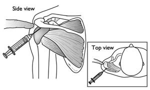
These treatments are conservative medical treatments, if these and physical therapy (see below) don’t work the patient will have to undergo surgery
The operative treatment is done mostly arthroscopically which is less invasive (than open/mini-open surgery) and leaves only a few small scars. Being less invasive the patients need to use less painkillers and will be able to start rehabilitation sooner. With time and development of better techniques the arthroscopic approach of rotator cuff tendon repair now even has a higher(20%) long term success rate. This was measured with the American Shoulder and Elbow Surgeons scores an reoccurrence of the supraspinatus tears. [4]
Depending on the severity of the tear (partial/complete) a different approach will be used.
If there is just a partial repair necessary the tendon and surrounding bone will be smoothed to avoid further damage and therefor allowing the tendon to heal mostly on its own.[5] In case of a complete tear in the middle of the tendon the surgeon will have to suture the two parts of the tendon back together. When the tear has occurred close or on its point of attachment on the head of the humerus another approach will be taken. This will be the case most of the times. The surgeon will attach the tendon back to its original place by way of an anchor(sometimes two). This anchor actually consists of a small screw that is bored into the head of the humerus with on the back surgical wires with witch to hold the tendon in place. [6]

 4
4
11. Physical Therapy Management[edit | edit source]
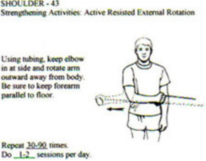
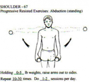
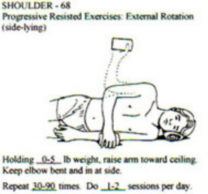
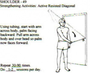
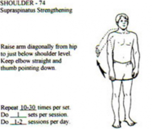
In case of a complete tear of the supraspinatus muscle, significant pain and dysfuction after six months of treatment or repeated dislocations surgery will be preferred as treatment. However if it is only a partial tear in most cases a conservative treatment will reduce pain(2-6weeks) and over time even allow the patient to regain function (up to three months). [11] The reduced pain is not just the direct effect of the pain reducing abilities of NSAID’s. The long term effects will mostly be attributable to a well preformed physical therapy. This will consist of different parts: reduce pain, manipulate blood flow (control inflammation and speed up healing), increase range of motion, increase control of muscles and their strength. Massage can be used to reduce pain, cryotherapy is useful to reduce pain, but only in the first 48 hours after injury. Corticosteroid injection may be useful to reduce pain as well, but only works on short term. . To increase range of motion one can use stretching exercises of the ruptured muscle (not to soon in recovery since premature stretching might aggravate the injury)(see below), passive- and active range of motion exercises such as pendulum exercises and symptom limited active-assisted range of motion exercises(see below). To increase control and strength the patient will also be prescribed strengthening exercises for the rotator cuff specifically the functions of the supraspinatus muscle (abduction and exorotation) explain SS and external rotation [3] [12] (see below for a few examples).
 [17]
[17]


Home exercises consisting of stretching and strengthening exercises prove to be effective, no matter what type of injury (partial defects, full thickness tears of the supraspinatus tendon or massive rotator cuff defects). Patients with rotator cuff defects do benefit from simple home exercises independent from the size of the defect. There is an improvement in range of motion and a downward trend for impingement. [7]
12. References[edit | edit source]
1. Heers G. et al. Efficacy of home exercises for symptomatic rotator cuff tears in correlation to the size of the defect. (2005) Klinik für Orthopädie der Universität Regensburg, Bad Abbach.
Level : 3B
2. Jobe F.W., Moynes D.R. Delineation of diagnostic criteria and a rehabilitation program for rotator cuff injuries. (1982) Am J Sports Med, 10, 336–339.
Level : 5
3. Orthop J. Rotator cuff tear: physical examination and conservative treatment
(2013) Department of Orthopaedic Surgery, Tohoku University, 18, 197–204.
Level : 5
4. Millar N.L. et al. Open versus two forms of arthroscopic rotator cuff repair. (2009) Clinical Orthopaedics and Related Research, 467, 966-78
5. American Academy of Orthopedic Surgeons. Rotator Cuff Tears: Surgical Treatment Options (2011)
Level: 5
6. Akpinar S. et al. Prospective evaluation of the functional and anatomical results of arthroscopic repair in small and medium-sized full-thickness tears of the supraspinatus tendon.(2011) Acta Orthop Traumatol Turc 45, 248-253
Level : 2B
7. Manhattan Orthopedic & Sports Medicine. Rotator Cuff Tendon Tears / Shoulder Arthroscopy. (2012) from: http://manhattanorthopedic.com/2010/09/rotator-cuff-tendon-tears-shoulder-arthroscopy/
Level: 5
8. Codsi M.J. The painful shoulder: When to inject and when to refer. (2007)Department of Orthopaedics, Cleveland Clinic, 74, 473-480
Level : 5
9. Bjorkenheim J.M. et al. Surgical repair of the rotator cuff and surrounding tissues. Factors influencing the results. (1988) Clinical Orthopaedic Relations, 236
10. Sarwark J.F. Essentials of Musculoskeletal Care. (2010) American Academy of Orthopaedic Surgeons, Rosemont
Level : 5
11. Dr. Romanski C., Schuldt J. Conservative Treatment of Rotator Cuff Injuries to Avoid Surgical Repair. (2009)
Level: 5
12. Tanaka M. et al. Factors related to successful outcome of conservative treatment for rotator cuff tears. (2010) Journal of Medical Sciences, Upsala, 115, 193–200
Level : 3B
13.
14.
15. Leggin B.J. et al. The Penn Shoulder Score: Reliability and Validity. (2006) from: http://www.jospt.org/issues/articleID.1021,type.14/article_detail.asp
Level : 3B
16. Kamper S.J. et al. Global Rating of Change Scales: A Review of Strengths and Weaknesses and Considerations for Design. (2009) from: level : 4
17. Kristian Berg. Prescriptive stretching; Human Kinetics; 2011
