Tibia: Difference between revisions
No edit summary |
No edit summary |
||
| Line 1: | Line 1: | ||
<div class="editorbox"> | <div class="editorbox"> | ||
'''Original Editor '''- | '''Original Editor '''- The [[Open Physio]] project. | ||
'''Lead Editors''' - Your name will be added here if you are a lead editor on this page. [[Physiopedia:Editors|Read more.]] | '''Lead Editors''' - Your name will be added here if you are a lead editor on this page. [[Physiopedia:Editors|Read more.]] | ||
</div> | </div> | ||
== Tibial plateau == | == Tibial plateau == | ||
== Shaft == | == Shaft == | ||
== Lateral Malleolus == | == Lateral Malleolus == | ||
== Medial Malleolus == | == Medial Malleolus == | ||
| Line 30: | Line 21: | ||
The ''medial surface'' of this process is convex and subcutaneous. Its ''lateral'' or ''articular surface'' is smooth and slightly concave, and articulates with the [[Talus]]. Its ''anterior border'' is rough, for the attachment of the anterior fibers of the deltoid ligament of the [[Ankle]] joint. Its ''posterior border'' presents a broad groove, the malleolar sulcus, directed obliquely downward and medially, and occasionally double; this sulcus lodges the tendons of the [[Tibialis posterior]] and [[Flexor digitorum longus]]. The ''summit'' of the medial malleolus is marked by a rough depression behind, for the attachment of the [[Deltoid ligament]]. | The ''medial surface'' of this process is convex and subcutaneous. Its ''lateral'' or ''articular surface'' is smooth and slightly concave, and articulates with the [[Talus]]. Its ''anterior border'' is rough, for the attachment of the anterior fibers of the deltoid ligament of the [[Ankle]] joint. Its ''posterior border'' presents a broad groove, the malleolar sulcus, directed obliquely downward and medially, and occasionally double; this sulcus lodges the tendons of the [[Tibialis posterior]] and [[Flexor digitorum longus]]. The ''summit'' of the medial malleolus is marked by a rough depression behind, for the attachment of the [[Deltoid ligament]]. | ||
< | == Recent Related Research (from [http://www.ncbi.nlm.nih.gov/pubmed/ Pubmed]) == | ||
<div class="researchbox"> | |||
<rss>http://eutils.ncbi.nlm.nih.gov/entrez/eutils/erss.cgi?rss_guid=1-cNR3TsrjMLfrmLd5Lpn3g-jxYIQVFP0ZisFBqYZdNl09Z9QE|charset=UTF-8|short|max=10</rss> | |||
</div> | |||
== References == | |||
References will automatically be added here, see [[Adding References|adding references tutorial]]. | |||
<references /> | |||
*Medial malleolus. (2008, September 22). In ''Wikipedia, The Free Encyclopedia''. Retrieved 21:12, September 26, 2008, from http://en.wikipedia.org/w/index.php?title=Medial_malleolus&oldid=240146811 | *Medial malleolus. (2008, September 22). In ''Wikipedia, The Free Encyclopedia''. Retrieved 21:12, September 26, 2008, from http://en.wikipedia.org/w/index.php?title=Medial_malleolus&oldid=240146811 | ||
Revision as of 22:58, 30 May 2011
Original Editor - The Open Physio project.
Lead Editors - Your name will be added here if you are a lead editor on this page. Read more.
Tibial plateau[edit | edit source]
Shaft[edit | edit source]
Lateral Malleolus[edit | edit source]
Medial Malleolus[edit | edit source]
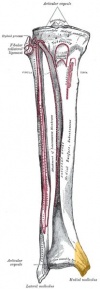
The medial malleolus is the medial surface of the Distal portion of the Tibia. It is prolonged downward to form a strong pyramidal process and flattened from without, inward.
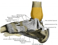
The medial surface of this process is convex and subcutaneous. Its lateral or articular surface is smooth and slightly concave, and articulates with the Talus. Its anterior border is rough, for the attachment of the anterior fibers of the deltoid ligament of the Ankle joint. Its posterior border presents a broad groove, the malleolar sulcus, directed obliquely downward and medially, and occasionally double; this sulcus lodges the tendons of the Tibialis posterior and Flexor digitorum longus. The summit of the medial malleolus is marked by a rough depression behind, for the attachment of the Deltoid ligament.
Recent Related Research (from Pubmed)[edit | edit source]
Failed to load RSS feed from http://eutils.ncbi.nlm.nih.gov/entrez/eutils/erss.cgi?rss_guid=1-cNR3TsrjMLfrmLd5Lpn3g-jxYIQVFP0ZisFBqYZdNl09Z9QE|charset=UTF-8|short|max=10: Error parsing XML for RSS
References[edit | edit source]
References will automatically be added here, see adding references tutorial.
- Medial malleolus. (2008, September 22). In Wikipedia, The Free Encyclopedia. Retrieved 21:12, September 26, 2008, from http://en.wikipedia.org/w/index.php?title=Medial_malleolus&oldid=240146811
- Osteology - the tibia. Grays anatomy (public domain version), downloaded on 26 September, 2008, from http://www.bartleby.com/107/61.html


