Chiari Malformations
Original Editor - Wendy Walker
Lead Editors - Wendy Walker, Laura Ritchie, WikiSysop, Kim Jackson, 127.0.0.1, Evan Thomas, Naomi O'Reilly and Rucha Gadgil
Clinically Relevant Anatomy[edit | edit source]
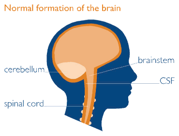
Chiari malformations are a group of defects associated with congenital caudal 'displacement' of the cerebellum and brainstem[1].
They are named after Hans Chiari, the pathologist who first described the whole group of malformations.
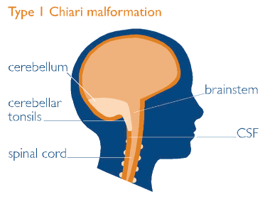
Illustrations provided by the Brain & Spine Foundation.
Types of Chiari Malformations[edit | edit source]
Chiari I[edit | edit source]
This is the most common variant.
Peg-like cerebellar tonsils are displaced into the upper cervical canal through the foramen magnum.
Radiologically if a person has a 5mm or more herniation below the foramen magnum it is classified as Chiari type I
Chiari 1.5[edit | edit source]
Described in the literature as both a condition in its own right as well as a variant of Chiari I malformation
Chiari II[edit | edit source]
AKA Arnold Chiari Malformation.
Displacement of the medulla, fourth ventricle and cerebellum through the foramen magnum.
Usually with associated with a lumbosacral spinal myelomeningocoele.
Chiari III[edit | edit source]
Features similar to Chiari II but with an occipital and/or high cervical encephalocoele.
Chiari IV[edit | edit source]
The most severe variant.
Severe cerebellar hypoplasia without displacement of the cerebellum through the foramen magnum.
Thought likely to be a variation of cerebellar hypoplasia.
Clinical Presentation[edit | edit source]
| [2] |
Type I
Symptoms of Chiari I develop as a result of 3 pathophysiological consequences of the disordered anatomy:
- compression of the medulla and upper spinal cord
- compression of cerebellum,
- disruption of CSF flow through foramen magnum.
Compression of cord and medulla may result in myelopathy and lower cranial nerve and nuclear dysfunction. Compression of cerebellum may result in ataxia, dysmetria, nystagmus, and dysequilibrium. Disruption of CSF flow through the foramen magnum probably accounts for the most common symptom, pain.
Type II
The pathophysiology of Chiari II is more complex. Although compressive mechanisms likely play a role, as in Chiari I, additional mechanisms may be operative in Chiari II. Stretching of abnormally oriented cranial nerves is believed to play a role.
Chiari II may become acutely symptomatic with shunt malfunction.
Intrinsic neuroembryological abnormalities in Chiari II are widespread and not limited to the posterior fossa (eg, heterotopias, gyral abnormalities, callosal and thalamic abnormalities, in addition to hydrocephalus and myelomeningocele), further complicating the pathophysiology of this disorder. Types III & IV
These more severe variants of Chiari malformations are almost always diagnosed in infancy as the child presents with severe neurological symptoms.
Diagnostic Procedures[edit | edit source]
The key tests for diagnosis of Arnold Chiari Malformation are MRI and CT scans[3].
MRI will show the abnormal CSF flow and configuration and position of the brain and spinal cord[4].
Antenatal ultrasound imaging shows an indentation of the frontal bone with an abnormally shaped cerebellum[5].
Differential Diagnosis[edit | edit source]
Pain in neck or headache made worse by coughing or the valsalva manoeuvre are suggestive of Chiari malformations, as a result of increased CSF pressure.
Physiotherapy Management[edit | edit source]
Physiotherapy can help with many of the symptoms caused by Chiari Malformations:
- Vestibular Rehabilitation is indicated when patients have vestibular problems
- Conservative physiotherapy soft tissue techniques may be of benefit for cervical pain
- Soft tissue techniques may be of benefit for headaches
Surgical Management[edit | edit source]
In some cases, surgical intervention is required.
Commonly decompression surgery is performed to reduce pressure at the base of the brain. An incision is made at the back of the head and a small piece of bone is removed from the base of the skull to widen the space in the foramen magnum. This relieves the pressure on the cerebellum and creates room for the CSF to flow normally. Occasionally, cervical fusion surgery may also be required.
Patients with associated hydrocephalus, or intracranial hypertension, may require a shunt to be surgically inserted.
Resources[edit | edit source]
The Brain & Spine Foundation has a very useful factsheet (suitable for patients) and website page on Chiari Malformations.
References[edit | edit source]
- ↑ Koehler PJ. Chiari's description of cerebellar ectopy (1891). With a summary of Cleland's and Arnold's contributions and some early observations on neural-tube defects. J Neurosurg. Nov 1991;75(5):823-6
- ↑ Mayfield Clinic. Chiari Malformation Diagnosis & Treatment Options. Available from: http://www.youtube.com/watch?v=ImWtvtSQx50 [last accessed 13/02/16]
- ↑ Curnes JT, Oakes WJ, Boyko OB. MR imaging of hindbrain deformity in Chiari II patients with and without symptoms of brainstem compression. AJNR Am J Neuroradiol. 10 (2): 293-302
- ↑ Pillay PK, Awad IA, Little JR, Hahn JF. Symptomatic Chiari malformation in adults: a new classification based on magnetic resonance imaging with clinical and prognostic significance. Neurosurgery. May 1991;28(5):639-45
- ↑ El gammal T, Mark EK, Brooks BS. MR imaging of Chiari II malformation. AJR Am J Roentgenol. 1988;150 (1): 163-70


