Ulna
Original Editor-Abbey Wright
Top Contributors - Abbey Wright, Kim Jackson, Chrysolite Jyothi Kommu, Rishika Babburu and Joao Costa
Description[edit | edit source]
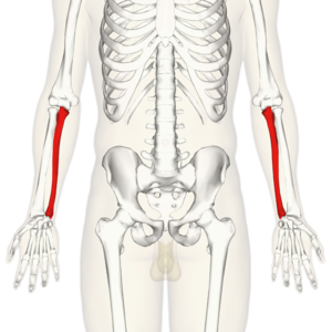
The ulna is one of two bones that make up the forearm, the other being the radius. It forms the elbow joint with the humerus and also articulates with the radius both proximally and distally. It is located in the medial forearm when the arm is in the anatomical position. It is the larger of the two forearm bones.[1]Ulna assists in pronation and supination of the forearm and hand.[2]
Structure[edit | edit source]
The ulna is a long bone larger proximally than distally.
Proximal ulna[edit | edit source]
The proximal ulna is hook-like in form which articulates with the trochlea of the humerus to create the hinge joint of the Elbow.[3]
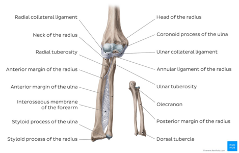
The articulation is formed of the olecranon and the coronoid process.
Image: Overview of the radius and ulna (anterior and posterior views)[4]
Olecranon[edit | edit source]
This is a large, curved bony prominence which is accepted into the olecranon fossa, located on the humerus, during elbow extension.
The olecranon forms the upper part of the semi-lunar notch which is a smooth, large depression and articulates with the humeral trochlea during elbow flexion and extension.
Coronoid process[edit | edit source]
The coronoid process is a horizontal, bony projection which attaches directly onto the ulnar shaft. It is received into the coronoid fossa of the humerus in elbow flexion. The coronoid process also forms the lower part of the semi-lunar notch.
On the lateral side of the coronoid process is the radial notch where the head of the radius sits.
Head of the ulna[edit | edit source]
The lateral, distal end of the ulna is the head of the ulna. It articulates with the ulnar notch on the radius and with the triangular articular disc in the Wrist Joint.
Shaft of the Ulna[edit | edit source]
The shaft of ulna is triangular in shape and it consists of three borders and three surfaces. It's width is decreased as it moves towards distal end.
The three surfaces are:
- Anterior surface
- Posterior Surface
- Medial Surface
The three borders are:
- Interosseous Border
- Anterior Border
- Posterior Border
Distal End[edit | edit source]
The head of Ulna has convex articular surface on its lateral side inorder to articulate with ulnar notch of radius.It forms the distal radio-ulnar joint.
The styloid process has attachment of ulnar collateral ligament.
Articulations[edit | edit source]
Elbow[edit | edit source]
The ulna articulates with the humerus at its most proximal point forming the elbow in a hinge joint. It is the trochlea of the humerus which sits in the semi-lunar notch of the ulna to form this joint.
Radio-ulnar joints[edit | edit source]
The ulna articulates with the radius proximally and distally to produce pronation (from the proximal joint) and supination (from the distal joint) of the forearm.
The ulna also articulates with the radius in a syndesmosis joint via its interosseous membrane. Which runs the length of the shaft of the ulna from the radial notch proximally to the head of the ulna. This also acts to compartmentalise dorsal and volar sides of the forearm. [1]
Muscle attachments[edit | edit source]
Olecranon process[edit | edit source]
Triceps - inserts onto the posterior of the olecranon process.[1]
Anconeus - inserts onto lateral aspect
Flexor Carpi Ulnaris - origin:posterior, also shares an origin from humeral medial epicondyle
Coronoid process[edit | edit source]
Brachialis - inserts to anterior, inferior coronoid process
Pronator teres - originates medial surface, also from humeral medial epicondyle
Flexor Digitorum Superficialis - originates medial surface, also from humeral medial epicondyle
Shaft of ulna[edit | edit source]
All of the following arise from the shaft of the ulna:
Pronator quadratus
Extensor carpi ulnaris - also shared with lateral epicondyle of humerus
Abductor pollicis longus - also originates from interosseous membrane
Extensor Pollicis Longus - also originates from interosseous membrane
Extensor indicis - also originates from interosseous membrane
Clinical relevance[edit | edit source]
Like any other joint in the body the ulna can be affected by osteoarthritis in the elbow joint.
Ulnar fractures[edit | edit source]
Typically occur as a result of fragility and normally following a fall such as a fall on an outstretched hand (FOOSH)[6]
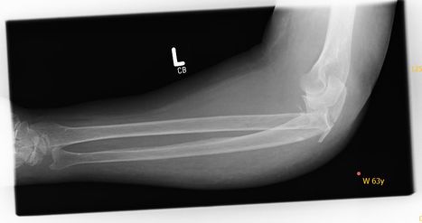
- Monteggia fracture - fracture of the proximal 1/3 of the ulnar shaft accompanied by the dislocation of the radial head.
- Hume fracture - fracture of the olecranon accompanied by anterior dislocation of the radial head.
- Galezzi's fracture-fracture to the distal radius accompanied by ulnar head dislocation at distal radio-ulna joint.[7]
References[edit | edit source]
- ↑ 1.0 1.1 1.2 Gray HFRS, Gray's Anatomy 15th edition, New York, NY: Barnes & Noble,2010. p120-126
- ↑ https://www.kenhub.com/en/library/anatomy/the-radius-and-the-ulna
- ↑ Palastanga N, Soames R. Anatomy and Human Movement, structure and function 6th edition, London, UK: Churchill Livingstone Elsevier ,2011. p45
- ↑ Overview of the radius and ulna (anterior and posterior views image - © Kenhub https://www.kenhub.com/en/library/anatomy/the-radius-and-the-ulna
- ↑ Simply AandP. Ulna. Available from: https://www.youtube.com/watch?v=-1oOpLsyHUA [last accessed: 11/10/2013]
- ↑ Duckworth AD, Clement ND, Aitken SA, Court-Brown CM, McQueen MM. The epidemiology of fractures of the proximal ulna. Injury. 2012 Mar 1;43(3):343-6.
- ↑ Oliver Jones,The Ulna-Proximal-Shaft-Distal(Updated January 20,2020).TeachMeAnatomy.Assessed June 30,2020


