Radius: Difference between revisions
Abbey Wright (talk | contribs) No edit summary |
Abbey Wright (talk | contribs) (content edit) |
||
| Line 35: | Line 35: | ||
==== Elbow ==== | ==== Elbow ==== | ||
The radius articulates with the ulna in a synovial pivot joint. The radial head rotates within the annular ligament and radial notch on the ulna to produce pronation | The radius articulates with the ulna in a synovial pivot joint. The radial head rotates within the annular ligament and radial notch on the ulna to produce pronation of the forearm.<ref name=":2">Gray HFRS, Gray's Anatomy 15th edition, New York, NY: Barnes & Noble,2010. p231- 235</ref> | ||
The radius and ulna also articulate distally in reverse to their articulation at the elbow to produce supination. This movement occurs with the head of the ulna being received into the ulnar notch of the radius. <ref name=":2" /> | |||
{{#ev:youtube|v=3F9d-qaXYp0}}<ref>Young Lae Moon. Forearm supination & pronation. Available from: https://www.youtube.com/watch?v=3F9d-qaXYp0 [Last accessed 26/03/2016]</ref> | {{#ev:youtube|v=3F9d-qaXYp0}}<ref>Young Lae Moon. Forearm supination & pronation. Available from: https://www.youtube.com/watch?v=3F9d-qaXYp0 [Last accessed 26/03/2016]</ref> | ||
==== Wrist ==== | ==== Wrist ==== | ||
The radius articulates with the first row of carpel bones: mainly scaphoid laterally and the lunate medially to form the radio-carpel joint. The radio-carpel joint is an ellipsoid joint | |||
=== Muscle attachments === | === Muscle attachments === | ||
Revision as of 22:34, 25 September 2018
Original Editor
Top Contributors - Abbey Wright, Kim Jackson, Joao Costa, Chrysolite Jyothi Kommu, Amanda Ager and Pacifique Dusabeyezu
Description[edit | edit source]
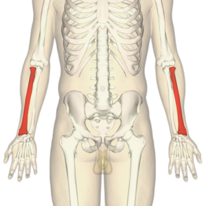
The radius is one of the two bones that make up the forearm, the other being the ulna. It forms the radio-carpel joint at the wrist and the radio-ulnar joint at the elbow. It is in the lateral forearm when in the anatomical position. It is the smaller of the two bones.
Structure[edit | edit source]
Proximal radius[edit | edit source]
The proximal radius consists of the radial head, neck and tuberosity.
The radial head is cylindrical which articulates with the capitellum of the humerus[1]. The head rotates within the annular ligament to produce supination and pronation of the forearm.[2]
The neck and tuberostiy support the head and provide points of attachments for supinator brevis and biceps bracii.[1]
Radial shaft[edit | edit source]
The shaft of the radius is slightly curved into convex from the body. The majority of the shaft has three borders: anterior, posterior and interosseous.
Distal radius[2][edit | edit source]
The distal radius has five surfaces:
- Lateral - which extends to form the styloid process
- Medial - consists of a concave ulnar notch to articulate with the ulnar head in pronation
- Posterior - convex and contains a prominent ridge called Lister's tubercle
- Anterior - smooth and forms a distinct margin
- Distal articular surface - articulates laterally with scaphoid and medially with lunate
Function[edit | edit source]
The radius' main functions are to articulate with the ulna and humerus at the elbow to provide supination and pronation. Then to articulate with the lunate and scaphoid to provide all the movements of the wrist.
Articulations[edit | edit source]
Elbow[edit | edit source]
The radius articulates with the ulna in a synovial pivot joint. The radial head rotates within the annular ligament and radial notch on the ulna to produce pronation of the forearm.[3]
The radius and ulna also articulate distally in reverse to their articulation at the elbow to produce supination. This movement occurs with the head of the ulna being received into the ulnar notch of the radius. [3]
Wrist[edit | edit source]
The radius articulates with the first row of carpel bones: mainly scaphoid laterally and the lunate medially to form the radio-carpel joint. The radio-carpel joint is an ellipsoid joint
Muscle attachments[edit | edit source]
Clinical relevance[edit | edit source]
Most commonly injured by fracture: fall on outstretched hand (FOOSH) commonly referred to as colles fracture.
Assessment[edit | edit source]
Treatment[edit | edit source]
Resources[edit | edit source]
References[edit | edit source]
- ↑ 1.0 1.1 Gray HFRS, Gray's Anatomy 15th edition, New York, NY: Barnes & Noble,2010. p126-128
- ↑ 2.0 2.1 Palastanga N, Soames R. Anatomy and Human Movement, structure and function 6th edition, London, UK: Churchill Livingstone Elsevier ,2011. p45
- ↑ 3.0 3.1 Gray HFRS, Gray's Anatomy 15th edition, New York, NY: Barnes & Noble,2010. p231- 235
- ↑ Young Lae Moon. Forearm supination & pronation. Available from: https://www.youtube.com/watch?v=3F9d-qaXYp0 [Last accessed 26/03/2016]


