Spondyloarthritis: Difference between revisions

No edit summary
m (Reverted edits by Kim Jackson (talk) to last revision by Rachael Lowe)
Line 1: Line 1:
<div class="editorbox">
'''Original Editors''' - [[User:Alicia Keefe|Alicia Keefe]] and [[User:Brenna Rutledge|Brenna Rutledge]] as part of the [[Temple University Evidence-Based Practice Project|Temple University EBP Project]]  
'''Original Editors ''' - [[User:Els Bernaers|Els Bernaers]]


<br> '''Top Contributors''' - {{Special:Contributors/{{FULLPAGENAME}}}} &nbsp;
'''Top Contributors''' - {{Special:Contributors/{{FULLPAGENAME}}}} &nbsp;  
</div>
</div>
== Definition/description ==
== Definition/Description ==


Skier's thumb is a acute partial or complete rupture of the ulnar collateral ligament (UCL) of the thumb’s metacarpophalangeal joint (MCPJ) due to a hyperabduction trauma of the thumb.  
Spondyloarthritis is a name for a group of diseases that is included in a larger term 'arthritis'.<ref name="p1"/><ref name="p2">Braun J., Sieper J., Spondyloarthritides., Z Rheumatol. 2010 Jul; 69(5):425-32 :4: 2C</ref><ref name="p3"/> Inflammation can occur in spine, sacroiliac and peripheral joints as well near the attachments of tendons and ligaments.<ref name="p3"/> This disease provokes to pain, stiffness and fatigue in back, legs and arms as in joints, ligaments and tendons.<ref name="p6">Rudwaleit M .Et al. The Assessment of SpondyloArthritis International Society classification criteria for peripheral spondyloarthritis and for spondyloarthritis in general. Ann Rheum Dis. 2011;70(1):25.: 4</ref> <ref name="p7">Burgos-Vargas R.The assessment of the spondyloarthritis international society concept and criteria for the classification of axialspondyloarthritis and peripheral spondyloarthritis: A critical appraisal for the pediatric rheumatologist. Pediatric Rheumatology 2012, 10:14  : 2C</ref>Eruption, eye and intestinal problems may also occur.<ref name="p1" /><ref name="p3"/><br>Spondyloarthritis in adults can be subdivided more specifically:<ref name="p1"/><ref name="p2"/><ref name="p8">Braun J, Sieper J. Early diagnosis of spondyloarthritis. Nature clinical practice rheumatology. october 2006 vol 2 no 10  : 2C</ref><ref name="p9">Jürgen Braun* and Joachim Sieper†, Early diagnosis of spondyloarthritis , 2006 : 2C</ref><ref name="p0">ozgur akgul, Classification criteria for spondyloarthropathies, , World J Orthop. 2011 December 18; 2(12): 107-115 : 2A</ref><ref name="p1"/><br>


Whilst both terms are often used interchangeably, skier’s thumb refers to the cause being acute injury. [[Gamekeeper’s Thumb|Gamekeeper’s thumb]] specifically refers to the cause being chronic injury to the UCL in which it became attenuated through repetitive stress. <br>UCL damage caused by Chronic injury may have a serious risk of disabling instability, pinch strength, and pain-free motion if not treated adequately .<ref name=“Ritting”>Ritting AW, Baldwin PC, Rodner CM. Ulnar collateral ligament injury of the thumb metacarpophalangeal joint. Clinical Journal of Sport Medicine. 2010 Mar 1;20(2):106-12.</ref><ref name=“Mandhkani”>Mandhkani Mahajan. Rupture of the ulnar collateral ligament of the thumb. Mahajan and Rhemrev International Journal of Emergency Medicine. 2013;6:31</ref><ref name=“Chrysi”>Chrysi Tsiour. Injury to the Ulnar Collateral Ligament of the Thumb. American Association for Hand Surgery;2008</ref><ref name=“Mandhkani M”>Mandhkani M, et al. Rupture of the ulnar collateral ligament of the thumb – a review. Int J Emerg Med. 2013; 6: 31. doi: 10.1186/1865-1380-6-31PMCID: PMC3765347</ref><ref name=“Fricker”> Fricker R1, et al.Skier’s thumb. Treatment, prevention and recommendations</ref><ref name=“Ritting et al”>Ritting et al., Ulnar Colletral Ligament Injury of the Thumb Metacarpophalangeal Joint. Sport Med; 2010; 20(2):106–112</ref> 
*ankylosing spondylitis or Bechterew disease
{{#ev:youtube|https://www.youtube.com/watch?v=0ZUtTT9v3r0|width}}<ref>Anna Pickens . Game keepers thumb. EM in 5. Available from: https://www.youtube.com/watch?v=0ZUtTT9v3r0 (last accessed 15.4.2019)</ref>
*psoriatic arthritis<ref name="p2"/>  
In 64–87% of total UCL tears, Stener lesion can occur. A Stener lesion occurs when the adductor aponeurosis becomes interposed between the ruptured UCL and its site of insertion at the base of the proximal phalanx. Hence making it impossible for the loosened ligament to reconnect with the site of insertion, necessitating surgery.<ref name=“Ebrahim”>Ebrahim FS et al. US diagnosis of UCL tears of the thumb and Stener lesions: technique, pattern-based approach, and differential diagnosis. Radiographics. 2006; 26(4): 1007-20 </ref>  
*reactive arthritis<ref name="p3"/>  
 
*enteric arthritis
Video illustration:<br>Stener lesion: https://www.youtube.com/watch?v=RLskrc7qifY<br>Skier’s thumb: https://www.youtube.com/watch?v=qFbxlgztK5U<br><br>
*undifferentiated arthritis


== Clinically Relevant Anatomy  ==


The metacarpophalangeal joint of the thumb is a diarthrodial joint, reinforced by a capsule and by other soft tissue structures. The surrounding soft tissue offers both dynamic and static stability.<ref name=“Patel”>Patel S, et al. Collateral ligament injuries of the metacarpophalangeal joint of the thumb: a treatment algorithm. Strat Trauma Limb Recon. 2010;5:1-10>/ref>
Spondyloarthritis is the overall name for a family of inflammatory rheumatic diseases. <ref name="p1"/><ref name="p2"/>  
 
The passive stability is provided by the following structures: <br>● Proper collateral ligament<br>● Accessory collateral ligament<br>● Volar plate<br>● Dorsal capsule[31]<br>
 
<br>
 
<br>
 
The proper collateral ligament extends from a point slightly dorsal to the mid-axis of the metacarpal head to the palmar aspect of the proximal phalanx (Fig. 1). The proper collateral ligament prevents palmar subluxation of the proximal phalanx and serves as the primary restraint to valgus stress with the metacarpophalangeal joint in flexion. The accessory collateral ligament courses palmarly to insert onto the volar plate. The accessory collateral ligament is contiguous with the proper collateral ligament proximally. The volar plate and the accessory collateral ligament function as the principal restraints to valgus stress with the metacarpophalangeal joint in extension. [31]
 
Dynamic stabilizers to valgus stress consist of the intrinsic and extrinsic muscles of the thumb:<br>● Extensor pollicis brevis<br>● Extensor pollicis longus<br>● Flexor pollicis longus<br>● Adductor pollicis<br>● Flexor pollicis brevis
 
The adductor mechanism presents as an aponeurosis superficial to the metacarpophalangeal joint capsule and ulnar collateral ligament. The adductor mechanism maintains dual insertions. The superficial insertion of the adductor mechanism is the extensor expansion via the adductor aponeurosis; the deep insertion extends to the palmar aspect of the proximal phalanx via the ulnar sesamoid of the metacarpophalangeal joint. <ref name=“Patel”/><ref name=“Chrysi”/> [31]
 
The range of motion is highly variable at the thumb metacarpophalangeal joint. [31]<br>The movement association with the thumb MCPJ include flexion, extension, rotation, abduction and adduction. <ref name=“Patel”/>
 
<br>
 
There are two main supporting ligaments traversing the MCPJ of the thumb:<br> 1) the UCL ligament<br> 2) the radial collateral ligament (RCL)<br>The UCL and RCL arise from the medial and lateral tubercles of the metacarpal condyles and insert into the base of the proximal phalanx on their respective sides, beneath the adductor aponeurosis (Figure 1b).<ref name=“Patel”/><ref name=“ASSH”>American Society for Surgery of the Hand. Thumb sprains. www.assh.org/Public/HandConditions/Pages/ThumbSprains.aspx (accessed 18 March 2011)</ref> The UCL prevents the thumb from pointing too far away from the hand.<ref name=“Ritting”/><br><br>
 
== Epidemyology/ etiology  ==
 
Skier’s thumb is caused by forced abduction and hyperextension of the thumb. <ref name=“PT”>Pediatric Trauma Care II: A clinical reference for physicians and nurses caring for the acutely injured child‬. AHC Media. LLC. 2014;6:52-53</ref> The UCL tears mostly find place at the distal attachment of the proximal phalange. But proximal avulsion, proximal and distal bony avulsion, isolated mid-substance tears and mid-substance tears with bony avulsion do also occur. <ref name=“Madan”>Madan, S. S., Injury to Ulnar Collateral Ligament of Thumb. Orthopaedic Surgery. 2014;6:1–7</ref>
 
<br>● Mechanism of injury. <br>It is important to note that this injury is not exclusive to skiers and can occur to anyone where there is an extreme valgus stress force applied to the thumb in abduction and extension. <ref name=“Engelhardt”>J. B. Engelhardt, Rupture of the ulnar collateral ligament of the metacarpophalangeal joint of the thumb.injury.1993;vol24:1:21-24</ref><br>An acute UCL injury occurs following a sudden, hyperabduction and hyperextension forces<ref name=“Madan”/> at the MCP joint, whereas a forced adduction movement would cause injury to the RCL (Figure 2A).<ref name=“Patel”/> With regards to skiing, the injury often occurs when a person lands on an outstretched hand while holding a ski pole, which causes forced abduction of the thumb with extension (Figure 2B).<ref name=“Anderson”>Anderson D. Skier’s thumb. Aust Fam Physician. 2010;39(8):575-577</ref> It is called skier’s thumb but can also occur in football, handball, basketball, rugby, soccer and even a handshake. <ref name=“Madan”/> If the injury to the UCL is not treated properly this can lead to chronic laxity, joint instability, pain, weakness and arthritis in the MCPJ.
 
<br>
 
● Frequency.<br>An often-encountered problem. It concerns 86% of all injuries to the base of the thumb. <br>Injuries to the ulnar collateral ligament of the thumb are the second most common ski-related injury. Prevalence of this injury during skiing varies from 7% up to as high as 32% of all skiing injuries and is the most frequent injury of the upper extremity that skiers experience.(figure 2C)<ref name=“Engelhardt”/><ref name=“Chuter”>G.S.J. Chuter,Ulnar collateral ligament injuries of the thumb: 10 years of surgical experience. Injury. June 2009;vol40:6:652-656.</ref><ref name=“Mandhkani M”/> <br>This type of injury is also seen in other sports, especially those that use a stick or ball, such as hockey or basketball. Only an estimated 10% of the patients diagnosed with a skier’s thumb had acquired this injury skiing. A fall on the hand, usually from a bicycle or motorcycle (in which the thumb gets stuck behind the handlebars), is a much more common cause of skier’s thumb, seen in approximately 40&nbsp;% of all patients(figure 2B). Other sports such as soccer or fighting are responsible for another estimated 30% of the causes(figure 2A). <br>In children, who still have an immature skeleton, hyperabduction trauma mostly leads to a Salter-Harris III avulsion of the UCL insertion and rarely to a true rupture of the UCL. <ref name=“Mandhkani M”/><br><br>
 
[[Image:Force.jpg|Mechanism of injury by falling.]]<br>
 
Figure 2A. Mechanism of injury by falling.<ref name="Zeigler T">Zeigler T. Thumb sprain also known as “skier’s thumb” or “gamekeeper’s thumb”. www.sportsmd.com/Articles/tabid/1010/id/50/Default.aspx?n=thumb_sprain_also_known_as_skier%E2%80%99s_thumb_or_gamekeeper%E2%80%99s_thumb (accessed 13 March 2011).</ref>  
 
[[Image:MOI.jpg|Mechanism of injury by skiing]]<br>
 
Figure 2B. Mechanism of injury by skiing.<ref name="Manhattan">Manhattan Orthopedic and Sports Medicine Group. Skier's thumb. manhattanorthopedic.com/2011/01/skier%E2%80%99s-thumb/ (accessed 13 March 2011).</ref>
 
<br>
 
● Prevention.<br>Preventive measures should include instruction in proper pole technique for powder skiing, avoidance of pole dragging and deep pole plants and downsizing baskets from the standard 4-inch diameter to 2.5-inches. Pole length should be 2 inches shorter than the recommended length for that skier. <ref name=“Palmer”>Palmer DH et al. Helicopter skiing wrist injuries. A case report of “bugaboo forearm”.; Am J Sports Med. 1994 Jan-Feb;22(1):148-9</ref>
 
Strapless poles do not reduce the chance of injuries, but if skiers are trained to discard the pole during a fall the risk might be reduced. <ref name=“Fricker”/>
 
Risk of injury can be further reduced by wearing a properly designed ski-glove which not only prevents extreme movement of the thumb, but also incorporates a mechanism for the ejection of the ski-pole. <ref name=“Fairclough”>J. A. Fairclough et al. Skier’s thumb-a method of prevention; Injury 1986; 17,203-204</ref><br>
 
== Characteristics/Clinical Presentation  ==
 
The most common presentation is pain over the ulnar aspect of the MCPJ of the thumb. If the injury is acute there will be bruising and inflammation (Figure 4). There may be tenderness with palpation, which localizes the injury to the ulnar aspect of the thumb where the UCL is lesioned. In more chronic cases the patients typically complain of pain and weakness when using a pincer grip. There also can be instability of the thumb while doing these tasks. <ref name=“Madan”/><ref name=“Patel”/> In the instance of a Stener lesion, there may also be a palpable mass proximal to the adductor aponeurosis.<ref name=“Anderson”/>
 
● Symtoms.<br>These symptoms may occur minutes to hours after the fall that created the injury:[29]<br>‒ Pain at the base of the thumb in the web space between thumb and index finger.<br>‒ Swelling of your thumb.<br>‒ Inability to grasp or weakness of grasp between your thumb and index finger.<br>‒ Tenderness to the touch along the index finger side of your thumb.<br>‒ Blue or black discoloration of the skin over the thumb.<br>‒ Thumb pain that worsens with movement in any or all directions.<br>‒ Pain in the wrist (which may be referred pain from your thumb).


● Grades of thumb sprains.<br>Thumb sprains are ranked by how much the ligament is pulled or torn away from the bone. [31]<br>‒ Grade 1: Ligaments are stretched, but not torn. This is a mild injury. It can improve with some light stretching.<br>‒ Grade 2: Ligaments are partially torn (less than 3mm) <ref name=“Milner”>Milner CS, et al., Gamekeeper’s thumb—a treatment – oriented magnetic resonance imaging classification. J Hand Surg. Am., 2015; 40(1): 90-5</ref>. This injury may require wearing a splint or a cast for 5 to 6 weeks.<br>‒ Grade 3: Ligaments are completely torn or more than 3mm<ref name=“Milner”/>. This is a severe injury that usually requires surgery.<br>‒ Grade 4: Failed immobilization and required surgery as did all of those with a Stener lesion<ref name=“Milner”/>.
Due to this fact, there is a large complexity. This is because there are several anatomic structures involved. We can assume that the inflammation can occur on all the joints of the spine. The facet joints, endplates, bone marrow, … every part of the spine can be affected by an inflammation.&nbsp;<ref name="p4">Walter P. Maksymowych, Frpc, Magnetic Resonance Imaging for Spondyloarthritis — Avoiding the Minefield (https://jrheum.com/subscribers/07/02/259.html) 4</ref> Sacroiliitis in SpA is characterized by involvement of different joint structures. Whereas the iliac and the sacral side of the sacroiliac joints are almost equally affected, the dorsocaudal synovial part of the joint is involved significantly more often than the ventral part, especially in early disease. Sacroiliac enthesitis is not a special feature of early sacroiliac inflammation. There is a difference between axial and peripheral spondyloarthitis, with axial spondyloarthitis back pain and inflammation of the sacroiliac joints are the main complaints. In peripheral spondyloarthritis, the inflammation of peripheral joint and tendons are the main complaints. Further, spondyloarthritis can show an inflammation of peripheral joints (for example, knees and ankles), and tendons (for example, the Achilles tendon).<ref name="p1">Braun J. et al, Spondyloarthritides, Internist., 2011 May 19: 5: 2C</ref><ref name="p2"/><ref name="p3"/><ref name="p4"/>


<br>
== Epidemiology /Etiology  ==


<br>  
Spondyloarthritis is a pathology that specifically strikes young people.<ref name="p5">Sieper J. Et al. Concepts and epidemiology of spondyloarthritis. Elsevier Ltd. 2006 : 2C</ref> The symptoms most frequently start before the age of 45.&nbsp;<ref name="p2"/> It affects more males than females. <ref name="p7"/><ref name="p6"/><br>Predisposition to spondyloarthritis, especially SpA, is determined largely by genetic factors. The incidence rate is higher in populations with a higher prevalence of HLA-B27.<ref name="p8"/> Psoriatic skin lesions and colitis due to inflammatory bowel disease (IBD) have been considered as both basic, subtype-defining entities with their own genetic background (distinct from HLA-B27 genotype), and as manifestations of spondyloarthritis.<ref name="p8"/> There is a strong need to diagnose patients with SpA in an earlier stage; currently there is a delay of 5–10 years between onset of the first symptoms and diagnosis.<ref name="p8"/><ref name="p6"/>


<br>
== Characteristics/Clinical Presentation  ==


<br><br>  
Symptoms that may occur with spondyloarthritis are pain, stiffness and fatigue in the back, legs and arms. There are no typical characteristics, because spondyloarthritis characterises with more than one symptom. We see that significantly more women have knee pain as presenting symptom.<ref name="Reveille" /><ref name="Sieper" /><ref name="Mease" /><ref name="VDBerg" /><ref name="Roussou" /><ref name="Slobodin" />  and we can assume that severity of symptoms can vary between individuals<ref name="Roussou" />. Here are the most common characteristics.<ref name="p2" /><ref name="p9" /><ref name="Mease" /><ref name="VDBerg" /><ref name="Slobodin" /><ref name="SlobodinG">Slobodin G., Recently diagnosed axial spondyloarthritis: gender differences and factors related to delay in diagnosis., Clin Rheumatol., 2011 Mar 1</ref><ref name="Colbert">Colbert R.A., Early axial spondyloarthritis., Curr Opin Rheumatol., 2010 Sep;22(5):603-7</ref>
 
* back pain
[[Image:Haley-resized.jpg|300x300px|Haley-resized.jpg]]<br>  
* osteoporosis
 
* spinal fractures
Figure 4. Presentation of an ulnar collateral ligament injury with an avulsion fracture. Photo courtesy H. Stevenson.<br>  
* peripheral arthritis, usually asymmetric, relatively more in the lower limbs.<ref name="p2" /><ref name="p3" /><ref name="p2" />  
* enteritis
* dactylitis
* inflammation of the heart valve – pneumonia
* extra articular disorders such as uveitis, skin porosiasis or inflammatory bowel disease
* strong familial aggregation of spondyloarthritis, psoriasis, IBD, uveitis
* association with HLA-B27
* no increased CRP and rheumatoid factor


== Differential Diagnosis  ==


----
The disease starts with hip or low back pain. The most common symptom is intermittent pain that progressively gets worse thoughout the day, in the morning, and following intensive activity. <ref name="p1"/> Most patients experience back pain in the sacroiliac joints. However, pain can involve all the parts of the spine. Pain relief is sometimes achieved by bending over. It is possible that a patient is not able to fully expand the chest due to the involvement of the joints between the ribs.
 
The injury can involve other structures such as the adductor aponeurosis, the accessory collateral ligament, bony structures, tendons and neurological tissues.<ref name=“Anderson”/> The injuries all present with pincer grasp weakness. However they may be differentiated by the location of tenderness.<br>For al thumb injuries, radiographs should be obtained of the patient suspected to have a skier’s thumb. It’s important to remember that a skier’s thumb may or may not be visible on X-ray and the most common radiographic finding is an avulsion fracture of the proximal thumb phalanx at the site of UCL attachment. MRI can be usefull because it has the highest spicificity and sensivisity.<ref name=“Mandhkani”/> <br>
 
<br>
 
*&nbsp;Skier’s thumb (UCL tear):
 
is characterized by point tenderness and instability at the thumb MCP joint, while
 
*&nbsp;Stener lesion:
 
is a particular type of UCL injury with palmar subluxation of the base of the proximal phalanx.
 
*&nbsp;Bennett or Rolando fracture:
 
Is an intra-acticular fracture luxation at the base of MC I in the CMC joint.<ref name=“Madan”/>
 
*&nbsp;Avulsion fracture:


An avulsion fracture is an injury to the bone in a location where a tendon or ligament attaches to the bone. When an avulsion fracture occurs, the tendon or ligament pulls off a piece of the bone. [37] In children, who still have an immature skeleton, hyperabduction trauma mostly leads to a Salter-Harris III avulsion of the UCL insertion and rarely to a true rupture of the UCL. <ref name=“Mandhkani M”/>
== Diagnostic Procedures  ==


*&nbsp;Wrist sprain:
Antecedents and physical examination are the major factors leading to diagnosis, although radiologic evidence of sacroiliitis is very helpful <ref name="p2"/><ref name="p9"/> In the early-1990s, two classification criteria, Amor and the European Spondyloarthropathy Study Group (ESSG), were proposed for diagnosing SpA <ref name="p2"/><ref name="p3"/>  All criteria developed so far (including the ESSG and Amor criteria) were developed as classification criteria, although they are often used as diagnostic criteria <ref name="p9" /><ref name="p0" />


When a wrist sprain injury occurs, the ligaments of the wrist are stretched beyond their normal limits. [35]<br>Wrist sprains are graded according to severity:<br> Grade 1 (mild) <br> Grade 2 (moderate)<br> Grade 3 (severe)<br>Grade 4 (surgery)
Amor criteria for spondyloarthritis <ref name="p7"/>:
{| width="565" cellspacing="1" cellpadding="1" border="1"
|-
! scope="col" | Paramters<br>  
! scope="col" | Scoring<br>  
| <br>
|-
| '''Clinical symptoms or past history of'''<br>  
| <br>
|-
|
Lumbar or dorsal pain at night or morning stiffness of lumbar or dorsal region


*&nbsp;Wrist fracture:
| 1<br>
|-
| Asymmetric oligoarthritis<br>
| 2<br>
|-
|
Buttock pain<br>Or if alternate buttock pain


A broken wrist (wrist fracture) can involve the small bones in the wrist or the ends of the forearm bones. [35]
|
1


*&nbsp;Dislocation of 1st MCP joint:
2<br>


A dislocation is an injury to a joint — a place where two or more of your bones come together — in which the ends of your bones are forced from their normal positions. [33] [34]
|-
| Sausage-like toe or digit<br>
| 2<br>
|-
| Heel pain or other well- defined enthesitis<br>
| 2<br>
|-
| Iritis<br>
| 2<br>
|-
| Non- gonococcal urethritis or cervicitis within 1 month before the onset of arthritis<br>
|
1<br>


*&nbsp;Chronic instability of the 1ste MCP joint:
|-
| Acute diarrhoea within 1 month befor the onset of arthritis <br>
| 1<br>
|-
| Psoriasis, balanitis or inflammatory bowel disease ( ulcerative colitis or chrohn’s disease)<br>
| 2<br>
|-
| '''Radiological findings'''<br>
| <br>
|-
| Sacroiliitis (bilateral grade 2 or unilaterale grade 3)
| 3<br>
|-
| '''Genetic background<br>'''
| <br>
|-
| Presence of HLA-B27 or family history of ankylosing spondylitis, reactieve arthritis, uveitis, psoriasis or inflammatory bowel disease<br>
| 2<br>
|-
| '''Response to treatment'''<br>
| <br>
|-
| Clear- cut improvement within 48 hours after non steroidal anti- inflammatory drug intake or rapid relapse of the pain after their discontinuation<br>
| 2<br>
|}


Injuries to the two main supporting ligaments traversing the metacarpophalangeal (MCP) joint of the thumb can lead to symptomatic joint instability with subsequent pain, weakness and arthritis if ignored. These two ligaments are the ulnar and radial collateral ligaments. [36]
A patient is considered to be suffering from spondyloarthritis if the sum is ≥ 6 


*&nbsp;Lunate dislocation:
The need for a standardized, evidence-based approach to spondyloarthritis classification led to the development of the European Spondyloarthropathy Study Group (ESSG)&nbsp;<ref name="p6"/> preliminary classification criteria for spondyloarthritis in 1991 <ref name="p5"/>:<br>Inflammatory spinal pain or synovitis (asymmetric, predominantly in lower limbs) and any one of the following:&nbsp;<ref name="p6"/>


A lunate dislocation is an injury to one of the small bones of the wrist. Lunate dislocations usually occur as part of a major injury such as a fall from a height or an automobile collision. When a lunate dislocation occurs, one of the small bones of the wrist, called the carpal bones, comes out of its normal position. [30]
*Positive family history
*Psoriasis
*Inflammatory bowel disease
*Acute diarrhea or urethritis or cervicitis preceding the arthritis
*Alternate buttock pain
*Enthesopathy
*Radiological sacroilits


*&nbsp;Neuropraxia of the radial nerve
Another is the concept of IBP (Low Back Pain), which is defined as the presence of at least four of the following five parameters <ref name="p1"/>, <ref name="p8"/>:


arises secondary to traction, swelling or stiffness.<br><br>
#Age at onset less than 40 years
#Insidious onset
#Improvement with exercise
#No improvement with rest
#Pain at night (with improvement upon getting up).


== Diagnostic procedures  ==
Studies are under way to define ASAS criteria for nonaxial (peripheral) SpA. <br>In the ASAS classification criteria, several SpA features are described. These features are called SpA features because they are frequently present in patients with SpA ,<ref name="p4"/><ref name="p8"/>


The type of lesion can be accurately derived by means of ultrasound ( approx. 90% accurate ) or MRI ( approx 100% accurate )<br>For non-displaced lesions, conservative treatment is possible and has yielded excellent results. However, misinterpretation and incorrect diagnosis can and have lead to unsatisfactory clinical results, leading many to favour surgery. <ref name=“Hergan”>Hergan K, et al. Pitfalls in sonography of the Gamekeeper’s thumb. Eur Radiol. 1997; 7: 65–69</ref> <br>Displaced lesions ( Stener lesions ) cannot be treated conservatively due to impaired healing and require surgical intervention in order to achieve full recovery.  
[[Image:ASAS classification.png]]


<br>In all instances, hand surgery is strongly recommended. Post-operatively a cast, brace, or splint to partially immobilise the hand is mandatory due to the likelihood of long-term complications if left mobile and to avoid stiffness that may result from complete immobilisation. <ref name=“Hergan”/>  
The main features of an early diagnosis of any rheumatic disease, including spondyloarthritis, are clinical history, clinical symptoms, clinical examination, laboratory parameters and imaging. <ref name="p5"/><br>Clinical symptoms:


X-rays<br>Anteroposterior and lateral X-ray films of the thumb are taken to rule out any associated bony injuries. Associated bony avulsion fractures are seen in 20%–30% of UCL ruptures. The position of an avulsed bony fragment usually indicates the position of the distal end of the UCL.<ref name=“Thirkannad”>Thirkannad S, et al. The “two fleck sign” for an occult Stener lesion. J Hand Surg Eur Vol, 2008; 33: 208–211</ref> Indications for surgical treatment based on imaging include avulsion fractures with displacement of greater than 5 mm or any fracture involving 25% or more of the MCP joint surface . Stress X-ray films of the thumb MCP joint have also been used for diagnosis.<ref name=“Abrahamsson”>Abrahamsson SO, et al. Diagnosis of displaced ulnar collateral ligament of the metacarpophalangeal joint of the thumb. J Hand Surg Am. 1990; 15: 457–460</ref> Local anaesthetic infiltration increases the accuracy of assessment of ulnar collateral ligament injuries RCT.<br>
*Inflammatory back pain
*Arthritis ( swelling, joint effusion, or detected by imaging)
*Accompanying features, including psoriasis, crohn-like colitis and anterior uveitis


Ultrasound<br>Ultrasound (US) is an excellent and cost-effective modality for diagnosing UCL tears because it allows direct visualization of the entire UCL and surrounding structures. It locates the torn end of the UCL in almost 90% of cases. <ref name=“Hergan”/>[28]The diagnosis should be done by US before conservative therapy is performed with a glove spica cast.<ref name=“Hergan”/> There are some limitations when applying, for example the ultrasound cannot be performed later than 1 week after the initial trauma because shrinking of the torn ligament and scar tissue can be confounding when making a diagnosis. <ref name=“Mandhkani”/> The sensitivity of US was 95.4% with a specificity of 80% for detection of Stener lesions. US, with the use of this specific dynamic maneuver is a reliable and reproducible tool for detecting Stener lesions.
Clinical history:


MRI<br>MRI can be seen as a gold standard with a sensitivity of 96%-100% and specificity of 95-100%. An alternative can be an ultrasound of the thumb. <ref name=“Mandhkani”/> It is considered by some as the best modality for evaluating UCL injuries. Like US, MRI allows direct visualization of the UCL and surrounding structures and is safe and non-invasive; however, it is more costly and less readily available. <ref name=“Lohman”>Lohman M, et al. MR imaging in chronic rupture of the ulnar collateral ligament of the thumb. Acta Radiol. 2001; 42: 10–14</ref>
*Family
*Rheumatic symptoms
*Accompanying features


Arthrography<br>Arthrography involves distension of the MCP joint by injecting contrast material and then visualizing the joint by X-ray or MRI (MR-arthrography). UCL injuries are diagnosed by direct visualization of any focal defect or by extravasation of contrast from the joint, suggesting rents in the ligaments. <ref name=“Madan”/> Another indirect finding on arthrography suggestive of UCL tear is demonstration of the heads of the adductor pollicis muscle. <ref name=“Lohman”>Ganel A, et al. “Gamekeeper’s thumb”. Injuries of the ulnar collateral ligament of the metacarpophalangeal joint. Br J Sports Med. 1980; 14: 92–96</ref> The various modalities used for diagnosis of UCL injury are presented in Table 2. <ref name=“Madan”/><br>Clinical and anatomical findings and the understanding of the injury mechanism show that stability testing (performed with the joint in full flexion) and additional standard radiographs remain the keystones in decision making in all MCPJ sprains<br>● Complications. <br>If the UCL is ruptured there is a possibility that the distal end may become interposed by the adductor aponeurosis, which is referred to as a Stener lesion (Figure 5). A Stener lesion is difficult to diagnose but leads to poor healing and usually indicates operative management. If left untreated, a torn UCL can lead to joint instability and a weak pinch grip.<ref name=“Leggit”>Leggit JC et al. Acute finger injuries: Part II. Fractures, dislocations, and thumb injuries. Am Fam Physician. 2006;73(5):827-834>/ref><br>Figure 5 Stener lesion.<ref name=“Leggit”/><br>
Clinical examination:  


<br>
*Lateral flexion of the lumbar spine (&lt;10cm)
*Chest expansion (&lt;4cm)
*Cervical rotation (&lt;70°)


[[Image:Stener lesion.gif|Image:Stener_lesion.gif]]<br>
Laboratory parameters:  


Figure 5. Stener lesion.<ref name="Leggit" />
*HLA-B27
*C- reactive protein
*Erythrocyte sedimentation rate


Outcome measures
Imaging:


== Outcome Measures&nbsp;  ==
*Radiography
*MRI
*Ultrasonography


There are many ways to manage both acute and chronic thumb UCL deficiency<br>and controversy persists as to the best treatment options. This systematic review<br>(http://www.medscape.com/viewarticle/807179_1) has demonstrated excellent<br>clinical outcomes (pain, strength, motion, and stability) after surgical treatment<br>(repair and autograft reconstruction) of both acute and chronic UCL injury,<br>without any significant difference between repair and reconstruction for acute and<br>chronic injury. Non-operative treatment of acute UCL injury (with or without a<br>Stener lesion) frequently fails. This leads to chronic pain, instability, weakness<br>and eventually prompting surgical intervention. Thus, a patient with delayed<br>presentation of UCL injury can still achieve predictably successful outcomes,<br>equivalent to acute repair, with autograft UCL reconstruction. No significant<br>difference in the outcome was demonstrated between different types of autograft<br>used for UCL reconstruction. Complications, failures and reoperations are rare<br>after surgical treatment of UCL injury. <ref name=“Samora”>Samora J.B. Outcomes After Injury to the Thumb Ulnar Collateral Ligament,, Clin J Sport Med. 2013;23(4):247-254</ref><br>
== Outcome Measures  ==


● Follow up.<br>The orthopedic surgeon will see the patient after surgical repair or after a period of<br>immobilization in a cast. The patient's thumb will be reexamined. The doctor will<br>decide if the patient need to continue to immobilize the thumb or if physical<br>therapy is needed to regain movement. The remainder of the rehabilitation and the<br>management of any chronic problems that may arise from the injury will be<br>addressed by your orthopedic or hand surgeon. [29]
*BASFI <ref name="p1"/>  
*BASDAI <ref name="p1"/>  
*BASMI&nbsp;<ref name="p1"/>&nbsp; [[The Bath Indices]]
*Pain <ref name="p1" />
*ASQoL <ref name="p1" />  
*Questionnaire Rasch model: <ref name="p0" />


== Examination  ==
== Examination  ==


Begin looking for deformities with observation of the hand at rest and in flexion. Then<br>test the sensation in the hand followed by active range of motion (AROM). AROM<br>should be followed by passive range of motion (PROM) and resisted movement to<br>assess tendon integrity, if possible. <ref name=“Patel”/><br>Clinical examination may occasionally reveal a tender swelling and a hematoma at the<br>ulnar side of the base of the thumb. <ref name=“Mandhkani M”/> Sometimes a mass can be felt in that area,<br>which suggests a Stener lesion; however, it is not pathognomonic.<br>If there is any concern about the possibility of fractures to the first metacarpal or<br>proximal phalanx of the thumb, plain radiographs are indicated prior to stress testing<br>of the UCL. If there is no associated fracture of the shaft, the thumb MCP joint<br>stability is tested by executing the following stress tests chronologically:
Patients with spondyloarthritis will complain about back pain, fatigue and stiffness. The pain will decrease when the patients exercise, but will persist at they rest. It is common for the patient to have pain at night, this pain can improve when the patients gets out of bed and moves around. (this should improve when they get up).<ref name="p2"/> <ref name="p0"/> The motion of the lumbar spine of the patients will be limited in both the sagittal and the frontal planes. <ref name="p2"/>  


<br>1) Testing of the UCL with MCP in extension (Figure…)<br>- Extend the MCP joint<br>- Stabilise the thumb metacarpal proximal to the joint to stop rotation and<br>radially angulating the thumb<br>- Apply a valgus stress by which the proper ulnar collateral ligament is<br>brought under tension<br>- Meaning: to assess the integrity of the volar plate and the accessory<br>collateral ligament
{{#ev:youtube|721_1vxzrb0}}


<br>2) Testing of the UCL with MCP in flexion (Figure…)<br>- Bring MCP joint in flexion of at least 25°.<br>- Stabilise the thumb metacarpal proximal to the joint to stop rotation and<br>radially angulating the thumb<br>- Apply a valgus stress by which the proper ulnar collateral ligament is<br>brought under tension<br>- Meaning: Testing the stability of the thumb MCP joint when the volar plate<br>is relaxed and the UCL is taut. When positive, it means the accessory ulnar<br>collateral ligament is also torn. <ref name=“Chrysi”/><br>These tests for accessing the laxity of the MCP joint, and thus the rupture of the<br>proper collateral ligament, are referred to as the Valgus Stress to UCL tests.<br>It is worth noting that it is impossible for this test to, when correctly executed,<br>accidently cause a Stener lesion if one is not already present. A valgus stress test can<br>only cause this when all stabilizing ligaments of the thumb have been severed, which<br>does not occur under natural circumstances. ( cadaver study stener lesion clinical test 39 )<br>If a Stener lesion is already present however, then applying a valgus stress test can<br>cause possible avulsed bone fragments to displace, further impending healing.<br>Therefore this test should not be executed if an RX has yet to be taken. <ref name=“Hergan”/><br><br>When the accessory UCL (or ACL) is still intact, a Stener lesion is less likely. It is<br>important to note that pain when examining can cause apprehension with subsequent<br>tensing of surrounding muscles and can lead to a false negative. Therefor the<br>investigation under local anesthesia can be useful. A study by Cooper et al. [Local<br>anaesthetic infiltration increases the accuracy of assessment of ulnar collateral igament injuries] described how Oberst<br>anesthesia (in which 1–2 ml of lidocaine is injected in the MCP joint) increases the<br>clinical accuracy from 28% to 98% after an average of one week after the initial<br>trauma [2, 38]<br>Inter-individual differences in normal range of motion of the MCP joint makes it<br>difficult to say when a true laxity of the joint is seen. In most of the literature the<br>following standard has been used for laxity of the MCP joint:<br>- lateral deviation more than 35° during valgus stress<br>OR<br>- more than a 15° difference compared to the uninjured/contralateral side<br>OR<br>The absence of a firm endpoint during testing is a more reliable criterion when<br>clinically diagnosing a complete rupture of the UCL<ref name=“Chrysi”/>,<ref name=“Mandhkani M”/><br>
Psoriasis, finger swelling, Crohn's disease or ulcerative colitis can be indicative for Spondyloarthritis.


[[Image:Valgus stress.gif|Image:Valgus_stress.gif]]
Sacroiliitis grade ≥ 2 bilaterally or grade 3 to 4 unilaterally is suggestive for SpA (grade 0: normal; grade I: some blurring of the joint margins - suspicious; grade II: minimal sclerosis with some erosion; grade III: definite sclerosis on both sides of joint 5 &amp; severe erosions with widening of joint space with or without ankylosis; grade IV: complete ankylosis)&nbsp; <ref name="p6" /><ref name="p7" />


Figure 6. Valgus stress to UCL - compare stability in injured thumb to uninjured thumb.<ref name="Leggit" />  
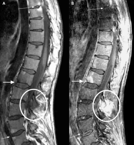
There are also active inflammatory and chronic lesions that can be found on a MRI-scan (see images).&nbsp;<ref name="p3"/>&nbsp;<ref name="p8"/> <ref name="p9"/><br>[[Image:MRI1.png]]<br>  


== Medical management  ==
<ref name="p3"/>Sieper et al. The Assessment of SpondyloArthritis international Society (ASAS) handbook: a guide to assess spondyloarthritis


A UCL injury may be managed conservatively or surgically depending on various factors that primarily include: timing of presentation (acute or chronic); grade (severity of injury); displacement (Stener lesion); location of tear (mid-substance or peripheral); associated or concomitant surrounding tissue injury (bone, volar plate, etc.); and patient related factors (occupational demands, etc.)<ref name=“Madan”/> <br>Indication for operative treatment of ulnar collateral ligament injuries of the thumb should be made on the basis of whether the ligament is displaced proximal to the adductor aponeurosis or not (Stener Leasion). Thus the diagnostic efforts should concentrate on ligament displacement rather than whether the ligament is ruptured totally or partially. <ref name=“Abrahamsson”/> <br>An injury that is not managed properly can lead to chronic instability of the MCPJ.<ref name=“Anderson”/>  
Laboratory testing
* Common presence of human leukocyte antigen-B27
* Elevated C-reactive protein
* Absence of rheumatoid factor <ref name="p2" />


Operative management depends on a timely diagnosis of the injury; chronic lesions become more difficult to repair with increased time since injury since remaining tissue becomes attenuated not robust enough to provide adequate support to the joint.<ref name=“Retting A et al”>Rettig A et al. Anatomic reconstruction of thumb metacarpophalangeal joint ulnar collateral ligament using an interference screw docking technique. Tech Hand Up Extrem Surg. 2009;13(1):7-10.>/ref> There are multiple methods of repair, which can be categorized into dynamic or static.<ref name=“Patel”/><ref name=“Retting A et al”/>
== Medical Management  ==


<br>● Dynamic:<br>- Extensor indicis proprius tendon transfer<br>- Extensor pollicis brevis tendon transfer<br>- Adductor pollicis brevis tendon transfer<br>Dynamic procedures are more likely to loosen with time while usually preserving a good mobility of the MCP joint. <ref name=“Fusetti”>C. Fusetti. The ECRL bone-tendon ligamentoplasty for chronic ulnar instability of the metacarpophalangeal joint of the thumb. Hand Surgery Unit, Chirurgie de la Main. 2005; Vol 24:5:217–221</ref>
Reliant on your symptoms and how severe your condition is, the doctor can decide what kind of treatment is the best option for the patient.  


<br>● Static:<br>- Figure-of-eight grafting<br>- Parallel configuration graft<br>- Triangular configuration with proximal apex graft<br>- Triangular configuration with distal apex graft<br>- Dually opposed biotenodesis fixation of tendon graft<br>- Tendon graft weaves<br>- Dually opposed suture anchor fixation <br>- Hybrid technique<br>- Free tendon graft: an insertion is made on the ulnar side of the thumb, exposing the ligament remnant. A tendon graft is used to connect the bones. The tendon graft replaces the ligament and is proximal bound to the ligament remnant and distal to the phalangeal bone. <ref name=“Mitsionis”>G.I Mitsionis. treatment of chronic injuries of the ulnar collateral ligament of the thumb using a free tendon graft and bone suture anchors. The Journal of Hand Surgery:British &amp; European volume. 2000; Vol 25:2:208–211</ref> (level of evidence: 2B)<br>There is a wide variety in static operations and there is still a lot of discussion which treatment is the best. Most treatments give positive results which lead to a lot of discussion.<ref name=“Fusetti”/> <ref name=“Basar”>Başar H. Comparison of results after surgical repair of acute and chronic ulnar collateral ligament injury of the thumb. Chirurgie de la Main. 2014;vol 33:6:384–389</ref>
Medicines such as:
* analgesics (pain-relievers, by example paracetamol)<ref name="p5" />  
* non-steroidal anti-inflammatory drugs (NSAIDS, by example naproxen, ibuprofen).<ref name="p4" />
* Anti-rheumatic drugs (DMARDs) have been proven effective in the treatment, but only for the arms and legs, not for the spine and sacroiliac joints.<ref name="p5" />
* Corticosteroids, given by the mouth or injections, can be effective. We must remind ourselves for the side effects, such as osteoporosis and infections.<ref name="p6" />  
* Injections of deposteroid in the joints of tendon sheaths are also used for symptomatic relief of the local flares.<ref name="p6" />  
* TNF alpha blockers are also effective in both spinal and peripheral joints.<ref name="p4" /><ref name="p5" />  
* There are three kind of TNF alpha blockers we can use:
** Infliximab (Remicade), given a dose of 5 mg/kg intravenously every sic to eight weeks.
**Etanercept (Enbrel), 25 mg given under de skin twice a week
**Adalimumab (Humira), 40 mg injected, every other week
TNF treatment is expensive and is not without complications, therefore, NSAIDs and DMARD should be tried first.  


Provided surgery has been performed timely ( quality of results drops when surgery is delayed, good results can still be achieved after 3–4 weeks. à<br>a period of usually 6 weeks of immobilization is applied, after which a new radiograph is made and physical therapy of the hand can be started.<br>When the pain has subsided and the range of motion has completely returned, the hand can be used again to full effect. Usually this takes about 3 months<br>Patients with worse outcomes are mostly patients with a delay in presentation. When repaired in a timely manner, complications are rare.
Surgery:


Different surgical techniques can be used. Which one applies depends on the anatomy of the lesion and can often only be decided upon during surgery. The UCL can be fixated with a suture anchor or with transosseous stitches. Small bone fragments can be removed; larger ones can be fixated with a Kirschner wire or a small screw. Results seem to be independent of the chosen technique, and successful recovery to the patient’s level before the initial trauma occurs in 90%-96% of all patients [8,21,22]. This means that the question remains whether the patients mentioned above (with persisting complaints after the first non-surgical and later surgical treatment) would have benefitted from initial surgical intervention
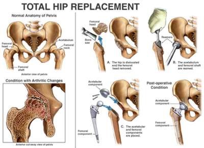
*Total hip replacement is also commonly done<ref name="p7" />


Not enough information is available on the chances for recurrence with this type of injury.<br>Also, no trials have even been set up to investigate whether a surgical intervention is really superior to a non-surgical treatment.<ref name=“Mandhkani M”/><br><br>  
[[Image:HIPREPLACEMENT.jpg]]<br>  


== Physical Therapy Management    ==
(Example of a total hip replacement, [http://ehealthmd.com/content/what-hip-replacement) ehealthmd.com/content/what-hip-replacement) ]


The treatment of skier’s thumb is different for partial and a complete ruptures. Partial ruptures are treated conservatively. The MCP joint is immobilized, with the MCP fixed and the IP joint remaining free to prevent unnecessary stiffness. A navicular cast or brace is usually used. Swelling can be controlled with elevation while supine and the use of cold compresses as needed.[7, LOE:3B]<br>The primary goal of rehabilitation is enhancing the patients' function and reducing the time of functional recovery, the reported treatment presents potential advantages in the management of this frequent acute hand injury.
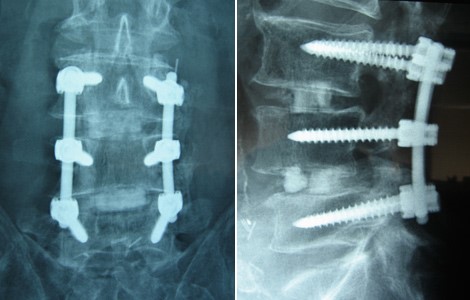
*Surgical spine fusion (when spinal cord or nerve function are compromised)&nbsp;<ref name="p0"/>


<br>  
[[Image:POSTOPERATIVE.jpg]]<br>  


● Conservative treatment<br>Partial UCL injuries like ligament strains, partial tears, low-demand patients and poor-operative candidates, including patients with degenerative MCP joint disease are effectively treated conservatively.[7, LOE:3B]<br>Controversy also exists about treating a bony skier’s thumb without surgery. The literature however shows that if the MCP joint is stable during testing and there is no dislocation of the fragment, this injury can be treated conservatively without reason for concern.  
(Postoperative x-rays anterior-posterior (A) and lateral (B) views demonstrate good pedicle screw placement and fusion at the patient’s six month follow-up., https://www.bnasurg.com/patient-resources-back-pain.php)


&nbsp; ❖ For patients with: <br>- less than 30 degrees of valgus laxity of extension of the MCPJ <br>- less than 15 degrees difference between sides and no signs of avulsion fracture on radiographs.[1, LOE:3B]
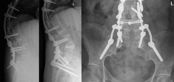
*Surgical correction of spinal deformities, this is called an osteotomy (2a) <ref name="p7"/><ref name="p8"/><ref name="p9"/>


<br>&nbsp; ❖ Immobilisation: <br>from 10 days up to 6 weeks, depending on the degree of laxity during the initial examination. Authors of a recent review on skier’s thumb agreed on a 4-week period. [38, LOE:1A]
[[Image:OSTEOTOMY.jpg]]<br>  


&nbsp; &nbsp; &nbsp; ■ suggestions:
( Example of a osteotomy on a women of 40, who has spinal deformities caused by spondyloarthritis,


- A short-arm thumb spica cast<br>- Thermoplastic splint:allows for the patient to begin movement of the interphalangeal joint.[1, LOE:3B] <br>- A hand-based removable thumb spica orthosis. The MCP joint is immobilized, with the MCP fixed and the IP joint remaining free to prevent unnecessary stiffness.(Figure 7)[1, LOE:3B] [38, LOE:1A][39, LOE:2B]<br>Wearing a splint will avoid putting radial stress on the thumb and gives the ligament time to heal.[1: LOE:3B] The optimal positioning for the splint involves holding the MCPJ in slight flexion with a slight ulnar deviation; the interphalangeal joints should not be immobilized in the splint.[1: LOE:3B]<br>
[http://www.scoliosisjournal.com/content/6/1/6/figure/F4)  http://www.scoliosisjournal.com/content/6/1/6/figure/F4) ]<br>No specific drugs is considered more superior than another for the treatment of spondyloartritis.


<br>
== Physical Therapy Management  ==


[[Image:Spica.jpg]]
Apart from a medication treatment, physiotherapy is recommended in spondyloarthritis. <ref name="p4"/><ref name="p3"/> This physical therapy generally focuses on the exercise regimens whose purpose is to maintain mobility and strength, relieve symptoms, prevent or decrease spinal deformity, and improve overall function and quality of life.&nbsp;<ref name="p1"/> The physiotherapy treatment consists mainly of exercise therapy. Evidence level of this therapy? The patients should perform daily special stretching and strengthening exercises to maintain the strength and mobility in the joints and reduce pain and stiffness.<ref name="p3">Reveille J.D., Americain college of Rheumatology, 2005 Jun 5: 5</ref><ref name="p3" /><ref name="p5" /> The strengthening exercises help to support and take pressure off sore joints. They also strengthen bones and improve balance. One can use weights or dumbbells for strengthening exercises.


Figure 7. Thumb spica splint.<ref name="Leggit" />LOE: 5
Flexibility training can maintain or even improve mobility of muscles and joints. Therefore major muscle groups such as erector spine, shoulder muscles, hip flexors, hamstrings and quadriceps should be stretched. This can also be done by partaking in yoga.<ref name="p1" /> <ref name="p4" />


<br>&nbsp; ❖ Exercise therapy: <br>After the period of immobilization is over, the therapy can be started. Most likely the patient will perform exercises that help strengthen and stretch the joint in order to regain full function in your thumb. (11) The patient should begin supervised hand therapy during the period of immobilization.<ref name=“Anderson”/> Gentle flexion and extension range of motion exercises can begin after about four weeks, with the patient continuing to wear the splint between therapy sessions. After 8 weeks progressive strengthening exercises may begin, but unrestricted activity is not allowed until after 12 weeks.<ref name=“Anderson”/> Gripping and pinching activities should not started until 10-12 weeks and should be advanced as tolerated; forceful gripping activities are typically not tolerated until about week 12.[7, LOE:3B]
Spa-exercise and balneotherapy programmes have short-term benefits in QoL outcomes; spa-exercise is superior in pain relief, while balneotherapy further improves disease activity.The balneotherapy interventions consist of mineral baths plus mud packs, radon-carbon dioxine baths, carbon dioxine baths, Dead Sea baths and tap water of 36°C.&nbsp;<ref name="p5" />  


● Treatment after surgery<br>The content of the physical therapy after surgery is the same as those of the conservative treatment, besides:<br>- Duration of Immobilisation: usually 6 weeks is applied <br>- Control radiograph after immobilisation <br> <br>Following surgery a splint is usually worn for four to five weeks. Immediate postoperative motion of the operated joint produced faster and better functional results. Therefor the use of a functional splint is preferred, as well as the early progressive start with moving within the boundaries of pain. [39] Athletes whose injuries require surgery can usually return to play in about three to four months. [11] The study of Derkash, considering pain, stability, muscular force (tweezers grip) and functionality in ADL, shows that less than 5% of the patients experience a weakened tweezers grip and stiffness. Pain was absent or mild in 99% of the cases. 96% of the treated patients were satisfied with the results of the operation. When a secondary operation is required results were less successful. [40]<br>When the pain has subsided and the range of motion has completely returned, the hand can be completely used again. Usually this takes about 3 months.[38, LOE:1A]
Unfortunately these benefits diminish or disappear over a period of 6 to 15 months.&nbsp;<ref name="p1" /> An addition of aerobic exercise to conventional stretching and mobility home exercise programmes results in superior functional fitness. Walking and swimming are examples of such aerobic exercise.&nbsp;<ref name="p1" /> <ref name="p4" />
* Swimming: three times a week for six weeks:
** 10 min warm-up + 5 min stretching
**30 min of swimming at a moderate intensity (60-70% heart rate [HR] reserve – 12 beats/minute)  
**10 min cooling down + 5 min stretching


<br>&nbsp; ❖ Exercises. [32, LOE:1A] <br>- Thumb active range of motion: With your palm flat on a table or other surface, move your thumb away from your palm as far you can. Hold this position for 5 seconds and bring it back to the starting position. Then rest your hand on the table in a handshake position. Move your thumb out to the side away from your palm as far as possible. Hold for 5 seconds. Return to the starting position. Next, bring your thumb across your palm toward your little finger. Hold this position for 5 seconds. Return to the starting position. Repeat this entire sequence 15 times. Do 2 sets of 15. [32,LOE:1A] <br>- Wrist range of motion <br>- Flexion: Gently bend your wrist forward. Hold for 5 seconds. Do 2 sets of 15. [32, LOE:1A] <br>- Extension: Gently bend your wrist backward. Hold this position 5 seconds. Do 2 sets of 15. [32, LOE:1A] <br>- Side to side: Gently move your wrist from side to side (a handshake motion). Hold for 5 seconds in each direction. Do 2 sets of 15. [32, LOE:1A] <br>- Thumb strengthening: Pick up small objects, such as paper clips, pencils, and coins, using your thumb and each of your other fingers, one at a time. Practice this exercise for about 5 minutes. [32, LOE:1A] <br>- Finger spring: Place a large rubber band around the outside of your thumb and fingers. Open your fingers to stretch the rubber band. Do 2 sets of 15. [32, LOE:1A] <br>- Grip strengthening: Squeeze a soft rubber ball and hold the squeeze for 5 seconds. Do 2 sets of 15. [32, LOE:1A] <br>- Wrist flexion: Hold a can or hammer handle in your hand with your palm facing up. Bend your wrist upward. Slowly lower the weight and return to the starting position. Do 2 sets of 15. Gradually increase the weight of the can or weight you are holding. [32, LOE:1A] <br>- Wrist extension: Hold a soup can or hammer handle in your hand with your palm facing down. Slowly bend your wrist up. Slowly lower the weight down into the starting position. Do 2 sets of 15. Gradually increase the weight of the object you are holding. [32, LOE:1A] <br><br>
* Walking: 30 minutes, three times a week for six weeks - Walking exercise should be performed at 60-70% of the pVO2, at a level of 13-15 on the Borg scale and 60-70% heart rate reserve.  
Supervised group exercise programs have better short-term outcomes than unsupervised home exercises.The chronic nature of SpA requires ongoing, regular exercise.&nbsp;<ref name="p1"/> <ref name="p1"/> <ref name="p3"/><br>Special attention should be given to a good posture of the patient.<ref name="p3"/> RAPIT (Rheumatoid Arthritis Patients In Training) is a training program for patients with rheumatoid arthritis. It is a biweekly, supervised groupsession that consists of bicycle training, an exercise circuit, and a sport or a game. The duration of each session varies from 60 to 75 minutes.


== Key evidence  ==
Cycle ergometer training (duration: 20 minutes)
* 1-2 minutes warm-up of at 40 watts (women) and 50 watts (men)
* 60–80 rounds per minute (rpm) and 60-80&nbsp;% of maximum heart rate (MHR=220/[226-age]) to increase aerobic capacity. Ratings of perceived exertion (0=“not at all exhausting” to 10=“maximal exhaustion”) should be at values of 5 to 6.
Exercise circuit (duration: 20-30 minutes) - The circuit training is a sequential training exercise to enhance muscle strength, strength endurance, mobility and coordination. Over 20 minutes a circuit of eight to ten single exercises is completed twice, each exercise lasting 60 to 90 seconds with 30-60 seconds resting time between each one.


== Recources  ==
Sport/games (duration: 20 minutes) - This section of the program consists of impact-delivering sporting activities such as badminton, volleyball, indoor soccer, and basketball.


Links to other physiopedia’s: Bennett fracture, ultrasound, MRI, X-rays and Valgus Stress to UCL
== References  ==


== Clinical bottom line  ==
see [[Adding References|adding references tutorial]].  
 
Skier’s thumb, also known as gamekeepers thumb, is an injury to the metacarpal phalangeal joint of the thumb. It can occur to the medial side of the thumb, but this is rare. The chance of an medial side injury is as small as 10-30%. <br>When there is abnormal movement like hyperabduction of the thumb or a sudden force on the thumb then the ligament can rupture (with or without an avulsion fracture). We diagnose an UCL rupture mostly with an ultrasound, which is the most reliable and not costly. We can also palpate an rupture by doing the stress test. <br>UCL rupture can be treated with an operation but this depends on different factors(see medical management). When an operation is needed, the treatment will likely be decided by the surgeon. This will also depend on some factors but most surgeons have a favourite treatment in the wide variety of operations. <br>Partial tears and strains are mostly not treated with an operation. It comes first with the immobilization of the MCP joint to give some rest and so that the body can recover. After that, the patients starts wearing a splint and gets six weeks of therapy sessions (this also includes for post-operative treatments). Where in the mobility and strength get back to the normal values compared with the contralateral thumb.<br>
 
<br> <br>
 
== Recent Related Research (from [http://www.ncbi.nlm.nih.gov/pubmed/ Pubmed])  ==
 
see tutorial on [[Adding PubMed Feed|Adding PubMed Feed]]  
<div class="researchbox">
<rss>http://eutils.ncbi.nlm.nih.gov/entrez/eutils/erss.cgi?rss_guid=1</rss>
 
Injuries in World Cup telemark skiing: a 5-year cohort study.<br>
</div>
== References  ==


1. ↑ 1.0 1.1 1.2 1.3 1.4 1.5 1.6 1.7 1.8 Anderson D. Skier’s thumb. Aust Fam Physician. 2010;39(8):575-577.<br>LEVEL OF EVIDENCE: 3B<br>2. ↑ 2.0 2.1 2.2 2.3 2.4 2.5 2.6 Patel S, et al. Collateral ligament injuries of the metacarpophalangeal joint of the thumb: a treatment algorithm. Strat Trauma Limb Recon. 2010;5:1-10.<br>LEVEL OF EVIDENCE: 3B<br>3. ↑ 3.0 3.1 American Society for Surgery of the Hand. Thumb sprains. www.assh.org/Public/HandConditions/Pages/ThumbSprains.aspx (accessed 18 March 2011).<br>LEVEL OF EVIDENCE: 5<br>4. ↑ Zeigler T. Thumb sprain also known as “skier’s thumb” or “gamekeeper’s thumb”. www.sportsmd.com/Articles/tabid/1010/id/50/Default.aspx?n=thumb_sprain_also_known_as_skier%E2%80%99s_thumb_or_gamekeeper%E2%80%99s_thumb (accessed 13 March 2011).<br>LEVEL OF EVIDENCE: 5<br>5. Milner CS, et al., Gamekeeper’s thumb—a treatment – oriented magnetic resonance imaging classification. J Hand Surg. Am., 2015; 40(1): 90-5.<br>LEVEL OF EVIDENCE: 1A<br>6. ↑ 6.0 6.1 6.2 6.3 Leggit JC et al. Acute finger injuries: Part II. Fractures, dislocations, and thumb injuries. Am Fam Physician. 2006;73(5):827-834.<br>LEVEL OF EVIDENCE: 4<br>7. ↑ 7.0 7.1 7.2 7.3 7.4 Rettig A et al. Anatomic reconstruction of thumb metacarpophalangeal joint ulnar collateral ligament using an interference screw docking technique. Tech Hand Up Extrem Surg. 2009;13(1):7-10.<br>LEVEL OF EVIDENCE: 3B<br>8. Madan, S. S., Injury to Ulnar Collateral Ligament of Thumb. Orthopaedic Surgery. 2014;6:1–7.<br>LEVEL OF EVIDENCE: 2A<br>9. Pediatric Trauma Care II: A clinical reference for physicians and nurses caring for the acutely injured child‬. AHC Media. LLC. 2014;6:52-53‬.<br>LEVEL OF EVIDENCE: 5 <br>10. Jonathan Cluett, Gamekeeper's Thumb. 2014<br>LEVEL OF EVIDENCE: 5 <br>11. Clifford D. Stark, Living with Sports Injuries. Infobase Publishing. 2010‬;6:84- 86‬‬‬‬‬‬‬‬‬‬‬‬‬‬‬‬‬‬‬‬‬‬‬‬‬‬<br>LEVEL OF EVIDENCE: 5<br>12. Mandhkani Mahajan. Rupture of the ulnar collateral ligament of the thumb. Mahajan and Rhemrev International Journal of Emergency Medicine. 2013;6:31.<br>LEVEL OF EVIDENCE: 1A <br>13. Chrysi Tsiour. Injury to the Ulnar Collateral Ligament of the Thumb. American Association for Hand Surgery;2008.<br>LEVEL OF EVIDENCE: 1A <br>14. G.I Mitsionis. treatment of chronic injuries of the ulnar collateral ligament of the thumb using a free tendon graft and bone suture anchors. The Journal of Hand Surgery:British &amp; European volume. 2000; Vol 25:2:208–211.<br>LEVEL OF EVIDENCE: 2B <br>15. C. Fusetti. The ECRL bone-tendon ligamentoplasty for chronic ulnar instability of the metacarpophalangeal joint of the thumb. Hand Surgery Unit, Chirurgie de la Main. 2005; Vol 24:5:217–221.<br>LEVEL OF EVIDENCE: 1B <br>16. Başar H. Comparison of results after surgical repair of acute and chronic ulnar collateral ligament injury of the thumb. Chirurgie de la Main. 2014;vol 33:6:384–389.<br>LEVEL OF EVIDENCE: 2B <br>17. Palmer DH et al. Helicopter skiing wrist injuries. A case report of "bugaboo forearm".; Am J Sports Med. 1994 Jan-Feb;22(1):148-9.<br>LEVEL OF EVIDENCE: 1A<br>18. J. B. Engelhardt, Rupture of the ulnar collateral ligament of the metacarpophalangeal joint of the thumb.injury.1993;vol24:1:21-24.<br>LEVEL OF EVIDENCE: 2B <br>19. G.S.J. Chuter,Ulnar collateral ligament injuries of the thumb: 10 years of surgical experience. Injury. June 2009;vol40:6:652-656.<br>LEVEL OF EVIDENCE: 2B <br>20. J. A. Fairclough et al. Skier’s thumb-a method of prevention; Injury 1986; 17,203-204<br>Level of evidence: 1A<br>21. Samora J.B. Outcomes After Injury to the Thumb Ulnar Collateral Ligament,, Clin J Sport Med. 2013;23(4):247-254.<br>LEVEL OF EVIDENCE: 3A <br>22. Schroeder NS.Thumb ulnar collateral and radial collateral ligament injuries.<br>Clin Sports Med. 2015;34(1):117-26.<br>Level of evidence: 2C<br>23.  .<br>LEVEL OF EVIDENCE: 3B <br>24. Abrahamsson SO, et al. Diagnosis of displaced ulnar collateral ligament of the metacarpophalangeal joint of the thumb. J Hand Surg Am. 1990; 15: 457–460.<br>LEVEL OF EVIDENCE: 2B <br>25. Hergan K, et al. Pitfalls in sonography of the Gamekeeper's thumb. Eur Radiol. 1997; 7: 65–69.<br>LEVEL OF EVIDENCE: 2B <br>26. Lohman M, et al. MR imaging in chronic rupture of the ulnar collateral ligament of the thumb. Acta Radiol. 2001; 42: 10–14.<br>LEVEL OF EVIDENCE: 3B<br>27. Ganel A, et al. “Gamekeeper's thumb”. Injuries of the ulnar collateral ligament of the metacarpophalangeal joint. Br J Sports Med. 1980; 14: 92–96.<br>LEVEL OF EVIDENCE: 4 <br>28. G.S.J. Chuter. Ulnar collateral ligament injuries of the thumb: 10 years of surgical experience. Departments of A&amp;E. 2009;Vol 40,Issue 6,:652–656<br>LEVEL OF EVIDENCE: 2B <br>29. Glickel SZ, et al. Dislocations and ligament injuries in the digits. Green DP, Hotchkiss RN, Pederson WC, eds. Green's Operative Hand Surgery. 4th ed. New York, NY: Churchill Livingstone. 1999; 772-807.<br>LEVEL OF EVIDENCE: 2C<br>30. Zemel NP. Metacarpophalangeal joint injuries in fingers. Hand Clin. 1992; 8(4):745-54.<br>LEVEL OF EVIDENCE: 2C<br>31. Sebastin S, et al. Overview of finger, hand and wrist fractures. http://www.uptodate.Foye PM et al, Skier's Thumb. Medscape. 2010<br>LEVEL OF EVIDENCE: 2C<br>32. Mayfield JK, et al. Carpal dislocations: pathomechanics and progressive perilunar instability. J Hand Surg Am. 1980;5 (3): 226-41. -Pubmed citation<br>LEVEL OF EVIDENCE: 3B<br>33. Michael A. et al, Evaluation and Treatment of Injuries of the Ulnar Collateral Ligament of the Thumb Metacarpophalangeal Joint, Bulletin of the NYU Hospital for Joint Diseases. 2009;67(1):68-74<br>LEVEL OF EVIDENCE: 1A<br>34. Hall ™, et al. Therapeutic Exercise Moving Toward Function. 3rd ed; Lippincott, Williams and Wilkins, 2010<br>LEVEL OF EVIDENCE: 2C<br>35. com/home. Accessed March 4, 2014.<br>LEVEL OF EVIDENCE: 2C<br>36. Shelain Patel, et al. Collateral ligament injuries of the metacarpophalangeal joint of the thumb: a treatment algorithm; Strategies Trauma Limb Reconstr. 2010; 5(1): 1–10.<br>LEVEL OF EVIDENCE: 2A<br>37. Laub Jr DR et al, Thumb Fractures and Dislocations, Medscape, Sep 2010<br>LEVEL OF EVIDENCE: 2C<br>38. Mandhkani M, et al. Rupture of the ulnar collateral ligament of the thumb – a review. Int J Emerg Med. 2013; 6: 31. doi: 10.1186/1865-1380-6-31PMCID: PMC3765347<br>LEVEL OF EVIDENCE: 1A <br>39. Rocchi L1, et al. A modified spica-splint in postoperative early-motion management of skier's thumb lesion: a randomized clinical trial. <br>LEVEL OF EVIDENCE: 2B<br>40. Derkash RS, et al. Acute surgical repair of the skier's thumb. Clin Orthop Relat Res 1987;(216):29-33.<br>LEVEL OF EVIDENCE: 2B<br>41. Fricker R1, et al.Skier's thumb. Treatment, prevention and recommendations.<br>LEVEL OF EVIDENCE:<br>42. Ritting et al., Ulnar Colletral Ligament Injury of the Thumb Metacarpophalangeal Joint. Sport Med; 2010; 20(2):106–112. <br>LEVEL OF EVIDENCE: <br>43. Ebrahim FS et al. US diagnosis of UCL tears of the thumb and Stener lesions: technique, pattern-based approach, and differential diagnosis. Radiographics. 2006; 26(4): 1007-20 <br>LEVEL OF EVIDENCE: 1B<br><br>
<references />  


[[Category:Primary Contact]]
[[Category:Vrije_Universiteit_Brussel_Project|Template:VUB]]
<references />
[[Category:Sports Medicine]]
[[Category:Sports Injuries]]

Revision as of 23:39, 20 April 2019

Definition/Description[edit | edit source]

Spondyloarthritis is a name for a group of diseases that is included in a larger term 'arthritis'.[1][2][3] Inflammation can occur in spine, sacroiliac and peripheral joints as well near the attachments of tendons and ligaments.[3] This disease provokes to pain, stiffness and fatigue in back, legs and arms as in joints, ligaments and tendons.[4] [5]Eruption, eye and intestinal problems may also occur.[1][3]
Spondyloarthritis in adults can be subdivided more specifically:[1][2][6][7][8][1]

  • ankylosing spondylitis or Bechterew disease
  • psoriatic arthritis[2]
  • reactive arthritis[3]
  • enteric arthritis
  • undifferentiated arthritis

Clinically Relevant Anatomy[edit | edit source]

Spondyloarthritis is the overall name for a family of inflammatory rheumatic diseases. [1][2]

Due to this fact, there is a large complexity. This is because there are several anatomic structures involved. We can assume that the inflammation can occur on all the joints of the spine. The facet joints, endplates, bone marrow, … every part of the spine can be affected by an inflammation. [9] Sacroiliitis in SpA is characterized by involvement of different joint structures. Whereas the iliac and the sacral side of the sacroiliac joints are almost equally affected, the dorsocaudal synovial part of the joint is involved significantly more often than the ventral part, especially in early disease. Sacroiliac enthesitis is not a special feature of early sacroiliac inflammation. There is a difference between axial and peripheral spondyloarthitis, with axial spondyloarthitis back pain and inflammation of the sacroiliac joints are the main complaints. In peripheral spondyloarthritis, the inflammation of peripheral joint and tendons are the main complaints. Further, spondyloarthritis can show an inflammation of peripheral joints (for example, knees and ankles), and tendons (for example, the Achilles tendon).[1][2][3][9]

Epidemiology /Etiology[edit | edit source]

Spondyloarthritis is a pathology that specifically strikes young people.[10] The symptoms most frequently start before the age of 45. [2] It affects more males than females. [5][4]
Predisposition to spondyloarthritis, especially SpA, is determined largely by genetic factors. The incidence rate is higher in populations with a higher prevalence of HLA-B27.[6] Psoriatic skin lesions and colitis due to inflammatory bowel disease (IBD) have been considered as both basic, subtype-defining entities with their own genetic background (distinct from HLA-B27 genotype), and as manifestations of spondyloarthritis.[6] There is a strong need to diagnose patients with SpA in an earlier stage; currently there is a delay of 5–10 years between onset of the first symptoms and diagnosis.[6][4]

Characteristics/Clinical Presentation[edit | edit source]

Symptoms that may occur with spondyloarthritis are pain, stiffness and fatigue in the back, legs and arms. There are no typical characteristics, because spondyloarthritis characterises with more than one symptom. We see that significantly more women have knee pain as presenting symptom.[11][12][13][14][15][16] and we can assume that severity of symptoms can vary between individuals[15]. Here are the most common characteristics.[2][7][13][14][16][17][18]

  • back pain
  • osteoporosis
  • spinal fractures
  • peripheral arthritis, usually asymmetric, relatively more in the lower limbs.[2][3][2]
  • enteritis
  • dactylitis
  • inflammation of the heart valve – pneumonia
  • extra articular disorders such as uveitis, skin porosiasis or inflammatory bowel disease
  • strong familial aggregation of spondyloarthritis, psoriasis, IBD, uveitis
  • association with HLA-B27
  • no increased CRP and rheumatoid factor

Differential Diagnosis[edit | edit source]

The disease starts with hip or low back pain. The most common symptom is intermittent pain that progressively gets worse thoughout the day, in the morning, and following intensive activity. [1] Most patients experience back pain in the sacroiliac joints. However, pain can involve all the parts of the spine. Pain relief is sometimes achieved by bending over. It is possible that a patient is not able to fully expand the chest due to the involvement of the joints between the ribs.

Diagnostic Procedures[edit | edit source]

Antecedents and physical examination are the major factors leading to diagnosis, although radiologic evidence of sacroiliitis is very helpful [2][7] In the early-1990s, two classification criteria, Amor and the European Spondyloarthropathy Study Group (ESSG), were proposed for diagnosing SpA [2][3] All criteria developed so far (including the ESSG and Amor criteria) were developed as classification criteria, although they are often used as diagnostic criteria [7][8]

Amor criteria for spondyloarthritis [5]:

Paramters
Scoring

Clinical symptoms or past history of

Lumbar or dorsal pain at night or morning stiffness of lumbar or dorsal region

1
Asymmetric oligoarthritis
2

Buttock pain
Or if alternate buttock pain

1

2

Sausage-like toe or digit
2
Heel pain or other well- defined enthesitis
2
Iritis
2
Non- gonococcal urethritis or cervicitis within 1 month before the onset of arthritis

1

Acute diarrhoea within 1 month befor the onset of arthritis
1
Psoriasis, balanitis or inflammatory bowel disease ( ulcerative colitis or chrohn’s disease)
2
Radiological findings

Sacroiliitis (bilateral grade 2 or unilaterale grade 3) 3
Genetic background

Presence of HLA-B27 or family history of ankylosing spondylitis, reactieve arthritis, uveitis, psoriasis or inflammatory bowel disease
2
Response to treatment

Clear- cut improvement within 48 hours after non steroidal anti- inflammatory drug intake or rapid relapse of the pain after their discontinuation
2

A patient is considered to be suffering from spondyloarthritis if the sum is ≥ 6

The need for a standardized, evidence-based approach to spondyloarthritis classification led to the development of the European Spondyloarthropathy Study Group (ESSG) [4] preliminary classification criteria for spondyloarthritis in 1991 [10]:
Inflammatory spinal pain or synovitis (asymmetric, predominantly in lower limbs) and any one of the following: [4]

  • Positive family history
  • Psoriasis
  • Inflammatory bowel disease
  • Acute diarrhea or urethritis or cervicitis preceding the arthritis
  • Alternate buttock pain
  • Enthesopathy
  • Radiological sacroilits

Another is the concept of IBP (Low Back Pain), which is defined as the presence of at least four of the following five parameters [1], [6]:

  1. Age at onset less than 40 years
  2. Insidious onset
  3. Improvement with exercise
  4. No improvement with rest
  5. Pain at night (with improvement upon getting up).

Studies are under way to define ASAS criteria for nonaxial (peripheral) SpA.
In the ASAS classification criteria, several SpA features are described. These features are called SpA features because they are frequently present in patients with SpA ,[9][6]

File:ASAS classification.png

The main features of an early diagnosis of any rheumatic disease, including spondyloarthritis, are clinical history, clinical symptoms, clinical examination, laboratory parameters and imaging. [10]
Clinical symptoms:

  • Inflammatory back pain
  • Arthritis ( swelling, joint effusion, or detected by imaging)
  • Accompanying features, including psoriasis, crohn-like colitis and anterior uveitis

Clinical history:

  • Family
  • Rheumatic symptoms
  • Accompanying features

Clinical examination:

  • Lateral flexion of the lumbar spine (<10cm)
  • Chest expansion (<4cm)
  • Cervical rotation (<70°)

Laboratory parameters:

  • HLA-B27
  • C- reactive protein
  • Erythrocyte sedimentation rate

Imaging:

  • Radiography
  • MRI
  • Ultrasonography

Outcome Measures[edit | edit source]

Examination[edit | edit source]

Patients with spondyloarthritis will complain about back pain, fatigue and stiffness. The pain will decrease when the patients exercise, but will persist at they rest. It is common for the patient to have pain at night, this pain can improve when the patients gets out of bed and moves around. (this should improve when they get up).[2] [8] The motion of the lumbar spine of the patients will be limited in both the sagittal and the frontal planes. [2]

Psoriasis, finger swelling, Crohn's disease or ulcerative colitis can be indicative for Spondyloarthritis.

Sacroiliitis grade ≥ 2 bilaterally or grade 3 to 4 unilaterally is suggestive for SpA (grade 0: normal; grade I: some blurring of the joint margins - suspicious; grade II: minimal sclerosis with some erosion; grade III: definite sclerosis on both sides of joint 5 & severe erosions with widening of joint space with or without ankylosis; grade IV: complete ankylosis)  [4][5]

There are also active inflammatory and chronic lesions that can be found on a MRI-scan (see images). [3] [6] [7]
MRI1.png

[3]Sieper et al. The Assessment of SpondyloArthritis international Society (ASAS) handbook: a guide to assess spondyloarthritis

Laboratory testing

  • Common presence of human leukocyte antigen-B27
  • Elevated C-reactive protein
  • Absence of rheumatoid factor [2]

Medical Management[edit | edit source]

Reliant on your symptoms and how severe your condition is, the doctor can decide what kind of treatment is the best option for the patient.

Medicines such as:

  • analgesics (pain-relievers, by example paracetamol)[10]
  • non-steroidal anti-inflammatory drugs (NSAIDS, by example naproxen, ibuprofen).[9]
  • Anti-rheumatic drugs (DMARDs) have been proven effective in the treatment, but only for the arms and legs, not for the spine and sacroiliac joints.[10]
  • Corticosteroids, given by the mouth or injections, can be effective. We must remind ourselves for the side effects, such as osteoporosis and infections.[4]
  • Injections of deposteroid in the joints of tendon sheaths are also used for symptomatic relief of the local flares.[4]
  • TNF alpha blockers are also effective in both spinal and peripheral joints.[9][10]
  • There are three kind of TNF alpha blockers we can use:
    • Infliximab (Remicade), given a dose of 5 mg/kg intravenously every sic to eight weeks.
    • Etanercept (Enbrel), 25 mg given under de skin twice a week
    • Adalimumab (Humira), 40 mg injected, every other week

TNF treatment is expensive and is not without complications, therefore, NSAIDs and DMARD should be tried first.

Surgery:

  • Total hip replacement is also commonly done[5]

HIPREPLACEMENT.jpg

(Example of a total hip replacement, ehealthmd.com/content/what-hip-replacement)

  • Surgical spine fusion (when spinal cord or nerve function are compromised) [8]

POSTOPERATIVE.jpg

(Postoperative x-rays anterior-posterior (A) and lateral (B) views demonstrate good pedicle screw placement and fusion at the patient’s six month follow-up., https://www.bnasurg.com/patient-resources-back-pain.php)

  • Surgical correction of spinal deformities, this is called an osteotomy (2a) [5][6][7]

OSTEOTOMY.jpg

( Example of a osteotomy on a women of 40, who has spinal deformities caused by spondyloarthritis,

http://www.scoliosisjournal.com/content/6/1/6/figure/F4)
No specific drugs is considered more superior than another for the treatment of spondyloartritis.

Physical Therapy Management[edit | edit source]

Apart from a medication treatment, physiotherapy is recommended in spondyloarthritis. [9][3] This physical therapy generally focuses on the exercise regimens whose purpose is to maintain mobility and strength, relieve symptoms, prevent or decrease spinal deformity, and improve overall function and quality of life. [1] The physiotherapy treatment consists mainly of exercise therapy. Evidence level of this therapy? The patients should perform daily special stretching and strengthening exercises to maintain the strength and mobility in the joints and reduce pain and stiffness.[3][3][10] The strengthening exercises help to support and take pressure off sore joints. They also strengthen bones and improve balance. One can use weights or dumbbells for strengthening exercises.

Flexibility training can maintain or even improve mobility of muscles and joints. Therefore major muscle groups such as erector spine, shoulder muscles, hip flexors, hamstrings and quadriceps should be stretched. This can also be done by partaking in yoga.[1] [9]

Spa-exercise and balneotherapy programmes have short-term benefits in QoL outcomes; spa-exercise is superior in pain relief, while balneotherapy further improves disease activity.The balneotherapy interventions consist of mineral baths plus mud packs, radon-carbon dioxine baths, carbon dioxine baths, Dead Sea baths and tap water of 36°C. [10]

Unfortunately these benefits diminish or disappear over a period of 6 to 15 months. [1] An addition of aerobic exercise to conventional stretching and mobility home exercise programmes results in superior functional fitness. Walking and swimming are examples of such aerobic exercise. [1] [9]

  • Swimming: three times a week for six weeks:
    • 10 min warm-up + 5 min stretching
    • 30 min of swimming at a moderate intensity (60-70% heart rate [HR] reserve – 12 beats/minute)
    • 10 min cooling down + 5 min stretching
  • Walking: 30 minutes, three times a week for six weeks - Walking exercise should be performed at 60-70% of the pVO2, at a level of 13-15 on the Borg scale and 60-70% heart rate reserve.

Supervised group exercise programs have better short-term outcomes than unsupervised home exercises.The chronic nature of SpA requires ongoing, regular exercise. [1] [1] [3]
Special attention should be given to a good posture of the patient.[3] RAPIT (Rheumatoid Arthritis Patients In Training) is a training program for patients with rheumatoid arthritis. It is a biweekly, supervised groupsession that consists of bicycle training, an exercise circuit, and a sport or a game. The duration of each session varies from 60 to 75 minutes.

Cycle ergometer training (duration: 20 minutes)

  • 1-2 minutes warm-up of at 40 watts (women) and 50 watts (men)
  • 60–80 rounds per minute (rpm) and 60-80 % of maximum heart rate (MHR=220/[226-age]) to increase aerobic capacity. Ratings of perceived exertion (0=“not at all exhausting” to 10=“maximal exhaustion”) should be at values of 5 to 6.

Exercise circuit (duration: 20-30 minutes) - The circuit training is a sequential training exercise to enhance muscle strength, strength endurance, mobility and coordination. Over 20 minutes a circuit of eight to ten single exercises is completed twice, each exercise lasting 60 to 90 seconds with 30-60 seconds resting time between each one.

Sport/games (duration: 20 minutes) - This section of the program consists of impact-delivering sporting activities such as badminton, volleyball, indoor soccer, and basketball.

References[edit | edit source]

see adding references tutorial.

  1. 1.00 1.01 1.02 1.03 1.04 1.05 1.06 1.07 1.08 1.09 1.10 1.11 1.12 1.13 1.14 1.15 1.16 1.17 1.18 Braun J. et al, Spondyloarthritides, Internist., 2011 May 19: 5: 2C
  2. 2.00 2.01 2.02 2.03 2.04 2.05 2.06 2.07 2.08 2.09 2.10 2.11 2.12 2.13 Braun J., Sieper J., Spondyloarthritides., Z Rheumatol. 2010 Jul; 69(5):425-32 :4: 2C
  3. 3.00 3.01 3.02 3.03 3.04 3.05 3.06 3.07 3.08 3.09 3.10 3.11 3.12 3.13 Reveille J.D., Americain college of Rheumatology, 2005 Jun 5: 5
  4. 4.0 4.1 4.2 4.3 4.4 4.5 4.6 4.7 Rudwaleit M .Et al. The Assessment of SpondyloArthritis International Society classification criteria for peripheral spondyloarthritis and for spondyloarthritis in general. Ann Rheum Dis. 2011;70(1):25.: 4
  5. 5.0 5.1 5.2 5.3 5.4 5.5 Burgos-Vargas R.The assessment of the spondyloarthritis international society concept and criteria for the classification of axialspondyloarthritis and peripheral spondyloarthritis: A critical appraisal for the pediatric rheumatologist. Pediatric Rheumatology 2012, 10:14  : 2C
  6. 6.0 6.1 6.2 6.3 6.4 6.5 6.6 6.7 Braun J, Sieper J. Early diagnosis of spondyloarthritis. Nature clinical practice rheumatology. october 2006 vol 2 no 10  : 2C
  7. 7.0 7.1 7.2 7.3 7.4 7.5 Jürgen Braun* and Joachim Sieper†, Early diagnosis of spondyloarthritis , 2006 : 2C
  8. 8.0 8.1 8.2 8.3 8.4 ozgur akgul, Classification criteria for spondyloarthropathies, , World J Orthop. 2011 December 18; 2(12): 107-115 : 2A
  9. 9.0 9.1 9.2 9.3 9.4 9.5 9.6 9.7 Walter P. Maksymowych, Frpc, Magnetic Resonance Imaging for Spondyloarthritis — Avoiding the Minefield (https://jrheum.com/subscribers/07/02/259.html) 4
  10. 10.0 10.1 10.2 10.3 10.4 10.5 10.6 10.7 Sieper J. Et al. Concepts and epidemiology of spondyloarthritis. Elsevier Ltd. 2006 : 2C
  11. Cite error: Invalid <ref> tag; no text was provided for refs named Reveille
  12. Cite error: Invalid <ref> tag; no text was provided for refs named Sieper
  13. 13.0 13.1 Cite error: Invalid <ref> tag; no text was provided for refs named Mease
  14. 14.0 14.1 Cite error: Invalid <ref> tag; no text was provided for refs named VDBerg
  15. 15.0 15.1 Cite error: Invalid <ref> tag; no text was provided for refs named Roussou
  16. 16.0 16.1 Cite error: Invalid <ref> tag; no text was provided for refs named Slobodin
  17. Slobodin G., Recently diagnosed axial spondyloarthritis: gender differences and factors related to delay in diagnosis., Clin Rheumatol., 2011 Mar 1
  18. Colbert R.A., Early axial spondyloarthritis., Curr Opin Rheumatol., 2010 Sep;22(5):603-7