Calcific Tendinopathy of the Shoulder: Difference between revisions
Mary Harris (talk | contribs) No edit summary |
No edit summary |
||
| (124 intermediate revisions by 15 users not shown) | |||
| Line 1: | Line 1: | ||
<div class="editorbox"> | |||
'''Original Editors''' | '''Original Editors''' [[User:Mary Harris|Mary Harris]], [[User:Thomas Lawlor|Tom Lawlor]], [[User:Patrick Bales|Patrick Bales]], [[User:Misty Hillin|Misty Hillin]], [[User:Rick Wetherald|Rick Wetherald]] as part of the Texas Evidence Based Practice Project. | ||
''' | '''Top Contributors''' - {{Special:Contributors/{{FULLPAGENAME}}}} | ||
</div> | </div> | ||
| |||
< | == Introduction == | ||
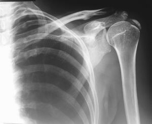
[[File:Dense calcification of the supraspinatus.jpeg|thumb|Calcification of the supraspinatus]] | |||
Calcific tendinopathy (CT) of the shoulder is a common, painful condition identified by the existence of calcium deposits in the rotator cuff tendons.<ref name=":2">Sansone V, Maiorano E, Galluzzo A, Pascale V. Calcific tendinopathy of the shoulder: clinical perspectives into the mechanisms, pathogenesis, and treatment. Orthopedic research and reviews. 2018;10:63.Available:https://www.dovepress.com/calcific-tendinopathy-of-the-shoulder-clinical-perspectives-into-the-m-peer-reviewed-fulltext-article-ORR (accessed 12.1.2023)</ref> It usually results in shoulder pain with decreased range of motion. Diagnosis is made by shoulder x-rays, with visible signs of calcium deposits overlying the rotator cuff insertion.Treatment consists of NSAIDs, physical therapy, corticosteroid injections and ultrasound-guided needle lavage. Those who fail conservative treatment may choose to have arthroscopic decompression of the calcium deposits.<ref name=":1">Orthobullets [https://www.orthobullets.com/shoulder-and-elbow/3042/calcific-tendonitis Calcific Tendonitis]Available:https://www.orthobullets.com/shoulder-and-elbow/3042/calcific-tendonitis (accessed 12.1.2023)</ref> | |||
"calcific | == Epidemiology == | ||
Usually occurs in middle-aged patients between the ages of 30 and 60, with a slight preference for females.<ref name=":0">Radiopedia [https://radiopaedia.org/articles/calcific-tendinitis?lang=gb Calcific Tendinitis] Available: https://radiopaedia.org/articles/calcific-tendinitis?lang=gb<nowiki/>(accessed 12.1.2023)</ref> | |||
== Pathogenesis == | |||
Current theories indicate that CT may be the result of a cell-mediated process in which calcium deposition occur followed by their spontaneous resorption. However, in a few cases, this self-healing process is disrupted, causing symptoms. Literature now is showing that biological and genetic factors may underlie CTs genesis.. These new finding may explain why most of the therapies currently in use provide partially satisfactory outcomes.<ref name=":2" /> | |||
== | |||
== Localisation == | |||
<sup></sup> | |||
*Supraspinatus tendon (80% of cases): critical zone - ''Most Common'' | |||
*Infraspinatus tendon (15% of cases): lower 1/3 | |||
*Subscapularis tendon (5%of cases): pre-insertional fibers<ref name="Serafini">Serafini G, Sconfienza L, Lacelli F, Silvestri E, Aliprandi A, Sardanelli F. Rotator cuff calcific tendonitis: short-term and 10-year outcomes after two-needle us-guided percutaneous treatment--nonrandomized controlled trial. Radiology [serial online]. July 2009;252(1):157-164. Available from: CINAHL Plus with Full Text, Ipswich, MA. Accessed September 20, 2011.</ref> | |||
{{#ev:youtube|ycphj08OJt0}} | |||
== Characteristics/Clinical Presentation == | == Characteristics/Clinical Presentation == | ||
The chief patient complaints to expect in calcific tendinopathy are: | |||
* Night pain, causing loss of sleep.<sup><ref name="Ebenbichler">Ebenbichler G R. et. al. Ultrasound therapy for calcific tendinitis of the shoulder. New England Journal of Medicine. 1999; Vol 340 (20): 1533-1538.</ref>, <ref name="Gimblett">Gimblett P, Saville J, Ebrall P. A conservative management protocol for calcific tendinitis of the shoulder. Journal Of Manipulative And Physiological Therapeutics [serial online]. November 1999;22(9):622-627.</ref>, <ref name="Alexander">Alexander L D., et. al. Exposure to Low Amounts of Ultrasound Energy Does Not Improve Soft Tissue Shoulder Pathology: A Systematic Review. Physical Therapy. 2010; vol 90 (1): 14-25.</ref>, <ref name="Wainner" />.</sup> | |||
* Constant dull ache<sup><ref name="Wainner" /></sup>. | |||
* Pain increases considerably with AROM<sup><ref name="Wainner" /></sup>. | |||
* Decrease in ROM, or complaint of stiffness <sup><ref name="Fusaro">Fusaro I, et. al. Functional results in calcific tendinitis of the shoulder treated with rehabilitation after ultrasonic-guided approach. Musculoskeletal Surgery. 2011 (95): S31–S36.</ref>, <ref name="Alexander" />, <ref name="Wainner" /></sup>. | |||
* Radiating pain up into the suboccipital region, or down into the fingers<ref name="Ebenbichler" />, <ref name="Gimblett" />, <ref name="Wainner" />.<br>The condition goes through 4 stage, see table below. | |||
{| width="400" cellspacing="1" cellpadding="1" border="1" | |||
{| width="400" | |||
|- | |- | ||
! | ! colspan="2" scope="col" bgcolor="#33cc00" | Stages<ref name="Wainner">Wainner R, Hasz M. Management of acute calcific tendinitis of the shoulder. Journal Of Orthopaedic & Sports Physical Therapy [serial online]. March 1998;27(3):231-237. ( LOE 4 )</ref> | ||
|- | |- | ||
| | | bgcolor="#66ff66" align="center" | Stage Name | ||
| | | bgcolor="#66ff66" align="center" | Presentation | ||
|- | |- | ||
| Chronic (Silent)<br> Phase | | Chronic (Silent)<br> Phase | ||
| | | | ||
*Presence of the calcific deposit <br>is asymptomatic and may be so for years. | *Presence of the calcific deposit <br>is asymptomatic and may be so for years. | ||
| Line 76: | Line 52: | ||
|- | |- | ||
| | | | ||
Mechanical Phase | Mechanical Phase | ||
| | | | ||
*Tendon impingement being a prominent finding | *Tendon impingement being a prominent finding | ||
*Pain of less severe nature than the acute phase | *Pain of less severe nature than the acute phase | ||
| Line 86: | Line 62: | ||
== Differential Diagnosis == | == Differential Diagnosis == | ||
* Incidental calcification: found in 2.5-20% of 'normal' healthy shoulders. | |||
* Degenerative calcification: found tendons with tear history; generally smaller; slightly older individuals | |||
* | * Loose bodies: associated chondral defect; associated secondary osteoarthritis<ref name=":0" /> | ||
* | |||
== Outcome Measures == | == Outcome Measures == | ||
*[https://www.physio-pedia.com/Visual_Analogue_Scale VAS Pain scale]<ref name="Cacchio">Cacchio A, Paoloni M, Spacca G, et al. Effectiveness of radial shock-wave therapy for calcific tendinitis of the shoulder: single-blind, randomized clinical study. Physical Therapy [serial online]. May 2006;86(5):672-682.( LOE 1b )</ref><br> | |||
*[[DASH Outcome Measure]] | |||
== Medical Management == | |||
Nonoperative | |||
# NSAIDs, physical therapy, stretching & strengthening, steroid injections | |||
# [[Tendinopathy Treatment Adjuncts|Extracorporeal shock-wave therapy as an adjunct treatment]]. Most useful in refractory calcific tendonitis in the formative and resting phases | |||
# Ultrasound-guided needle lavage vs. needle barbotage (needle to break up calcium deposit) | |||
< | Operative: surgical decompression of calcium deposit.<ref name=":1" /> | ||
== | == Physiotherapy == | ||
Physiotherapy techniques include | |||
* Range of motion exercises to avoid articular stiffness | |||
* Strength exercises to restore normal shoulder/scapular mechanics. | |||
* [[Scapular Dyskinesia|Scapular dyskinesia]] can cause subacromial impingement and a rehabilitation program that addresses this issue has been shown to reduce shoulder pain<ref name=":2" />. See link for detail. | |||
See [[Therapeutic Exercise for the Shoulder]] | |||
There is evidence supporting the use of extracorporeal shock wave therapy (ESWT) as a potentially effective treatment of calcific tendinopathy. See above link.<ref>Lee SY<sup>1</sup>, Cheng B, Grimmer-Somers K. | |||
The midterm effectiveness of extracorporeal shockwave therapy in the management of chronic calcific shoulder tendinitis. ( LOE 2a ) | |||
</ref> But ECSW is not free from complications, that included transient bone marrow edema and even reported cases of humeral head necrosis.<ref>Humeral head osteonecrosis after extracorporeal shock-wave treatment for rotator cuff tendinopathy. A case report. | |||
Liu HM, Chao CM, Hsieh JY, Jiang CC | |||
J Bone Joint Surg Am. 2006 Jun; 88(6):1353-6. ( LOE 4 ) | |||
</ref><ref>Osteonecrosis of the humeral head after extracorporeal shock-wave lithotripsy. | |||
Durst HB, Blatter G, Kuster MS | |||
J Bone Joint Surg Br. 2002 Jul; 84(5):744-6. ( LOE 4 ) | |||
</ref>See [[Tendinopathy Treatment Adjuncts]] | |||
== References == | |||
<references /> | |||
| |||
[[Category:Texas_State_University_EBP_Project]] | |||
[[Category:Musculoskeletal/Orthopaedics|Orthopaedics]] | |||
[[Category:Shoulder]] | |||
[[Category:Conditions]] | |||
[[Category:Conditions]] | |||
[[Category: | [[Category:Shoulder - Conditions]] | ||
[[Category:Sports Medicine]] | |||
[[Category:Tendinopathy]] | |||
Latest revision as of 09:53, 12 January 2023
Original Editors Mary Harris, Tom Lawlor, Patrick Bales, Misty Hillin, Rick Wetherald as part of the Texas Evidence Based Practice Project.
Top Contributors - Mary Harris, Thomas Lawlor, Karina Leahy, Rick Wetherald, Patrick Bales, Admin, Lucinda hampton, Fasuba Ayobami, Kim Jackson, Tony Lowe, Wanda van Niekerk, 127.0.0.1, Anthony Mertens, Naomi O'Reilly and WikiSysop
Introduction[edit | edit source]
Calcific tendinopathy (CT) of the shoulder is a common, painful condition identified by the existence of calcium deposits in the rotator cuff tendons.[1] It usually results in shoulder pain with decreased range of motion. Diagnosis is made by shoulder x-rays, with visible signs of calcium deposits overlying the rotator cuff insertion.Treatment consists of NSAIDs, physical therapy, corticosteroid injections and ultrasound-guided needle lavage. Those who fail conservative treatment may choose to have arthroscopic decompression of the calcium deposits.[2]
Epidemiology[edit | edit source]
Usually occurs in middle-aged patients between the ages of 30 and 60, with a slight preference for females.[3]
Pathogenesis[edit | edit source]
Current theories indicate that CT may be the result of a cell-mediated process in which calcium deposition occur followed by their spontaneous resorption. However, in a few cases, this self-healing process is disrupted, causing symptoms. Literature now is showing that biological and genetic factors may underlie CTs genesis.. These new finding may explain why most of the therapies currently in use provide partially satisfactory outcomes.[1]
Localisation[edit | edit source]
- Supraspinatus tendon (80% of cases): critical zone - Most Common
- Infraspinatus tendon (15% of cases): lower 1/3
- Subscapularis tendon (5%of cases): pre-insertional fibers[4]
Characteristics/Clinical Presentation[edit | edit source]
The chief patient complaints to expect in calcific tendinopathy are:
- Night pain, causing loss of sleep.[5], [6], [7], [8].
- Constant dull ache[8].
- Pain increases considerably with AROM[8].
- Decrease in ROM, or complaint of stiffness [9], [7], [8].
- Radiating pain up into the suboccipital region, or down into the fingers[5], [6], [8].
The condition goes through 4 stage, see table below.
| Stages[8] | |
|---|---|
| Stage Name | Presentation |
| Chronic (Silent) Phase |
|
|
Acute Painful Phase |
|
|
Mechanical Phase |
|
Differential Diagnosis[edit | edit source]
- Incidental calcification: found in 2.5-20% of 'normal' healthy shoulders.
- Degenerative calcification: found tendons with tear history; generally smaller; slightly older individuals
- Loose bodies: associated chondral defect; associated secondary osteoarthritis[3]
Outcome Measures[edit | edit source]
Medical Management[edit | edit source]
Nonoperative
- NSAIDs, physical therapy, stretching & strengthening, steroid injections
- Extracorporeal shock-wave therapy as an adjunct treatment. Most useful in refractory calcific tendonitis in the formative and resting phases
- Ultrasound-guided needle lavage vs. needle barbotage (needle to break up calcium deposit)
Operative: surgical decompression of calcium deposit.[2]
Physiotherapy[edit | edit source]
Physiotherapy techniques include
- Range of motion exercises to avoid articular stiffness
- Strength exercises to restore normal shoulder/scapular mechanics.
- Scapular dyskinesia can cause subacromial impingement and a rehabilitation program that addresses this issue has been shown to reduce shoulder pain[1]. See link for detail.
See Therapeutic Exercise for the Shoulder
There is evidence supporting the use of extracorporeal shock wave therapy (ESWT) as a potentially effective treatment of calcific tendinopathy. See above link.[11] But ECSW is not free from complications, that included transient bone marrow edema and even reported cases of humeral head necrosis.[12][13]See Tendinopathy Treatment Adjuncts
References[edit | edit source]
- ↑ 1.0 1.1 1.2 Sansone V, Maiorano E, Galluzzo A, Pascale V. Calcific tendinopathy of the shoulder: clinical perspectives into the mechanisms, pathogenesis, and treatment. Orthopedic research and reviews. 2018;10:63.Available:https://www.dovepress.com/calcific-tendinopathy-of-the-shoulder-clinical-perspectives-into-the-m-peer-reviewed-fulltext-article-ORR (accessed 12.1.2023)
- ↑ 2.0 2.1 Orthobullets Calcific TendonitisAvailable:https://www.orthobullets.com/shoulder-and-elbow/3042/calcific-tendonitis (accessed 12.1.2023)
- ↑ 3.0 3.1 Radiopedia Calcific Tendinitis Available: https://radiopaedia.org/articles/calcific-tendinitis?lang=gb(accessed 12.1.2023)
- ↑ Serafini G, Sconfienza L, Lacelli F, Silvestri E, Aliprandi A, Sardanelli F. Rotator cuff calcific tendonitis: short-term and 10-year outcomes after two-needle us-guided percutaneous treatment--nonrandomized controlled trial. Radiology [serial online]. July 2009;252(1):157-164. Available from: CINAHL Plus with Full Text, Ipswich, MA. Accessed September 20, 2011.
- ↑ 5.0 5.1 Ebenbichler G R. et. al. Ultrasound therapy for calcific tendinitis of the shoulder. New England Journal of Medicine. 1999; Vol 340 (20): 1533-1538.
- ↑ 6.0 6.1 Gimblett P, Saville J, Ebrall P. A conservative management protocol for calcific tendinitis of the shoulder. Journal Of Manipulative And Physiological Therapeutics [serial online]. November 1999;22(9):622-627.
- ↑ 7.0 7.1 Alexander L D., et. al. Exposure to Low Amounts of Ultrasound Energy Does Not Improve Soft Tissue Shoulder Pathology: A Systematic Review. Physical Therapy. 2010; vol 90 (1): 14-25.
- ↑ 8.0 8.1 8.2 8.3 8.4 8.5 Wainner R, Hasz M. Management of acute calcific tendinitis of the shoulder. Journal Of Orthopaedic & Sports Physical Therapy [serial online]. March 1998;27(3):231-237. ( LOE 4 )
- ↑ Fusaro I, et. al. Functional results in calcific tendinitis of the shoulder treated with rehabilitation after ultrasonic-guided approach. Musculoskeletal Surgery. 2011 (95): S31–S36.
- ↑ Cacchio A, Paoloni M, Spacca G, et al. Effectiveness of radial shock-wave therapy for calcific tendinitis of the shoulder: single-blind, randomized clinical study. Physical Therapy [serial online]. May 2006;86(5):672-682.( LOE 1b )
- ↑ Lee SY1, Cheng B, Grimmer-Somers K. The midterm effectiveness of extracorporeal shockwave therapy in the management of chronic calcific shoulder tendinitis. ( LOE 2a )
- ↑ Humeral head osteonecrosis after extracorporeal shock-wave treatment for rotator cuff tendinopathy. A case report. Liu HM, Chao CM, Hsieh JY, Jiang CC J Bone Joint Surg Am. 2006 Jun; 88(6):1353-6. ( LOE 4 )
- ↑ Osteonecrosis of the humeral head after extracorporeal shock-wave lithotripsy. Durst HB, Blatter G, Kuster MS J Bone Joint Surg Br. 2002 Jul; 84(5):744-6. ( LOE 4 )


