Pineal Gland: Difference between revisions
No edit summary |
No edit summary |
||
| (2 intermediate revisions by the same user not shown) | |||
| Line 9: | Line 9: | ||
== Anatomy and Function == | == Anatomy and Function == | ||
The human pineal gland is a tiny (weighs 0.1 g), highly vascularized, secretory neuroendocrine organ. It is found in the mid-line of the brain, connected to the roof of the third ventricle by a short stalk. The main role of the pineal gland is to produces melatonin, a derivative of serotonin, which affects the modulation of wake/sleep patterns and photoperiodic (seasonal) functions. In humans, the main cell types are pinealocytes (95%) along with scattered [[Glial Cells|glial cells]] (astrocytic and phagocytic subtypes). Pinealocytes synthesis and secrete melatonin.<ref>Arendt J, Aulinas A. [https://www.ncbi.nlm.nih.gov/books/NBK550972/ Physiology of the pineal gland and melatonin.] Endotext [Internet]. 2022 Oct 30. Available:https://www.ncbi.nlm.nih.gov/books/NBK550972/ (accessed 20.1.2023)</ref> Melatonin production is upregulated by darkness and decreases production when exposed to light. Melatonin also protects against neurodegeneration.<ref name=":0">Ilahi S, Beriwal N, Ilahi TB. [https://www.statpearls.com/articlelibrary/viewarticle/27230/ Physiology, pineal gland]. InStatPearls [Internet] 2022 Apr 28. StatPearls Publishing. Available;https://www.statpearls.com/articlelibrary/viewarticle/27230/ (accessed 20.1.2023)</ref> | [[File:Light, suprachiasmatic nuclei (SCN), and the pinealmelatonin circuit.jpeg|thumb|575x575px|Light and the pineal melatonin circuit]] | ||
The human pineal gland is a tiny (weighs 0.1 g), highly vascularized, secretory neuroendocrine organ. It is found in the mid-line of the brain, connected to the roof of the third ventricle by a short stalk. The main role of the pineal gland is to produces melatonin, a derivative of serotonin, which affects the modulation of wake/[[Sleep: Theory, Function and Physiology|sleep]] patterns and photoperiodic (seasonal) functions. In humans, the main cell types are pinealocytes (95%) along with scattered [[Glial Cells|glial cells]] (astrocytic and phagocytic subtypes). Pinealocytes synthesis and secrete melatonin.<ref>Arendt J, Aulinas A. [https://www.ncbi.nlm.nih.gov/books/NBK550972/ Physiology of the pineal gland and melatonin.] Endotext [Internet]. 2022 Oct 30. Available:https://www.ncbi.nlm.nih.gov/books/NBK550972/ (accessed 20.1.2023)</ref> Melatonin production is upregulated by darkness and decreases production when exposed to light. Melatonin also protects against [[Neurodegenerative Disease|neurodegeneration]].<ref name=":0">Ilahi S, Beriwal N, Ilahi TB. [https://www.statpearls.com/articlelibrary/viewarticle/27230/ Physiology, pineal gland]. InStatPearls [Internet] 2022 Apr 28. StatPearls Publishing. Available;https://www.statpearls.com/articlelibrary/viewarticle/27230/ (accessed 20.1.2023)</ref> | |||
== Pathophysiology == | == Pathophysiology == | ||
[[File:Tumour-pineal.jpeg|thumb|Pineal Tumour]] | |||
Due to the pineal gland's location, any pineal region mass leads to the compression of the aqueduct of Sylvius ( an important structure allowing the [[CSF Cerebrospinal Fluid|cerebrospinal fluid]] to circulate out. When blocked by an abnormal pineal gland, the passage of the duct is blocked, and CSF pressure increases, leading to [[Hydrocephalus|hydrocephalus.]] Symptoms of this include nausea, vomiting, visual changes, headaches, seizures, and memory changes. This can be life threatening.<ref name=":0" /> | Due to the pineal gland's location, any pineal region mass leads to the compression of the aqueduct of Sylvius ( an important structure allowing the [[CSF Cerebrospinal Fluid|cerebrospinal fluid]] to circulate out. When blocked by an abnormal pineal gland, the passage of the duct is blocked, and CSF pressure increases, leading to [[Hydrocephalus|hydrocephalus.]] Symptoms of this include nausea, vomiting, visual changes, headaches, seizures, and memory changes. This can be life threatening.<ref name=":0" /> | ||
== Sleep == | == Sleep == | ||
For more on the importance of melatonin and sleep, see [[Sleep: Theory, Function and Physiology]] and complete the Sleep Programme | For more on the importance of melatonin and sleep, see [[Sleep: Theory, Function and Physiology]] and complete the [https://members.physio-pedia.com/learn/sleep-programme-promopage/?utm_source=physiopedia&utm_medium=related_courses_normal&utm_campaign=ongoing_internal Sleep Programme]. | ||
== References == | == References == | ||
Latest revision as of 08:06, 20 January 2023
Original Editor - Lucinda hampton
Top Contributors - Lucinda hampton
Introduction[edit | edit source]
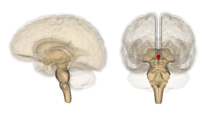
The pineal gland, a small, pine-cone shaped structure, is an endocrine gland located in the posterior aspect of the cranial fossa in the brain. The pineal glands main role is to control the circadian cycle of sleep and wakefulness by secreting melatonin. It is also has a role in reproductive function, being associated with the onset of puberty.[1]
Anatomy and Function[edit | edit source]
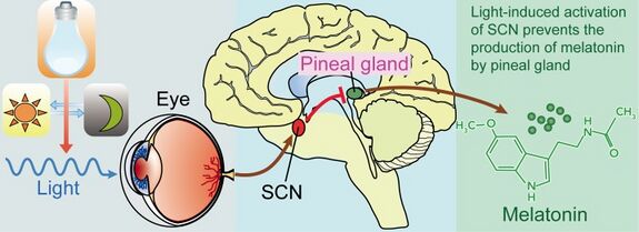
The human pineal gland is a tiny (weighs 0.1 g), highly vascularized, secretory neuroendocrine organ. It is found in the mid-line of the brain, connected to the roof of the third ventricle by a short stalk. The main role of the pineal gland is to produces melatonin, a derivative of serotonin, which affects the modulation of wake/sleep patterns and photoperiodic (seasonal) functions. In humans, the main cell types are pinealocytes (95%) along with scattered glial cells (astrocytic and phagocytic subtypes). Pinealocytes synthesis and secrete melatonin.[2] Melatonin production is upregulated by darkness and decreases production when exposed to light. Melatonin also protects against neurodegeneration.[3]
Pathophysiology[edit | edit source]
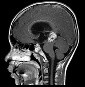
Due to the pineal gland's location, any pineal region mass leads to the compression of the aqueduct of Sylvius ( an important structure allowing the cerebrospinal fluid to circulate out. When blocked by an abnormal pineal gland, the passage of the duct is blocked, and CSF pressure increases, leading to hydrocephalus. Symptoms of this include nausea, vomiting, visual changes, headaches, seizures, and memory changes. This can be life threatening.[3]
Sleep[edit | edit source]
For more on the importance of melatonin and sleep, see Sleep: Theory, Function and Physiology and complete the Sleep Programme.
References[edit | edit source]
- ↑ Radiopedia Pineal Gland Available:https://radiopaedia.org/articles/pineal-gland?lang=gb (accessed 20.1.2023)
- ↑ Arendt J, Aulinas A. Physiology of the pineal gland and melatonin. Endotext [Internet]. 2022 Oct 30. Available:https://www.ncbi.nlm.nih.gov/books/NBK550972/ (accessed 20.1.2023)
- ↑ 3.0 3.1 Ilahi S, Beriwal N, Ilahi TB. Physiology, pineal gland. InStatPearls [Internet] 2022 Apr 28. StatPearls Publishing. Available;https://www.statpearls.com/articlelibrary/viewarticle/27230/ (accessed 20.1.2023)


