Gracilis: Difference between revisions
No edit summary |
No edit summary |
||
| (13 intermediate revisions by 3 users not shown) | |||
| Line 5: | Line 5: | ||
</div> | </div> | ||
== Introduction == | == Introduction == | ||
The gracilis is a spiral unipennate muscle in the medial thigh compartment. | [[File:Gracilis muscle adductor.gif|thumb|Gracilis Muscle|alt=|250x250px]] | ||
The gracilis is a spiral unipennate [[muscle]] in the [[Hip Adductors|medial thigh compartment]]. | |||
The gracilis | The gracilis: | ||
* Assists with hip adduction, knee flexion, and knee internal rotation. | * Assists with hip adduction, knee flexion, and knee internal rotation. | ||
* It is innervated by the anterior branch of the obturator nerve. | * It is innervated by the anterior branch of the [[Obturator Nerve|obturator nerve]]. | ||
* Crosses at both the [[hip]] and [[knee]] joints. | * Crosses at both the [[hip]] and [[knee]] joints. | ||
* The gracilis may | * The gracilis may is prone to strain injuries, resulting in [[Adductor Tendinopathy|adductor tendinopathy]]<nowiki/>eg common in soccer, hockey, football, and basketball athletes<ref name=":2">Bordoni B, Varacallo M. Anatomy, bony pelvis and lower limb, thigh quadriceps muscle. Available:https://www.statpearls.com/ArticleLibrary/viewarticle/22380 (accessed 19.1.2022)</ref>. | ||
=== | === Anatomy === | ||
[[File:Gracilis muscle - Kenhub.png|alt=Gracilis muscle (highlighted in green) - anterior view|right|frameless|500x500px|Gracilis muscle (highlighted in green) - anterior view]] | |||
'''Origin''' | |||
The gracilis muscle originates from the inferior ischiopubic ramus, and body of [[pubis]].<ref name=":0">Marieb EN, Hoehn K. Human anatomy & physiology. 10th ed. Boston, Ma: Pearson; 2016.</ref> | |||
The gracilis muscle | |||
'''Insertion''' | |||
=== Artery | The gracilis muscle decends almost vertically down the leg and inserts on the medial [[tibia]] at the [[Pes Anserinus Bursitis|Pes anserinus]].<ref name=":0" /> The pes anserinus is also the attachment site of the [[Sartorius]] and [[Semitendinosus]]. | ||
The gracilis muscle receives blood supply from the medial circumflex femoral artery.<ref name=":0" /> | |||
'''Nerve''' | |||
The gracilis muscles is innervated by the anterior branch of the obturator nerve (L2-L4). The anterior branch of the obturator nerve also innervates the [[Adductor Longus|adductor longus]], and sometimes [[Adductor Brevis|adductor brevis]].<ref name=":0" /> | |||
There are 5 to 7 muscle fiber bundle compartments in the gracilis muscle, with nerve branches coursing along each compartment, indicating possible independent [[Neuromuscular Adaptations to Exercise|neuromuscular]] compartment functioning.<ref name=":2" /> | |||
'''Artery''' | |||
The gracilis muscle receives [[Blood Physiology|blood]] supply from the medial circumflex [[Femoral Artery|femoral artery]].<ref name=":0" /> | |||
Image: Gracilis muscle (highlighted in green) - anterior view<ref >Gracilis muscle (highlighted in green) - anterior view image - © Kenhub https://www.kenhub.com/en/library/anatomy/gracilis-muscle</ref> | |||
== Function == | == Function == | ||
Due to | Due to its attachment on the tibia, the gracilis flexes the knee, adducts the thigh, and medially rotate the tibia on the femur.<ref>Gracilis. Available from: https://rad.washington.edu/muscle-atlas/gracilis/ (accessed 15 May 2018). </ref> | ||
== Physiotherapy Relevance == | |||
== | # [[File:Baseball.jpeg|thumb|Baseball - potential Gracilis injury]] [[Groin Strain|Groin strain]]/adductor tendinopathy commonly occur in high impact sports that involve ballistic movements or stretching eg soccer, hockey, baseball. Tearing of the muscles usually occur at proximal region near bony attachments at the pelvis<ref name=":1">Teach Me Anatomy. Muscles in the Medial Compartment of the Thigh. Available from: https://teachmeanatomy.info/lower-limb/muscles/thigh/medial-compartment/ (Accessed on 19 May 2020) | ||
</ref> | |||
# In women, the muscular volume of the medial vastus is greater than in men, while the volume of the gracilis muscle (and the sartorius muscle) is smaller. This predisposes females to [[Anterior Cruciate Ligament (ACL) Reconstruction|ACL injury]] compared to males.<ref name=":2" /> | |||
# Surgeons may use the gracilis tendon in reconstruction surgery of the ACL<ref name=":2" /> | |||
# [[Pes Anserinus Bursitis|Pes anserine bursitis]] is an inflammatory condition of bursa of the conjoined insertion of the sartorius, gracilis and semitendinosus. Pes anserine bursitis is commonly seen in patients who have obese, have osteoarthritis, and female.<ref name=":2" /> | |||
== Assessment and Treatment == | |||
See [[Adductor Tendinopathy]] [[Groin Strain|Groin Injuries]] | |||
This 1 minute video is titled "Applied Kinesiology - Manual Muscle Testing: Gracilis" | |||
{{#ev:youtube|DVfJW72bEAE}} | {{#ev:youtube|DVfJW72bEAE}} | ||
== | == Resources == | ||
This video from KenHub on anatomy of gracilis.<ref >Gracilis muscle video - © Kenhub https://www.kenhub.com/en/library/anatomy/gracilis-muscle</ref> | |||
{{#ev:youtube|265T4ywr6uY|300}} | |||
== References == | |||
[[Category:Anatomy]] | [[Category:Anatomy]] | ||
[[Category:Muscles]] | [[Category:Muscles]] | ||
Latest revision as of 09:09, 15 March 2023
Original Editor - Eric Henderson
Top Contributors - Eric Henderson, Lucinda hampton, Candace Goh, Joao Costa and Vidya Acharya
Introduction[edit | edit source]
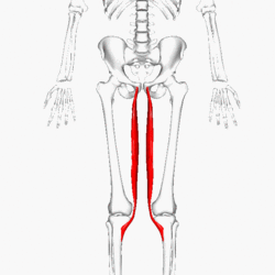
The gracilis is a spiral unipennate muscle in the medial thigh compartment.
The gracilis:
- Assists with hip adduction, knee flexion, and knee internal rotation.
- It is innervated by the anterior branch of the obturator nerve.
- Crosses at both the hip and knee joints.
- The gracilis may is prone to strain injuries, resulting in adductor tendinopathyeg common in soccer, hockey, football, and basketball athletes[1].
Anatomy[edit | edit source]
Origin
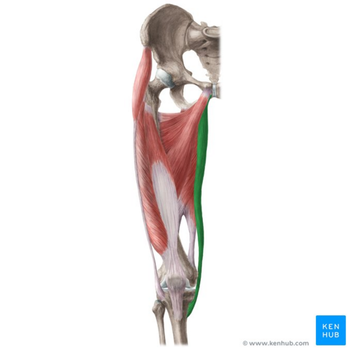
The gracilis muscle originates from the inferior ischiopubic ramus, and body of pubis.[2]
Insertion
The gracilis muscle decends almost vertically down the leg and inserts on the medial tibia at the Pes anserinus.[2] The pes anserinus is also the attachment site of the Sartorius and Semitendinosus.
Nerve
The gracilis muscles is innervated by the anterior branch of the obturator nerve (L2-L4). The anterior branch of the obturator nerve also innervates the adductor longus, and sometimes adductor brevis.[2]
There are 5 to 7 muscle fiber bundle compartments in the gracilis muscle, with nerve branches coursing along each compartment, indicating possible independent neuromuscular compartment functioning.[1]
Artery
The gracilis muscle receives blood supply from the medial circumflex femoral artery.[2]
Image: Gracilis muscle (highlighted in green) - anterior view[3]
Function[edit | edit source]
Due to its attachment on the tibia, the gracilis flexes the knee, adducts the thigh, and medially rotate the tibia on the femur.[4]
Physiotherapy Relevance[edit | edit source]
- Groin strain/adductor tendinopathy commonly occur in high impact sports that involve ballistic movements or stretching eg soccer, hockey, baseball. Tearing of the muscles usually occur at proximal region near bony attachments at the pelvis[5]
- In women, the muscular volume of the medial vastus is greater than in men, while the volume of the gracilis muscle (and the sartorius muscle) is smaller. This predisposes females to ACL injury compared to males.[1]
- Surgeons may use the gracilis tendon in reconstruction surgery of the ACL[1]
- Pes anserine bursitis is an inflammatory condition of bursa of the conjoined insertion of the sartorius, gracilis and semitendinosus. Pes anserine bursitis is commonly seen in patients who have obese, have osteoarthritis, and female.[1]
Assessment and Treatment[edit | edit source]
See Adductor Tendinopathy Groin Injuries
This 1 minute video is titled "Applied Kinesiology - Manual Muscle Testing: Gracilis"
Resources[edit | edit source]
This video from KenHub on anatomy of gracilis.[6]
References[edit | edit source]
- ↑ 1.0 1.1 1.2 1.3 1.4 Bordoni B, Varacallo M. Anatomy, bony pelvis and lower limb, thigh quadriceps muscle. Available:https://www.statpearls.com/ArticleLibrary/viewarticle/22380 (accessed 19.1.2022)
- ↑ 2.0 2.1 2.2 2.3 Marieb EN, Hoehn K. Human anatomy & physiology. 10th ed. Boston, Ma: Pearson; 2016.
- ↑ Gracilis muscle (highlighted in green) - anterior view image - © Kenhub https://www.kenhub.com/en/library/anatomy/gracilis-muscle
- ↑ Gracilis. Available from: https://rad.washington.edu/muscle-atlas/gracilis/ (accessed 15 May 2018).
- ↑ Teach Me Anatomy. Muscles in the Medial Compartment of the Thigh. Available from: https://teachmeanatomy.info/lower-limb/muscles/thigh/medial-compartment/ (Accessed on 19 May 2020)
- ↑ Gracilis muscle video - © Kenhub https://www.kenhub.com/en/library/anatomy/gracilis-muscle


