Plagiocephaly: Difference between revisions
mNo edit summary |
No edit summary |
||
| (35 intermediate revisions by 9 users not shown) | |||
| Line 1: | Line 1: | ||
<div class="editorbox"> | <div class="editorbox">'''Original Editor '''- [[User:Admin|Admin]] '''Top Contributors'''- {{Special:Contributors/{{FULLPAGENAME}}}}</div> | ||
'''Original Editor '''- | == Introduction == | ||
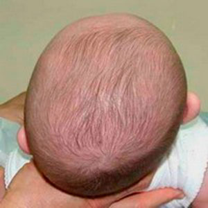
[[File:Plagiocephaly.jpg|Example of plagiocephaly in infant. | |||
Credit: [https://commons.wikimedia.org/wiki/File:Плагиоцефалия.jpg#file Medical advises, Плагиоцефалия,] [https://creativecommons.org/licenses/by-sa/3.0/legalcode CC BY-SA 3.0]. | |||
|alt=|right|frameless]] | |||
Positional plagiocephaly is increasingly common in infants. Positional plagiocephaly is an asymmetric deformation of the skull. It has a number of potential causes, including: first birth, assisted labour, multiple births (e.g. twins, triplets etc), prematurity, [[Congenital torticollis|congenital muscular torticollis]], position of the head and lying in the same position for prolonged periods.<ref name=":4">Jung BK, Yun IS. Diagnosis and treatment of positional plagiocephaly. Archives of craniofacial surgery. 2020 Apr;21(2):80.</ref><ref name=":2">Childerens health qld gov. Plagiocephaly Available:https://www.childrens.health.qld.gov.au/fact-sheet-plagiocephaly/ (accessed 8.10.2021)</ref> | |||
There are two types of plagiocephaly: 1. ''plagiocephaly followed by craniosynostosis'' (a birth defect in which the bones in a baby's skull fuse prematurely) and 2. ''positional plagiocephaly without craniosynostosis''.<ref name=":4" /><ref>Unnithan AK, De Jesus O. [https://www.statpearls.com/ArticleLibrary/viewarticle/126820 Plagiocephaly]. InStatPearls [Internet] 2022 May 1. StatPearls Publishing.</ref> | |||
* When plagiocephaly occurs with craniosynostosis, the skull deforms due to premature fusion of the sutures in the skull. This condition often requires surgical management and helmet therapy may be used.<ref name=":4" /> | |||
</ | |||
= | * In plagiocephaly without craniosynostosis, the sutures between the bones are normal, so skull growth is not affected. However, the shape can be altered - most commonly the head is flattened on one side of the posterior aspect.<ref name=":4" /> | ||
== Clinically Relevant Anatomy == | |||
The 2 minute video below explains the skull and fontanels of a newborn.{{#ev:youtube|G1XhXvrWmAE|300}}<ref>Dr. J. Baby Skull. Available from https://www.youtube.com/watch?v=G1XhXvrWmAE&t= [Accessed 14/6/2018]</ref> | |||
The skull covers and protects the brain. It consists of several bony plates connected together by fibrous material called sutures. Sutures allow movement of the bones necessary to accommodate brain growth and allow moulding of the head during birth.<ref>University of Rochester Medical Centre. Anatomy of the newborn skull. https://www.urmc.rochester.edu/encyclopedia/content.aspx?contenttypeid=90&contentid=p01840 (accessed 13 June 2018).</ref> As a result, the infant skull is vulnerable to deformation. | |||
== Mechanism of Injury / Pathological Process == | |||
== | It is not uncommon for newborns to have "unusually" shaped heads - depending on the cause, most of these cases resolve within around six weeks of birth.<ref name=":3">RCHM Plagiocephaly – misshapen head Available:https://www.rch.org.au/kidsinfo/fact_sheets/Plagiocephaly_misshapen_head/ (accessed 8.10.2021)</ref> Reasons for a change in head shape include the baby's position in the uterus and "moulding" of head during labour, including if the delivery is assisted (i.e. ventouse, forceps).<ref name=":3" /> | ||
''Positional plagiocephaly'' is caused by pressure on the developing infant skull from an external force. This can occur in the womb (particularly with first birth, multiple births), but more commonly develops postnatally. | |||
* Babies in many areas spend significant amounts of time on their backs (in a cot, car seat, buggy etc) - the external forces from these firm surfaces can cause positional plagiocephaly | |||
** please note that "Back to Sleep" approaches are recommended to reduce the risk of sudden infant death syndrome (SIDS)<ref name=":1">Great Ormond Street Hospital for Children. Positional Plagiocephaly. https://www.gosh.nhs.uk/conditions-and-treatments/conditions-we-treat-index-page-group/positional-plagiocephaly (Accessed 14 June 2018)</ref> | |||
* [[Congenital torticollis|Congenital muscular torticollis (CMT)]] can co-exist with positional plagiocephaly in as many as 30% of cases<ref>Ellenbogen RG, Abdulrauf SI, Sekhar LN Principles of Neurological Surgery. Philedelphia: Elsevier, 2018.</ref> (see Differential Diagnosis section for more information) | |||
== Diagnostic Procedures == | == Diagnostic Procedures == | ||
Positional plagiocephaly is diagnosed from the child's history and clinical presentation and does not usually require any imaging. However, a skull x-ray may be required to rule out craniosynostosis.<ref>Reece A, Cohn A. Clinical Cases in Pediatrics: A trainee handbook. London: JP Medical Ltd, 2014.</ref> | |||
== Outcome Measures == | == Outcome Measures == | ||
As diagnosis is largely based on observation, it is helpful to record observations from different views. This can be supplemented with photography. Clinically, where no equipment is available it may be useful for parents/ carers to take photographs periodically to identify change. | |||
== Management / Interventions< | == Physiotherapy Management / Interventions == | ||
Physiotherapy treatments include:<ref>Eskay K. Torticollis and Plagiocephaly Course. Plus, 2023.</ref><ref>Hohendahl L, Hohendahl J, Lemhöfer C, Best N. The Effect of Pediatric Physiotherapy on Positional Plagiocephaly: A Retrospective Trial. Physikalische Medizin, Rehabilitationsmedizin, Kurortmedizin. 2022 Sep 8.</ref><ref>Di Chiara A, La Rosa E, Ramieri V, Vellone V, Cascone P. Treatment of deformational plagiocephaly with physiotherapy. Journal of Craniofacial Surgery. 2019 Oct 1;30(7):2008-13.</ref> | |||
* caregiver education, including preventative counselling | |||
** positioning for babies<ref>Burmeister S, Kayne AN, Yazdanyar AR, Hagstrom JN, Burmeister DB. Plagiocephaly Perception and Prevention: A Need to Intervene Early to Educate Parents. The Open Journal of Occupational Therapy. 2021;9(3):1-1.</ref> | |||
** avoid baby being in containers all day (e.g. rockers, hammocks etc) | |||
** avoid baby lying on back all day (but always sleep on back) | |||
** encourage early tummy time | |||
* aggressive repositioning - i.e. position the infant to decrease the pressure on the flattened area of the head | |||
** during feeding reduce pressure on the affected occiput; switch sides if there is a rotation preference | |||
** place a blanket roll under the shoulder and hip to help offload the flattened area of the head | |||
** sometimes transport the baby in a front carrier or an upright stroller to offload the back of the head | |||
* developmental facilitation | |||
** positioning in side-lying/propping | |||
** strengthen symmetrically in the midline | |||
* helmet therapy | |||
** less than one in ten babies with plagiocephaly have a severe and persistent deformity, and they may need to be treated with helmet therapy<ref name=":3" /><ref>Watt A, Alabdulkarim A, Lee J, Gilardino M. Practical Review of the Cost of Diagnosis and Management of Positional Plagiocephaly. Plastic and Reconstructive Surgery Global Open. 2022 May;10(5).</ref> | |||
** there is no evidence supporting the use of cranial remodelling helmets in healthy babies who are typically developing<ref name=":2" /> | |||
== Differential Diagnosis | The following video outlines the concept of Tummy Time.{{#ev:youtube|M3rCtW9DMD4|300}}<ref>Pathways. Five essential Tummy Time moves. Available from: https://www.youtube.com/watch?v=M3rCtW9DMD4 [accessed 14/6/2018]</ref> | ||
== Differential Diagnosis == | |||
==== Congenital Muscular Torticollis (CMT) ==== | |||
A shortened sternocleidomastoid muscle causes [[Congenital torticollis|congenital muscular torticollis (CMT)]], which can flatten the occiput on the contralateral side. A child with left-sided CMT has right-sided positional plagiocephaly. Active and passive neck movements and head tilt need to be assessed to determine if CMT is causing the plagiocephaly. Early physiotherapy input is required to restore neck range of motion and improve the plagiocephaly.<ref name=":0">BC Children's Hospital. A Clinician's Guide to Positional Plagiocephaly<nowiki/>http://www.bcchildrens.ca/neurosciences-site/Documents/BCCH034PlagiocephalyCliniciansGuideWeb1.pdf (accessed 14 June 2018)</ref> | |||
== | ==== Unilateral Lambdoid Synostosis ==== | ||
A rare condition where there is premature fusion of one lambdoid suture, which results in plagiocephaly. It is identified by retraction of the '''ipsilateral''' ear and forehead and a trapezoid shape of the head when viewed from above.<ref name=":0" /> | |||
==== Unilateral Coronal Synostosis ==== | |||
Premature fusion of a coronal suture, which causes assymetry of the forehead. It is diagnosed by examining orbital symmetry. Looking from the front, the ipsilateral orbit will be higher and wider than the contralateral orbit; when viewed from above, the eyeball on the side of the forehead flattening will protrude.<ref name=":0" /> | |||
== References == | == References == | ||
<references /> | <references /> | ||
[[Category:Paediatrics]] | |||
[[Category:Musculoskeletal/Orthopaedics]] | |||
[[Category:Conditions]] | |||
[[Category:Primary Contact]] | |||
[[Category:Paediatrics - Conditions]] | |||
[[Category:Congenital Conditions]] | |||
[[Category:Course Pages]] | |||
Latest revision as of 08:03, 2 April 2023
Introduction[edit | edit source]
Positional plagiocephaly is increasingly common in infants. Positional plagiocephaly is an asymmetric deformation of the skull. It has a number of potential causes, including: first birth, assisted labour, multiple births (e.g. twins, triplets etc), prematurity, congenital muscular torticollis, position of the head and lying in the same position for prolonged periods.[1][2]
There are two types of plagiocephaly: 1. plagiocephaly followed by craniosynostosis (a birth defect in which the bones in a baby's skull fuse prematurely) and 2. positional plagiocephaly without craniosynostosis.[1][3]
- When plagiocephaly occurs with craniosynostosis, the skull deforms due to premature fusion of the sutures in the skull. This condition often requires surgical management and helmet therapy may be used.[1]
- In plagiocephaly without craniosynostosis, the sutures between the bones are normal, so skull growth is not affected. However, the shape can be altered - most commonly the head is flattened on one side of the posterior aspect.[1]
Clinically Relevant Anatomy[edit | edit source]
The 2 minute video below explains the skull and fontanels of a newborn.
The skull covers and protects the brain. It consists of several bony plates connected together by fibrous material called sutures. Sutures allow movement of the bones necessary to accommodate brain growth and allow moulding of the head during birth.[5] As a result, the infant skull is vulnerable to deformation.
Mechanism of Injury / Pathological Process[edit | edit source]
It is not uncommon for newborns to have "unusually" shaped heads - depending on the cause, most of these cases resolve within around six weeks of birth.[6] Reasons for a change in head shape include the baby's position in the uterus and "moulding" of head during labour, including if the delivery is assisted (i.e. ventouse, forceps).[6]
Positional plagiocephaly is caused by pressure on the developing infant skull from an external force. This can occur in the womb (particularly with first birth, multiple births), but more commonly develops postnatally.
- Babies in many areas spend significant amounts of time on their backs (in a cot, car seat, buggy etc) - the external forces from these firm surfaces can cause positional plagiocephaly
- please note that "Back to Sleep" approaches are recommended to reduce the risk of sudden infant death syndrome (SIDS)[7]
- Congenital muscular torticollis (CMT) can co-exist with positional plagiocephaly in as many as 30% of cases[8] (see Differential Diagnosis section for more information)
Diagnostic Procedures[edit | edit source]
Positional plagiocephaly is diagnosed from the child's history and clinical presentation and does not usually require any imaging. However, a skull x-ray may be required to rule out craniosynostosis.[9]
Outcome Measures[edit | edit source]
As diagnosis is largely based on observation, it is helpful to record observations from different views. This can be supplemented with photography. Clinically, where no equipment is available it may be useful for parents/ carers to take photographs periodically to identify change.
Physiotherapy Management / Interventions[edit | edit source]
Physiotherapy treatments include:[10][11][12]
- caregiver education, including preventative counselling
- positioning for babies[13]
- avoid baby being in containers all day (e.g. rockers, hammocks etc)
- avoid baby lying on back all day (but always sleep on back)
- encourage early tummy time
- aggressive repositioning - i.e. position the infant to decrease the pressure on the flattened area of the head
- during feeding reduce pressure on the affected occiput; switch sides if there is a rotation preference
- place a blanket roll under the shoulder and hip to help offload the flattened area of the head
- sometimes transport the baby in a front carrier or an upright stroller to offload the back of the head
- developmental facilitation
- positioning in side-lying/propping
- strengthen symmetrically in the midline
- helmet therapy
The following video outlines the concept of Tummy Time.
Differential Diagnosis[edit | edit source]
Congenital Muscular Torticollis (CMT)[edit | edit source]
A shortened sternocleidomastoid muscle causes congenital muscular torticollis (CMT), which can flatten the occiput on the contralateral side. A child with left-sided CMT has right-sided positional plagiocephaly. Active and passive neck movements and head tilt need to be assessed to determine if CMT is causing the plagiocephaly. Early physiotherapy input is required to restore neck range of motion and improve the plagiocephaly.[16]
Unilateral Lambdoid Synostosis[edit | edit source]
A rare condition where there is premature fusion of one lambdoid suture, which results in plagiocephaly. It is identified by retraction of the ipsilateral ear and forehead and a trapezoid shape of the head when viewed from above.[16]
Unilateral Coronal Synostosis[edit | edit source]
Premature fusion of a coronal suture, which causes assymetry of the forehead. It is diagnosed by examining orbital symmetry. Looking from the front, the ipsilateral orbit will be higher and wider than the contralateral orbit; when viewed from above, the eyeball on the side of the forehead flattening will protrude.[16]
References[edit | edit source]
- ↑ 1.0 1.1 1.2 1.3 Jung BK, Yun IS. Diagnosis and treatment of positional plagiocephaly. Archives of craniofacial surgery. 2020 Apr;21(2):80.
- ↑ 2.0 2.1 Childerens health qld gov. Plagiocephaly Available:https://www.childrens.health.qld.gov.au/fact-sheet-plagiocephaly/ (accessed 8.10.2021)
- ↑ Unnithan AK, De Jesus O. Plagiocephaly. InStatPearls [Internet] 2022 May 1. StatPearls Publishing.
- ↑ Dr. J. Baby Skull. Available from https://www.youtube.com/watch?v=G1XhXvrWmAE&t= [Accessed 14/6/2018]
- ↑ University of Rochester Medical Centre. Anatomy of the newborn skull. https://www.urmc.rochester.edu/encyclopedia/content.aspx?contenttypeid=90&contentid=p01840 (accessed 13 June 2018).
- ↑ 6.0 6.1 6.2 RCHM Plagiocephaly – misshapen head Available:https://www.rch.org.au/kidsinfo/fact_sheets/Plagiocephaly_misshapen_head/ (accessed 8.10.2021)
- ↑ Great Ormond Street Hospital for Children. Positional Plagiocephaly. https://www.gosh.nhs.uk/conditions-and-treatments/conditions-we-treat-index-page-group/positional-plagiocephaly (Accessed 14 June 2018)
- ↑ Ellenbogen RG, Abdulrauf SI, Sekhar LN Principles of Neurological Surgery. Philedelphia: Elsevier, 2018.
- ↑ Reece A, Cohn A. Clinical Cases in Pediatrics: A trainee handbook. London: JP Medical Ltd, 2014.
- ↑ Eskay K. Torticollis and Plagiocephaly Course. Plus, 2023.
- ↑ Hohendahl L, Hohendahl J, Lemhöfer C, Best N. The Effect of Pediatric Physiotherapy on Positional Plagiocephaly: A Retrospective Trial. Physikalische Medizin, Rehabilitationsmedizin, Kurortmedizin. 2022 Sep 8.
- ↑ Di Chiara A, La Rosa E, Ramieri V, Vellone V, Cascone P. Treatment of deformational plagiocephaly with physiotherapy. Journal of Craniofacial Surgery. 2019 Oct 1;30(7):2008-13.
- ↑ Burmeister S, Kayne AN, Yazdanyar AR, Hagstrom JN, Burmeister DB. Plagiocephaly Perception and Prevention: A Need to Intervene Early to Educate Parents. The Open Journal of Occupational Therapy. 2021;9(3):1-1.
- ↑ Watt A, Alabdulkarim A, Lee J, Gilardino M. Practical Review of the Cost of Diagnosis and Management of Positional Plagiocephaly. Plastic and Reconstructive Surgery Global Open. 2022 May;10(5).
- ↑ Pathways. Five essential Tummy Time moves. Available from: https://www.youtube.com/watch?v=M3rCtW9DMD4 [accessed 14/6/2018]
- ↑ 16.0 16.1 16.2 BC Children's Hospital. A Clinician's Guide to Positional Plagiocephalyhttp://www.bcchildrens.ca/neurosciences-site/Documents/BCCH034PlagiocephalyCliniciansGuideWeb1.pdf (accessed 14 June 2018)


