Thalamus: Difference between revisions
No edit summary |
No edit summary |
||
| (10 intermediate revisions by one other user not shown) | |||
| Line 5: | Line 5: | ||
</div> | </div> | ||
== Thalamus Structure == | == Thalamus Structure == | ||
[[File:Brain stem and thalamus.jpeg|thumb|The thalamus is located centrally in the brain, just above the brainstem.]]The thalamus is located medial to the cerebral hemispheres and consists of two oval-shaped masses connected by the intermediate mass. Each mass consists of several groups of nuclei that serve different functions. Motor and sensory pathways (except olfaction) pass through this central structure. | |||
The thalamus can be divided into approximately 60 regions known as '''thalamic nuclei'''. Each nucleus has unique pathways as inputs and various projections as outputs, most of which send information to the '''[[Cerebral Cortex|cerebral cortex]]'''.<ref name=":1" /> | The thalamus can be divided into approximately 60 regions known as '''thalamic nuclei'''. Each nucleus has unique pathways as inputs and various projections as outputs, most of which send information to the '''[[Cerebral Cortex|cerebral cortex]]'''.<ref name=":1">Torrico TJ, Munakomi S. [https://www.ncbi.nlm.nih.gov/books/NBK542184/ Neuroanatomy, Thalamus]. [Updated 2023 Jul 24]. In: StatPearls [Internet].</ref> | ||
=== | === Hypothalamus === | ||
* Located inferior to the thalamus | * Located inferior to the thalamus | ||
| Line 18: | Line 15: | ||
* Anteriorly, the hypothalamus is connected to the pituitary gland by a long stalk called the '''infundibulum''', also known as the '''pituitary stalk''' | * Anteriorly, the hypothalamus is connected to the pituitary gland by a long stalk called the '''infundibulum''', also known as the '''pituitary stalk''' | ||
The hypothalamus plays a major role in maintaining homeostasis via the autonomic nervous system, the neuroendocrine system, and the limbic system. It regulates emotions, hormone production from the pituitary gland, and | The hypothalamus plays a major role in maintaining homeostasis via the [[Autonomic Nervous System|autonomic nervous system]], the [[Endocrine System|neuroendocrine system]], and the [[Limbic System|limbic system]]. It regulates emotions, hormone production from the pituitary gland, and bodily functions such as appetite, body temperature, reproduction, and circadian rhythms. | ||
=== | === Epithalamus === | ||
* Located behind the thalamus | * Located behind the thalamus | ||
* Includes the '''pineal gland''', which secretes the hormone melatonin in response to darkness, regulating our circadian rhythms and sleep-wake cycles | |||
=== | === Subthalamus === | ||
* Located beneath the thalamus | * Located beneath the thalamus | ||
* Includes the '''subthalamic nucleus''', which is functionally considered part of the [[Basal Ganglia|basal ganglia]] | |||
== | == Function of the Thalamus == | ||
[[File:Brain cross section with subcortical structures.jpeg|thumb|500x500px|Note the central location of the thalamus in relation to other subcortical structures and the cerebral cortex.]] | <blockquote>The '''thalamus''' functions as a relay station between the brain and the body, filtering through various types of sensory and motor information.</blockquote>[[File:Brain cross section with subcortical structures.jpeg|thumb|500x500px|Note the central location of the thalamus in relation to other subcortical structures and the cerebral cortex.]] | ||
The thalamus | * The thalamus has many connections with the cerebral cortex,<ref name=":1" /> which are known as '''thalamacortical loops''' | ||
* It transmits nearly all sensory information to the cortex, including vision, taste, touch and balance, but excludes olfactory information | |||
* It conducts motor signals from the cerebral cortex to the spinal cord and ultimately to the peripheral nervous system | |||
* The thalamus also modulates arousal mechanisms, maintains alertness, and directs attention to sensory events<ref name=":0">Halassa, M.M., Kastner, S. Thalamic functions in distributed cognitive control. ''Nat Neurosci, 2017;'' 20, 1669–1679. </ref> | |||
* The thalamus has connections with a number of structures of the [[Limbic System|limbic system]], including the hippocampus, mammillary bodies, and fornix, so it has a role in learning and episodic memory<ref name=":1" /> | |||
The thalamus can be divided into five major functional components.<ref name=":1" /> | |||
# '''Reticular and intralaminar nuclei''': involved in arousal and pain regulation | |||
# '''Reticular and intralaminar nuclei''' | # '''Sensory nuclei''': regulate sensory domains, apart from olfaction | ||
# '''Effector nuclei''': govern motor and language functions | |||
# '''Associative nuclei''': involved in cognitive functions | |||
# '''Limbic nuclei:''' manage mood and motivation | |||
# '''Sensory nuclei''' | These specific nuclei help to scan the cerebral cortex and determine active brain regions, relaying this information to the rest of the thalamus.<ref name=":0" /> | ||
# '''Effector nuclei''' | |||
# '''Associative nuclei''' | |||
# '''Limbic nuclei''' | |||
These specific nuclei | |||
== Thalamus and Injury == | == Thalamus and Injury == | ||
The thalamus | The thalamus is involved in many critical functions and injury to the thalamus can cause a range of issues, including: | ||
* sensory issues (e.g. pain, paraesthesia, numbness, hypersensitivity) | |||
* vision impairment and light sensitivity | |||
* | * motor impairment | ||
* | * tremor | ||
* | * issues with attention | ||
* | * memory impairment | ||
* | * sleep difficulties | ||
* | * proprioception impairment | ||
* | |||
* | |||
'''The following presentations are unique to thalamic injury''': | |||
== | * '''thalamic pain syndrome''': an excruciating sensation of pain that does not respond to narcotics. Once called Dejerine-Roussy Syndrome, this condition is commonly associated with infarction of the ventroposterolateral thalamus.<ref>Jahngir MU, Qureshi AI. [https://www.ncbi.nlm.nih.gov/books/NBK519047/ Dejerine roussy syndrome]. 2023.In: StatPearls [Internet]. </ref> <ref>Dydyk AM, Munakomi S. [https://www.ncbi.nlm.nih.gov/books/NBK554490/ Thalamic Pain Syndrome].[Updated 2023 Aug 13]. StatPearls [Internet]. Treasure Island (FL): StatPearls Publishing. 2023.</ref> | ||
* [[Pusher Syndrome|'''Pusher syndrome''']] (also referred to as "persons who push"): a lesion to the posterior thalamus interrupts the connection to the vestibular nuclei, leading to lateropulsion in the direction of the affected side.<ref>Rosenzopf H, Klingbeil J, Wawrzyniak M, Röhrig L, Sperber C, Saur D, Karnath HO. [https://academic.oup.com/brain/article-abstract/146/9/3648/7082493?redirectedFrom=fulltext&login=false Thalamocortical disconnection involved in pusher syndrome.] Brain. 2023;146(9):3648-3661.</ref> | |||
* '''vegetative state and coma''': a lesion to the non-specific (intralaminar and reticular) nuclei.<ref>Adams JH, Graham DI, Jennett B. [https://academic.oup.com/brain/article/123/7/1327/380151?login=false The neuropathology of the vegetative state after an acute brain insult.] Brain. 2000 Jul;123 (Pt 7):1327-38.</ref> Coma may occur due to the role of the thalamus in sleep and arousal. | |||
For more information, please read the following articles: | == Additional Resources == | ||
For more information on treatment options, please read the following articles: | |||
* [[Overview of Traumatic Brain Injury|Traumatic Brain Injury]] | * [[Overview of Traumatic Brain Injury|Traumatic Brain Injury]] | ||
| Line 83: | Line 75: | ||
* [[Mirror Therapy]] | * [[Mirror Therapy]] | ||
* [[Pusher Syndrome#Management / Interventions|Management of pushing tendencies]] | * [[Pusher Syndrome#Management / Interventions|Management of pushing tendencies]] | ||
* [[Desensitization]] | * [[Desensitization|Desensitisation]] for thalamic pain | ||
== References == | == References == | ||
Latest revision as of 19:41, 1 July 2024
Original Editor - Lucinda hampton
Top Contributors - Lucinda hampton, Jess Bell, Stacy Schiurring, Kirenga Bamurange Liliane, Uchechukwu Chukwuemeka, Vidya Acharya, Rucha Gadgil and Mason Trauger
Thalamus Structure[edit | edit source]
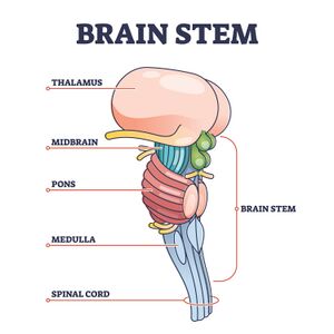
The thalamus is located medial to the cerebral hemispheres and consists of two oval-shaped masses connected by the intermediate mass. Each mass consists of several groups of nuclei that serve different functions. Motor and sensory pathways (except olfaction) pass through this central structure.
The thalamus can be divided into approximately 60 regions known as thalamic nuclei. Each nucleus has unique pathways as inputs and various projections as outputs, most of which send information to the cerebral cortex.[1]
Hypothalamus[edit | edit source]
- Located inferior to the thalamus
- Two rounded eminences protrude from the back, called mammillary bodies
- Anteriorly, the hypothalamus is connected to the pituitary gland by a long stalk called the infundibulum, also known as the pituitary stalk
The hypothalamus plays a major role in maintaining homeostasis via the autonomic nervous system, the neuroendocrine system, and the limbic system. It regulates emotions, hormone production from the pituitary gland, and bodily functions such as appetite, body temperature, reproduction, and circadian rhythms.
Epithalamus[edit | edit source]
- Located behind the thalamus
- Includes the pineal gland, which secretes the hormone melatonin in response to darkness, regulating our circadian rhythms and sleep-wake cycles
Subthalamus[edit | edit source]
- Located beneath the thalamus
- Includes the subthalamic nucleus, which is functionally considered part of the basal ganglia
Function of the Thalamus[edit | edit source]
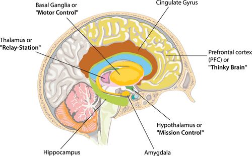
The thalamus functions as a relay station between the brain and the body, filtering through various types of sensory and motor information.
- The thalamus has many connections with the cerebral cortex,[1] which are known as thalamacortical loops
- It transmits nearly all sensory information to the cortex, including vision, taste, touch and balance, but excludes olfactory information
- It conducts motor signals from the cerebral cortex to the spinal cord and ultimately to the peripheral nervous system
- The thalamus also modulates arousal mechanisms, maintains alertness, and directs attention to sensory events[2]
- The thalamus has connections with a number of structures of the limbic system, including the hippocampus, mammillary bodies, and fornix, so it has a role in learning and episodic memory[1]
The thalamus can be divided into five major functional components.[1]
- Reticular and intralaminar nuclei: involved in arousal and pain regulation
- Sensory nuclei: regulate sensory domains, apart from olfaction
- Effector nuclei: govern motor and language functions
- Associative nuclei: involved in cognitive functions
- Limbic nuclei: manage mood and motivation
These specific nuclei help to scan the cerebral cortex and determine active brain regions, relaying this information to the rest of the thalamus.[2]
Thalamus and Injury[edit | edit source]
The thalamus is involved in many critical functions and injury to the thalamus can cause a range of issues, including:
- sensory issues (e.g. pain, paraesthesia, numbness, hypersensitivity)
- vision impairment and light sensitivity
- motor impairment
- tremor
- issues with attention
- memory impairment
- sleep difficulties
- proprioception impairment
The following presentations are unique to thalamic injury:
- thalamic pain syndrome: an excruciating sensation of pain that does not respond to narcotics. Once called Dejerine-Roussy Syndrome, this condition is commonly associated with infarction of the ventroposterolateral thalamus.[3] [4]
- Pusher syndrome (also referred to as "persons who push"): a lesion to the posterior thalamus interrupts the connection to the vestibular nuclei, leading to lateropulsion in the direction of the affected side.[5]
- vegetative state and coma: a lesion to the non-specific (intralaminar and reticular) nuclei.[6] Coma may occur due to the role of the thalamus in sleep and arousal.
Additional Resources[edit | edit source]
For more information on treatment options, please read the following articles:
- Traumatic Brain Injury
- Traumatic Brain Injury Clinical Guidelines
- Physiotherapy Management of Traumatic Brain Injury
- Physical Activity Guidelines for Traumatic Brain Injury
- Stroke: The Role of Physical Activity
- Stroke: Clinical Guidelines
- Post-Stroke Pain
- Mirror Therapy
- Management of pushing tendencies
- Desensitisation for thalamic pain
References[edit | edit source]
- ↑ 1.0 1.1 1.2 1.3 Torrico TJ, Munakomi S. Neuroanatomy, Thalamus. [Updated 2023 Jul 24]. In: StatPearls [Internet].
- ↑ 2.0 2.1 Halassa, M.M., Kastner, S. Thalamic functions in distributed cognitive control. Nat Neurosci, 2017; 20, 1669–1679.
- ↑ Jahngir MU, Qureshi AI. Dejerine roussy syndrome. 2023.In: StatPearls [Internet].
- ↑ Dydyk AM, Munakomi S. Thalamic Pain Syndrome.[Updated 2023 Aug 13]. StatPearls [Internet]. Treasure Island (FL): StatPearls Publishing. 2023.
- ↑ Rosenzopf H, Klingbeil J, Wawrzyniak M, Röhrig L, Sperber C, Saur D, Karnath HO. Thalamocortical disconnection involved in pusher syndrome. Brain. 2023;146(9):3648-3661.
- ↑ Adams JH, Graham DI, Jennett B. The neuropathology of the vegetative state after an acute brain insult. Brain. 2000 Jul;123 (Pt 7):1327-38.


