Limbic System
Original Editor - Lucinda hampton
Top Contributors - Lucinda hampton, Jess Bell, Stacy Schiurring, Kim Jackson and Rucha Gadgil
Introduction to the Limbic System[edit | edit source]
The limbic system is involved in essential survival behaviours, such as feeding, reproduction, caring for offspring and our fight or flight response.[1]
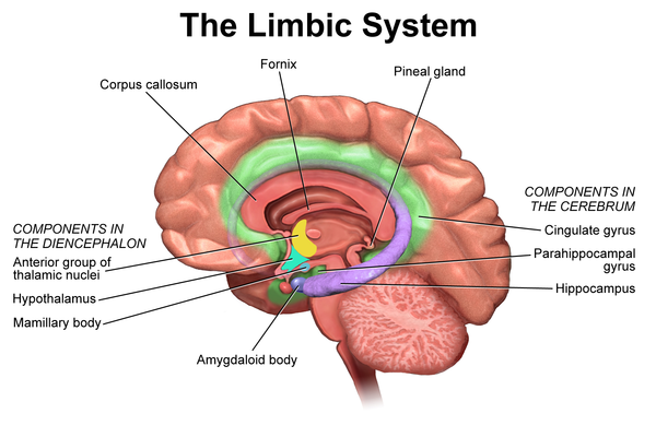
It is a complex network of brain of structures that are located lateral to the thalamus, beneath the cerebral cortex and above the brainstem.[2] The limbic system supports various functions, including (1) emotion, (2) behaviour, (3) motivation, (4) long-term memory, (5) olfaction and (6) stress response.[3]
In evolutionary terms, it is one of the oldest parts of the brain and it "constitutes a functional concept, and is characterized by dense afferent projections from brainstem and forebrain nuclei, which contributes to behavioral modulation"[4]
Important Limbic Structures[edit | edit source]
There is still no consensus on all the structures that make up the limbic system,[4] but key structures include the amygdala, hippocampus, thalamus, hypothalamus, basal ganglia, and cingulate gyrus[2]
Amygdala[edit | edit source]
The amygdala is a small almond-shaped structure located in each temporal lobe, just below the uncus.[5] We have two amygdalae, one in each hemisphere of the brain:
- the amygdalae play an imprtant role in processing information between the prefrontal-temporal association cortices and the hypothalamus[5]
- they are involved in regulating anxiety, aggression, fear conditioning, emotional memory and social cognition, behavioural and physiological stress responses[5]
Hippocampus[edit | edit source]
The hippocampus is a paired structure[6] located in the parahippocampal gyrus, near the inferior temporal horn of the lateral ventricle.[7] It has three regions: (1) dentate gyrus, (2) hippocampus proper, (3) subiculum:[7]
- the hippocampus plays an essential role in forming new memories, particularly consolidated short- and long-term memories, spatial navigation, regulating emotions, motivation, hormones and autonomic activity[6][7][8]
- with the prefrontal cortex, the hippocampus is involved in episodic memory[9]
Thalamus and Hypothalamus[edit | edit source]
The thalamus functions as a relay station between the brain and the body, filtering through various types of sensory and motor information.
The hypothalamus plays a major role in maintaining homeostasis via the autonomic nervous system, the neuroendocrine system, and the limbic system.
If you would like to learn more about these structures, please see:
Cingulate Gyrus[edit | edit source]
The cingulate gyrus is a paired structure that is located on the medial surface of each cerebral hemisphere, wrapping around the corpus callosum.[10] It has four regions (1) anterior cingulate cortex (ACC) (2) midcingulate cortex, (3) posterior cingulate cortex (PCC) and (4) retrosplenial cortex.[10]
Key functions include:[10]
- processing emotions and regulating emotional responses
- cognitive processing (particularly reward-based decision-making)
- processing information from internal and external states to help guide motor responses
- imagination
- episodic memory formation and consolitation
The cingulate cortex can be described as "a connecting hub of emotions, sensations, and action."[10]
Basal Ganglia[edit | edit source]
The basal ganglia are a group of subcortical nuclei[11] that are involved in motor control and learning, executive functions, emotional behaviours, reward and reinforcement, addictive behaviours and habit formation.[12]
For more informatin, please see Basal Ganglia.
Limbic System Disorders[edit | edit source]
Disorders or behaviours that are related to limbic system dysfunction/damage (e.g. traumatic brain injuries or aging) include:
- Disinhibited behaviour: Person does not consider the risk of behaviours and ignores social conventions/rules.
- Increased anger and violence: Commonly tied to amygdala damage
- Hyperarousal: Amygdala damage, or damage to parts of the brain connected to the amygdala, can cause increased fear and anxiety. Anxiety disorders are sometimes treated with drugs that target areas of the amygdala to decrease fear-based emotions.
- Hypoarousal: Can cause low energy or lack of drive and motivation.
- Hyperorality/Kluver-Bucy Syndrome: This is characterized by amygdala damage that can lead to increased drive for pleasure, hypersexuality, disinhibited behaviour and insertion of inappropriate objects in the mouth.
- Appetite dysregulation: Destructive behaviours tied to hyperorality or thalamus dysfunction can include overeating, binge eating or emotional eating.
- Trouble forming memories: Hippocampal damage can include short-term or long-term memory loss. Learning is often greatly impacted by hippocampal damage, as learning it depends on memory.
- Alzheimer’s disease: Research shows that people with Alzheimer’s and memory loss usually have experienced damage to the hippocampus[13]. This causes memory loss, disorientation and changes in moods. Some of the ways that the hippocampus can become damaged include free radical damage/oxidative stress, oxygen starvation (hypoxia), strokes or seizures/epilepsy.
Puberty and the Limbic System[edit | edit source]
Puberty is the beginning of major changes in the limbic system[14].
- The amygdala is thought to connect sensory information to emotional responses. Its development, along with hormonal changes, may give rise to newly intense experiences of rage, fear, aggression (including toward oneself), excitement and sexual attraction[3].
- Over the course of adolescence, the limbic system gradually comes under greater control of the prefrontal cortex (an area associated with planning, impulse control and higher order thought).
- As additional areas of the brain start to help process emotion, older teens gain some equilibrium and have an easier time interpreting others. But until then, they often misread others e.g. teachers and parents.
References[edit | edit source]
- ↑ Queensland Brain Institute. The limbic system. Available from: https://qbi.uq.edu.au/brain/brain-anatomy/limbic-system (last accessed 30 June 2024).
- ↑ 2.0 2.1 Torrico TJ, Abdijadid S. Neuroanatomy, Limbic System. [Updated 2023 Jul 17]. In: StatPearls [Internet]. Treasure Island (FL): StatPearls Publishing; 2024 Jan-. Available from: https://www.ncbi.nlm.nih.gov/books/NBK538491/
- ↑ 3.0 3.1 Martin JH. The limbic system and cerebral circuits for reward, emotions, and memory. In: Neuroanatomy: Text and Atlas. 5th ed. McGraw Hill, 2021.
- ↑ 4.0 4.1 Banwinkler M, Theis H, Prange S, van Eimeren T. Imaging the limbic system in Parkinson's disease-a review of limbic pathology and clinical symptoms. Brain Sci. 2022 Sep 15;12(9):1248.
- ↑ 5.0 5.1 5.2 AbuHasan Q, Reddy V, Siddiqui W. Neuroanatomy, Amygdala. [Updated 2023 Jul 17]. In: StatPearls [Internet]. Treasure Island (FL): StatPearls Publishing; 2024 Jan-. Available from: https://www.ncbi.nlm.nih.gov/books/NBK537102/
- ↑ 6.0 6.1 Kenhub. Hippocampus: Anatomy and functions. Available from: https://www.kenhub.com/en/library/anatomy/hippocampus-structure-and-functions (last accessed 30 June 2024).
- ↑ 7.0 7.1 7.2 Fogwe LA, Reddy V, Mesfin FB. Neuroanatomy, Hippocampus. [Updated 2023 Jul 20]. In: StatPearls [Internet]. Treasure Island (FL): StatPearls Publishing; 2024 Jan-. Available from: https://www.ncbi.nlm.nih.gov/books/NBK482171/
- ↑ Moini J, Logalbo A, Schnellmann JG. Review of the central nervous system. In: Moini J, Logalbo A, Schnellmann JG, editors. Neuropsychopharmacology, Academic Press, 2023. p153-79.
- ↑ Eichenbaum H. Prefrontal-hippocampal interactions in episodic memory. Nat Rev Neurosci. 2017 Sep;18(9):547-58.
- ↑ 10.0 10.1 10.2 10.3 Jumah FR, Dossani RH. Neuroanatomy, Cingulate Cortex. [Updated 2022 Dec 6]. In: StatPearls [Internet]. Treasure Island (FL): StatPearls Publishing; 2024 Jan-. Available from: https://www.ncbi.nlm.nih.gov/books/NBK537077/
- ↑ Yanagisawa N. Functions and dysfunctions of the basal ganglia in humans. Proc Jpn Acad Ser B Phys Biol Sci. 2018;94(7):275-304.
- ↑ Lanciego JL, Luquin N, Obeso JA. Functional neuroanatomy of the basal ganglia. Cold Spring Harbor perspectives in medicine. 2012 Dec 1;2(12):a009621.
- ↑ Rao YL, Ganaraja B, Murlimanju BV, Joy T, Krishnamurthy A, Agrawal A. Hippocampus and its involvement in Alzheimer's disease: a review. 3 Biotech. 2022;12(2):55. doi:10.1007/s13205-022-03123-4
- ↑ Casey BJ, Jones RM, Hare TA. The adolescent brain. Ann N Y Acad Sci. 2008;1124:111-126. doi:10.1196/annals.1440.010


