Anterior Cerebral Artery: Difference between revisions
No edit summary |
No edit summary |
||
| Line 25: | Line 25: | ||
== Assessment == | == Assessment == | ||
It`s important to recognise the stroke symptoms (figure 8.) and to call immediately for emergency assistance. Note the time of onset of the symptoms to give this information to the ambulance team. | |||
== Resources == | == Resources == | ||
Revision as of 21:50, 8 November 2016
Original Editor - Nadja Thöner
Top Contributors - George Prudden, Daniele Barilla, Kim Jackson, Evan Thomas, WikiSysop and Rewan Elsayed Elkanafany
Description[edit | edit source]
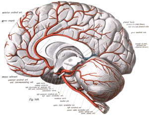
The anterior cerebral artery (ACA) is one of a pair of arteries on the brain that supplies oxygenated blood to most midline portions of the frontal lobes and superior medial parietal lobes.
The ACA has five segments. A1 originates from the internal carotid artery and extends to the anterior communicating artery.
A2 extends from the anterior communicating artery to the bifurcation forming the pericallosal and callosomarginal arteries.
A3 is one of the main terminal branches of the ACA, which extends posteriorly to form the internal parietal arteries and the precuneal artery.
A4 and A5 are the smallest branches and are known as callosal arteries.
Function[edit | edit source]
ACA supplies the frontal, pre-frontal and supplementary motor cortex, as well as parts of the primary motor and primary sensory cortex.
Clinical relevance[edit | edit source]
ACA infarcts are rare because of the collateral circulation provided by the anterior communicating artery.
Assessment[edit | edit source]
It`s important to recognise the stroke symptoms (figure 8.) and to call immediately for emergency assistance. Note the time of onset of the symptoms to give this information to the ambulance team.


