Anterior Cerebral Artery: Difference between revisions
No edit summary |
No edit summary |
||
| Line 6: | Line 6: | ||
== Description == | == Description == | ||
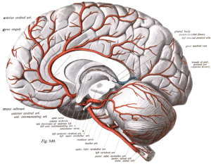
[[Image:ACA2.png|thumb|right|300px]] The anterior cerebral artery (ACA) | [[Image:ACA2.png|thumb|right|300px]] The anterior cerebral artery (ACA) arises from the internal carotid, at the medial extremity of the lateral cerebral fissure. It passes forward and medialward across the anterior perforated substance, above the optic nerve, to the commencement of longitudinal fissure<ref name="Gray">Gray, Henry, Lewis, Warren Harmon. Anatomy of the human body. Philadelphia: Lea & Febiger, 1825-1861</ref>. Is one of a pair of arteries on the brain that supplies oxygenated blood to most midline portions of the frontal lobes and superior medial parietal lobes. | ||
The ACA has five segments. A1 originates from the internal carotid artery and extends to the anterior communicating artery. | The ACA has five segments. A1 originates from the internal carotid artery and extends to the anterior communicating artery. | ||
| Line 18: | Line 18: | ||
== Function == | == Function == | ||
ACA supplies the frontal, pre-frontal and supplementary motor cortex, as well as parts of the primary motor and primary sensory cortex.<ref name="tre" /> | ACA supplies the frontal, pre-frontal and supplementary motor cortex, as well as parts of the primary motor and primary sensory cortex.<ref name="tre" /> | ||
== Clinical relevance == | == Clinical relevance == | ||
ACA infarcts are rare because of the collateral circulation provided by the anterior communicating artery. ACA infarct can present as contralateral hemiparesis with loss of sensibility in the foot and lower extremity, sometimes with urinary incontinence. This is due to the involvement of the medial paracentral gyrus. If the lesion is very proximal, it is possible that there may be cognitive impairment due to lesions in the prefrontal cortex.<ref name="tre">Trepel,M. Blutversorgung des Gehirns (Blood supply oft he brain). In: Trepel,M. Neuroanatomie (Neuroanatomy), 5. Edition. München: Urban&amp;amp;Fischer, 2012. P 280-283.</ref> | ACA infarcts are rare because of the collateral circulation provided by the anterior communicating artery. ACA infarct can present as contralateral hemiparesis with loss of sensibility in the foot and lower extremity, sometimes with urinary incontinence. This is due to the involvement of the medial paracentral gyrus. If the lesion is very proximal, it is possible that there may be cognitive impairment due to lesions in the prefrontal cortex.<ref name="tre">Trepel,M. Blutversorgung des Gehirns (Blood supply oft he brain). In: Trepel,M. Neuroanatomie (Neuroanatomy), 5. Edition. München: Urban&amp;amp;amp;Fischer, 2012. P 280-283.</ref> | ||
== Assessment == | == Assessment == | ||
It`s important to recognise the stroke symptoms and to immediately seek medical assistance. FAST (Face, Arm, Speech, Time) has a Sensitivity of 82% and a Specifity of 83%. Assess for Facial Palsy, Arm Weakness and Speech Impairment. The test is positive if ≥ 1 is present.<ref>Gbinigie, I.I., Reckless, I.P., Buchan, A.M. Stroke: management and prevention. Stroke, 2016; 521- 530.</ref> | It`s important to recognise the stroke symptoms and to immediately seek medical assistance. FAST (Face, Arm, Speech, Time) has a Sensitivity of 82% and a Specifity of 83%. Assess for Facial Palsy, Arm Weakness and Speech Impairment. The test is positive if ≥ 1 is present.<ref>Gbinigie, I.I., Reckless, I.P., Buchan, A.M. Stroke: management and prevention. Stroke, 2016; 521- 530.</ref> | ||
== Resources == | |||
{| width="100%" cellspacing="1" cellpadding="1" | {| width="100%" cellspacing="1" cellpadding="1" | ||
|- | |- | ||
| Line 38: | Line 39: | ||
== See also == | == See also == | ||
*[http://www.physio-pedia.com/Stroke Stroke] | *[http://www.physio-pedia.com/Stroke Stroke] | ||
*[[ | *[[Frontal Lobe Brain Injury|Frontal lobe brain injury]] | ||
*[[ | *[[Introduction to Neuroanatomy|Neuroanatomy]] | ||
== Recent Related Research (from Pubmed) == | == Recent Related Research (from Pubmed) == | ||
<div class="researchbox"> | <div class="researchbox"> | ||
<rss>https://eutils.ncbi.nlm.nih.gov/entrez/eutils/erss.cgi?rss_guid=1F5sQ8LSxW_sDvCDCttU1aUuA8nWjjgEtZCb7iLLI0YD5uL99t|charset=UTF-8|short|max=10</rss> | <rss>https://eutils.ncbi.nlm.nih.gov/entrez/eutils/erss.cgi?rss_guid=1F5sQ8LSxW_sDvCDCttU1aUuA8nWjjgEtZCb7iLLI0YD5uL99t|charset=UTF-8|short|max=10</rss> | ||
</div> | </div> | ||
== References == | == References == | ||
Revision as of 14:14, 6 March 2017
Original Editor - Nadja Thöner
Top Contributors - George Prudden, Daniele Barilla, Kim Jackson, Evan Thomas, WikiSysop and Rewan Elsayed Elkanafany
Description[edit | edit source]
The anterior cerebral artery (ACA) arises from the internal carotid, at the medial extremity of the lateral cerebral fissure. It passes forward and medialward across the anterior perforated substance, above the optic nerve, to the commencement of longitudinal fissure[1]. Is one of a pair of arteries on the brain that supplies oxygenated blood to most midline portions of the frontal lobes and superior medial parietal lobes.
The ACA has five segments. A1 originates from the internal carotid artery and extends to the anterior communicating artery.
A2 extends from the anterior communicating artery to the bifurcation forming the pericallosal and callosomarginal arteries.
A3 is one of the main terminal branches of the ACA, which extends posteriorly to form the internal parietal arteries and the precuneal artery.
A4 and A5 are the smallest branches and are known as callosal arteries.
Function[edit | edit source]
ACA supplies the frontal, pre-frontal and supplementary motor cortex, as well as parts of the primary motor and primary sensory cortex.[2]
Clinical relevance[edit | edit source]
ACA infarcts are rare because of the collateral circulation provided by the anterior communicating artery. ACA infarct can present as contralateral hemiparesis with loss of sensibility in the foot and lower extremity, sometimes with urinary incontinence. This is due to the involvement of the medial paracentral gyrus. If the lesion is very proximal, it is possible that there may be cognitive impairment due to lesions in the prefrontal cortex.[2]
Assessment[edit | edit source]
It`s important to recognise the stroke symptoms and to immediately seek medical assistance. FAST (Face, Arm, Speech, Time) has a Sensitivity of 82% and a Specifity of 83%. Assess for Facial Palsy, Arm Weakness and Speech Impairment. The test is positive if ≥ 1 is present.[3]
Resources[edit | edit source]
See also[edit | edit source]
Recent Related Research (from Pubmed)[edit | edit source]
Failed to load RSS feed from https://eutils.ncbi.nlm.nih.gov/entrez/eutils/erss.cgi?rss_guid=1F5sQ8LSxW_sDvCDCttU1aUuA8nWjjgEtZCb7iLLI0YD5uL99t|charset=UTF-8|short|max=10: There was a problem during the HTTP request: 422 Unprocessable Entity
References[edit | edit source]
- ↑ Gray, Henry, Lewis, Warren Harmon. Anatomy of the human body. Philadelphia: Lea & Febiger, 1825-1861
- ↑ 2.0 2.1 Trepel,M. Blutversorgung des Gehirns (Blood supply oft he brain). In: Trepel,M. Neuroanatomie (Neuroanatomy), 5. Edition. München: Urban&amp;amp;Fischer, 2012. P 280-283.
- ↑ Gbinigie, I.I., Reckless, I.P., Buchan, A.M. Stroke: management and prevention. Stroke, 2016; 521- 530.


