Articular Cartilage Lesions of the Knee: Difference between revisions
Rotimi Alao (talk | contribs) (content) |
Rotimi Alao (talk | contribs) (content) |
||
| Line 44: | Line 44: | ||
| Instability (ACL, PCL, etc.) | | Instability (ACL, PCL, etc.) | ||
|- | |- | ||
| | | Osteochondritis dissecans<br> | ||
|- | |- | ||
| Osteoarthritis | | Osteoarthritis | ||
| Line 71: | Line 71: | ||
<br>Because of this there will be a vicious cycle ensues, only exacerbated by further mechanical trauma to the joint, mostly in the form of sporting activity. Enzymatic breakdown of articular cartilage can lead to capsular distension and synovitis, worsening symptoms that causes the presence of an effusion . These pathologic changes create a feeling of achiness deep in the joint. After checking the patient's medical history, several key points on the physical examination should be specifically noted. | <br>Because of this there will be a vicious cycle ensues, only exacerbated by further mechanical trauma to the joint, mostly in the form of sporting activity. Enzymatic breakdown of articular cartilage can lead to capsular distension and synovitis, worsening symptoms that causes the presence of an effusion . These pathologic changes create a feeling of achiness deep in the joint. After checking the patient's medical history, several key points on the physical examination should be specifically noted. | ||
The medial and lateral palpation of a loose body confirms the presence of an articular cartilage injury but other reproducible and consistent findings are lacking. Pain might not be localized correctly, and crepitus ( Crackling noise when moving over each other rough surfaces, eg. Bone on cartilage. Consider also a tendon in a tendon sheath)may be present. Hearing the sound of crepitus may be alarming to patients, but it is not a bad signal and the patient should be reassured. However, painful crepitus and other mechanical symptoms should be carefully noted and, if possible, localized. Atrophy of the quadriceps and nonarticularpatellofemoral pain may be the cause of knee pain and must be examined.<ref name="Cole et al" /> <ref name="Serban and Oana et al">Serban, Oana, et al. "Pain in bilateral knee osteoarthritis–correlations between clinical examination, radiological, and ultrasonographical findings."&nbsp;Medical Ultrasonography&nbsp;18.3 (2016): 318-325.</ref><ref name="Wright and Huston et al">MARS GroupRick W.&nbsp;Wright,&nbsp;MD,&nbsp;Laura J.&nbsp;Huston, et al. "Meniscal and Articular Cartilage Predictors of Clinical Outcome After Revision Anterior Cruciate Ligament Reconstruction" The american journal of sports medicine" (2016): 1671</ref | The medial and lateral palpation of a loose body confirms the presence of an articular cartilage injury but other reproducible and consistent findings are lacking. Pain might not be localized correctly, and crepitus ( Crackling noise when moving over each other rough surfaces, eg. Bone on cartilage. Consider also a tendon in a tendon sheath)may be present. Hearing the sound of crepitus may be alarming to patients, but it is not a bad signal and the patient should be reassured. However, painful crepitus and other mechanical symptoms should be carefully noted and, if possible, localized. Atrophy of the quadriceps and nonarticularpatellofemoral pain may be the cause of knee pain and must be examined.<ref name="Cole et al" /> <ref name="Serban and Oana et al">Serban, Oana, et al. "Pain in bilateral knee osteoarthritis–correlations between clinical examination, radiological, and ultrasonographical findings."&nbsp;Medical Ultrasonography&nbsp;18.3 (2016): 318-325.</ref><ref name="Wright and Huston et al">MARS GroupRick W.&nbsp;Wright,&nbsp;MD,&nbsp;Laura J.&nbsp;Huston, et al. "Meniscal and Articular Cartilage Predictors of Clinical Outcome After Revision Anterior Cruciate Ligament Reconstruction" The american journal of sports medicine" (2016): 1671</ref> | ||
== Differential diagnosis == | == Differential diagnosis == | ||
Articular cartilage damage is often associated with meniscal and ligament abnormalities. There are different injuries of articular cartilage, such as osteoarthritis, focal articular cartilage defects and osteochondral injuries. <br>MR techniques can allow detection of articular cartilage damage with high accuracy. It can also improve accuracy for determining the integrity of overlying articular cartilage and the extent of detachment of the underlying bone fragment. <ref name="McCauley et al">McCauley, Thomas R., Michael P. Recht, and David G. Disler. "Clinical imaging of articular cartilage in the knee." Seminars in musculoskeletal radiology. Vol. 5. No. 04. Copyright© 2001 by Thieme Medical Publishers, Inc., 333 Seventh Avenue, New York, NY 10001, USA. Tel.:+ 1 (212) 584-4662, 2001.</ref> | Articular cartilage damage is often associated with meniscal and ligament abnormalities. There are different injuries of articular cartilage, such as osteoarthritis, focal articular cartilage defects and osteochondral injuries. <br>MR techniques can allow detection of articular cartilage damage with high accuracy. It can also improve accuracy for determining the integrity of overlying articular cartilage and the extent of detachment of the underlying bone fragment. <ref name="McCauley et al">McCauley, Thomas R., Michael P. Recht, and David G. Disler. "Clinical imaging of articular cartilage in the knee." Seminars in musculoskeletal radiology. Vol. 5. No. 04. Copyright© 2001 by Thieme Medical Publishers, Inc., 333 Seventh Avenue, New York, NY 10001, USA. Tel.:+ 1 (212) 584-4662, 2001.</ref> | ||
== Diagnostic Procedures == | == Diagnostic Procedures == | ||
| Line 96: | Line 92: | ||
*Subchondralpseudocystic sclerotic areas | *Subchondralpseudocystic sclerotic areas | ||
*Altered shape of bony ends | *Altered shape of bony ends | ||
<u>'''Magnetic Resonance Imaging (MRI)'''</u> | <u>'''Magnetic Resonance Imaging (MRI)'''</u> | ||
Revision as of 12:00, 24 October 2018
Description[edit | edit source]
Articular cartilage lesion is a collective term for injuries where the articular cartilage of the knee joint is affected, such as chondromalacia, tears in the articular cartilage, etc. They occur in patients of varying ages. Articular cartilage lesions in weight-bearing joints often fail to heal on their own and may be associated with pain, loss of function and long-term complications such as osteoarthritis.[1]
Articular cartilage lesions can be subdivided in various ways and into different groups. You may divide according to the place where the injury is located or depending on the number of places that are affected. In the first subdivision, we distinguish damage of the femoral condyles or tibial plateaus and cartilage damage to the deep surface of the patella. In the other subdivision, we mark off focal lesions located on one aspect of the tibiofemoral or patellofemoral joint and extremely large lesions or lesions that involve multiple compartments of the knee joint, these are often referred as osteoarthritis.
Articular cartilage lesions have a poor capacity for intrinsic repair.
The recognition of these lesions is one of the most difficult diagnostic problems in knee joint injuries.[2][3][4]
Clinically Relevant Anatomy[edit | edit source]
The knee is the central joint of the lower extremity.
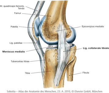
It is constructed by 4 bones (femur, tibia, patella and fibula) and an extensive network of ligaments and muscles. (figure 1)
The knee joint consists of 3 articulations: the articulatiotibiofemoralis located between the convex femoral condyles and the concave tibial condyles, the articulatiopatellofemoralis where the patella lies in the intercondylar groove of the femur, and the articulatiotibiofibularis located between the tibia and fibula.[5]
The tibia, femur and patella are covered in articular cartilage. The normal function of the knee joint depends upon this.
Figure 1:the knee joint
Histology
Hyaline articular cartilage is an avascular, alymphatic, anisotropic tissue. It is not innervated and does not have a basal membrane. Depending on the topography, it varies in thickness and its nutrition is solely based on diffusion. On the articular surface of the patella, the cartilage can reach a thickness of up to 7-8mm. The shock-absorbing properties, as well as the ability of nearly frictionless gliding, are provided by a complex matrix. Elasticity mainly results from a high level of proteoglycans, whereas stiffness and tear strength are enhanced by a network of collagen fibres.[6]
On a histological level we see chondrocytes, collagen fibres, proteoglycans and water.
The superficial collagen fibres are thin and closely packed.They are arranged parallel to the joint surface and lie perpendicular to the principal axis of motion for a particular joint. They allow for near frictionless joint motion. Collagen fibres in the middle or transitional zone are intermediate in size and obliquely oriented.The proteoglycans are trapped and contained within their framework. The largest collagen fibres in the deep or radial zone are arranged perpendicular to the joint surface.They penetrate the calcified layer to enter the subchondral bone and anchor the cartilage to the underlying bone.
Proteoglycans account for the filling pressure in articular cartilage.
Chondrocytes are the power source behind the proteoglycan pump. They balance matrix synthesis and degradation by responding to their chemical and biomechanical environments.[7]
Epidemiology / Etiology[edit | edit source]
Epidemiology
A large retrospective study has aimed to provide data on the prevalence, epidemiology and etiology of the knee articular cartilage lesions, on the ground of analysis of a large database of arthroscopies (25.124 arthroscopies performed from 1989 to 2004).[8]
These data are along the same lines as data from several other studies. [9] [10]
Chondral lesions were found in 60% of patients.They were classified as localized focal osteochondral or chondral lesions in 67%, osteoarthritis in 29%, osteochondritisdissecans in 2% and other types in 1%.
Non-isolated cartilage lesions accounted for 70% and isolated lesions for 30%.
The patellar articular surface (36%) and the medial femoral condyle (34%) were the most frequent localisation of the cartilage lesions, while medial tibial plateau (6%) was the least frequent one. Grade II according to outerbridge classification was the most frequent grade of the cartilage lesion (42%).
The most common associated articular lesions were meniscus tear (37%) and injury of the anterior cruciate ligament (36%).
The analysis of the onset of symptoms revealed that in 58% it was a traumatic non-contact onset, usually connected with a day living activity (45%) and with sports participation (46%, especially football and skiing).
Etiology
Most articular cartilage defects are caused by trauma, which can either be one single impact injury or repeated micro trauma. A specific group of cartilage damage is 'osteochondritisdissecans' where a well-demarcated small area of cartilage and underlying bone loses its blood supply, dies and eventually fragments and separates into the joint. Axial malalignment of the knee can also cause articular cartilage defects this can lead to local overload in certain compartments of the joint. Following cartilage degeneration and focal, compartmental osteoarthritis. Axial malalignments of the knee is usually multifactorial. It can be changes of the condylar level or lateral and medial sloping of the tibia plateau.
Rheumatoid arthritis causes progressive joint erosion and polyarthric strike. Most genetic disorders have an increase of chondromalacia-like cartilage lesions mostly combined with ligamentary laxity. This can have its origin in genetic defaults of the collagen synthesis. Being overweight can also be a risk factor in developing cartilage lesions because of the increasing pressure put upon the joint. [11] [12]
| Aetiology of articular cartilage lesions of the knee Trauma (blunt impacts, traumatic patellar dislocation, polytraumatic injuries) |
|---|
| Axial malalignment of the knee |
| Partial or total meniscectomy |
| Instability (ACL, PCL, etc.) |
| Osteochondritis dissecans |
| Osteoarthritis |
| Rheumatoid arthritis |
| Genetic factors |
| Obesity |
| Cartilage tumours |
| Microtrauma |
Characteristics / Clinical Presentation[edit | edit source]
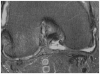
Lower activity levels, decreased sports perticipation, pain, stiffness and functional limitations are some characteristics of articular cartilage damage. The clinical presentation encompasses the gamut from incidentally noted, asymptomatic lesions to dramatic, disabling arthritic symptoms. Knowing the patient his medical history, and the patient should be queried regarding any previous treatment. There may be a history of ligament injury (often the ACL), patellar dislocation, or a traumatic "dashboard" injury to the knee. Hemarthroses are seen in almost all acute injuries that create a full thickness chondral injury. Loose-body symptoms are other common scenarios that often connote a full-thickness articular injury. Pain is mostly worse when the patient does physical activity, and swelling is often intermittent and activity related in chronic cases. Pain with prolonged sitting, stair climbing, and kneeling may localize the pain to the patella or femoral trochlea. Activities that provoke pain should be specifically queried. To assess physical disability, work requirements and limitations the patient is currently experiencing should be noted. It is critical to note Athletic participation and patient expectations regarding return to sports or activity because they are often unrealistic. Previous treatments, including both surgical and nonsurgical interventions must be discussed as well as the patients' clinical responses. Important interventions that must be recorded are medications, including viscosupplementation and chondrosupplementation with glucosamine or chondroitin or both. The loss of the cushioning function of the articular cartilage causes the presence of pain. If the subchondral bone in the knee is experiences increased pressure, pain fibers in this region will be stimulated. Increased venous flow accompanies bony sclerosis and congestion of the cancellous bone occurs (Figure 2).
Figure 2: Subacromial edema (bone bruise) secondary to degenerative joint disease (DJD). See the loss of overlying articular cartilage
Because of this there will be a vicious cycle ensues, only exacerbated by further mechanical trauma to the joint, mostly in the form of sporting activity. Enzymatic breakdown of articular cartilage can lead to capsular distension and synovitis, worsening symptoms that causes the presence of an effusion . These pathologic changes create a feeling of achiness deep in the joint. After checking the patient's medical history, several key points on the physical examination should be specifically noted.
The medial and lateral palpation of a loose body confirms the presence of an articular cartilage injury but other reproducible and consistent findings are lacking. Pain might not be localized correctly, and crepitus ( Crackling noise when moving over each other rough surfaces, eg. Bone on cartilage. Consider also a tendon in a tendon sheath)may be present. Hearing the sound of crepitus may be alarming to patients, but it is not a bad signal and the patient should be reassured. However, painful crepitus and other mechanical symptoms should be carefully noted and, if possible, localized. Atrophy of the quadriceps and nonarticularpatellofemoral pain may be the cause of knee pain and must be examined.[3] [13][14]
Differential diagnosis[edit | edit source]
Articular cartilage damage is often associated with meniscal and ligament abnormalities. There are different injuries of articular cartilage, such as osteoarthritis, focal articular cartilage defects and osteochondral injuries.
MR techniques can allow detection of articular cartilage damage with high accuracy. It can also improve accuracy for determining the integrity of overlying articular cartilage and the extent of detachment of the underlying bone fragment. [15]
Diagnostic Procedures[edit | edit source]
X-ray examination
The X-ray is frequently applied in daily practice to diagnose articular cartilage damage. Although cartilage is invisible on plain radiography, but it can be used to determine some characteristics.[16][6]
Hallmarks of cartilage degeneration by X-ray:
- Joint space narrowing
- Calcification of cartilage
- Bone spurs (osteophytes)
- Patella maltracking or tilt
- Malalignments (varus or valgus deformities)
- Signs of inflammatory diseases
- Periarticularossicles
- Subchondralpseudocystic sclerotic areas
- Altered shape of bony ends
Magnetic Resonance Imaging (MRI)
Modern magnetic resonance tomographs give a detailed view of the articular cartilage itself and can bring out even smaller lesions. Several MRI scoring techniques have been focusing on size and location of the lesions and subchondral, cartilaginous, bone and meniscal abnormalities.
The conventional MRI sequences are a good way to diagnose articular cartilage lesions.They offer a good analysis of all the structures of the joints. But they are only useful when there is actually a lesion present. MRI remains a useful tool for a detailed therapy planning.[16][17]
MRI imaging should focus on the following pathologies:
- Cartilage defects (where? How large? How deep?)
- Injuries to the subchondral plate
- Bony lesions (OD)
- Secondary damages (meniscus tears etc)
Arthroscopy
Diagnostic arthroscopy is indicated on suspicion of an articular cartilage defect or in persistent, unclear disorders of the knee. With arthroscopy there is a direct visualization of the actual cartilage damage. It enables a direct view of the cartilage surface and palpation of its stiffness. Softening, partial delamination and fibrillation can be discovered that way. It seems to be a good method of grading severe focal cartilage lesions, but may not be suitable for quantitative assessment of early cartilage damage.
Only arthroscopic examination allows exact determination of defect size and quality and it is essential in terms of differential diagnosis and classification of a cartilage lesion. [16] [6]
Outcome Measures[edit | edit source]
We studied the outcome measurements of a experimental study about the effect of ultrasound immediatley after osteochondral grafting surgery.
Osteochondral grafting surgery has been used for treatment of articular cartilage lesions. In this surgery, cylindrical plugs incorporating both articular cartilage and the underlying subchondral bone are harvested from the donor site and grafted to the lesion.
From August 2002 to November 2005, 37 patients with knee disorders who were attending the Department of Orthopaedic Surgery, Kyoto University Hospital, were screened for eligibility.
The inclusion criteria were:
- osteochondral grafting surgery,
- focal traumatic cartilage lesion of the knee of the trochlea or femoral condyles,
- osteonecrosis,
- cartilage damage grade 3 (ICRS),
- no upper age limit, and
- informed consent.
The exclusion criteria were:
- osteochondritisdissecans,
- early stage of osteoarthritis,
- grade 1 and grade 2 cartilage lesions (ICRS).
Here, ICRS describes cartilage standard evaluation form as follows:
o grade 0, normal cartilage
o grade 1, near-normal cartilage with superficial lesions
o grade 2, cartilage with lesions extending to less than 50% of the depth of the cartilage
o grade 3, cartilage with defects that extend to more than 50% of the depth of the cartilage
o grade 4, severely abnormal cartilage that the cartilage defects reaches to subchondral bone
High-frequency pulse-echo ultrasound is sensitive for detecting the degeneration of superficial collagen-rich cartilage zone. Ultrasound measurements are related to changes in the extracellular matrix collagen and fibrillar network organization. Ultrasound can detect microstructural changes up to a depth of 500 μm. Therefore, signal intensity provides information on superficial collagen integrity of cartilage.
In the study we examined, the signal intensities of the damaged cartilage in the traumatic cartilage lesions and in the osteonecrosis were 21% (0.40 vs. 1.91) and 24% (0.30 vs. 1.25) of the adjacent intact cartilage.
Lower signal intensity represents lower superficial collagen integrity of cartilage.
It is well known that articular cartilage has limited intrinsic healing potential, lower signal intensity may be a critical sign of cartilage.
The signal intensities of plug cartilage in the traumatic cartilage lesions and in the osteonecrosis were 533% (2.13 vs. 0.40) and 463 % (1.39 vs. 0.30) of the damaged cartilage.
Therefore, these results indicate that the osteochondral grafting surgery replaced the lesion with cartilage of greater signal intensity, which means greater superficial collagen integrity of cartilage and cartilage with greater interval between signals, means greater thickness of the cartilage. [11]
Examination[edit | edit source]
Physical examination should focus on the following pathologies:
- Malalignments (valgus/varus deformities)
- Limited range of motion
- Effusion
- Instability
- Clicking, grinding or any other pathological sounds
- Catching or locking
- Maltracking or tilt of the patella
Physical examination:
knee pain can be appreciated during medial and lateral palpation of each knee joint and using visual analogue scale (VAS) and the western ontario and McMaster universities osteoarthritis index (WOMAC) [13]
Retropatellar cartilage
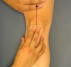
Pain can be provoked by bending the knee on one leg, the patient will often refuse doing this because he probably knows this will be painful. This mechanism can be simulated by the examiner by giving pressure over the patella when the quadriceps are relaxed, then request the patient to tighten his quadriceps muscle. The patella s drawn into axial stress which leads to pain
(Figure 3). It is often more easy to feel retropatellar crepitus then to hear. The mobility of the patella will also be tested horizontally and vertically. The standard examination of the knee joint, including testing for an effusion, ligament stability, and meniscal integrity are also needed. Mostly retropatellar cartilage does not affect the entire surface, the medial and lateral facets are examined separately. The examiner will shift the patella medially, that way the medial protrudes from the intercondylar fossa and can be palpated directly. The lateral facet will be pressed against the condyle (Figure 4)
When there is a lateral displacement of the patella, the medial facet is compressed and the lateral facet becomes palpable (Figure 5). It is possible to differentiate the involvement of the facets by determining the point of maximum tenderness. Reaching an overall diagnosis of retropatellararthrosis and determine the condition of major patellar changes is allowed by this examination technique. To clarify the etiology and extent of the complaint, further investigation is necessary. It is important to inquire as to any history of fracture, contusion, injections, or possible infections in addition to documenting the nature of the pain. [18]
Figure 3: the examination of the sensitivity of the patellar gliding
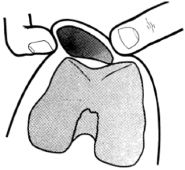
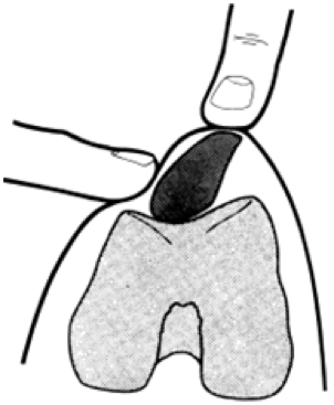
Figure 4:Palpation of the medial facet Figure 5: Palpation of the lateral facet
Medical Management[edit | edit source]
NSAID per os
NSAID’s are non-steroid anti-inflammatory drugs that show rapid effects in a short time but they aren’t always as efficient in a long time process. By inhibition of prostaglandin syntheses they show both analgesic as anti-inflammatory effects. The downside of NSAID’s is that they usually show severe side effects that can evolve in fatality. The use of 2nd and 3th generation of NSAID’s can reduce the risks of those side effects. ( Level of evidence 3A) [12]
Neuroceticals
Glucosamines per os, intra-articular cortisone, intra-articular hyaluronic acid. One of the main components of the hyaline cartilage is chondroitine-sulfate. In osteoarthritic cartilage ass well as in degenerative cartilage are both proteoglycan and chondroitine-sulfate deficiencies notable. There still remain some doubts whether or not the intake of these nutrients plays a specific role in the deficiency. Medication with glucosamines has not yet shown effective results on the regeneration of the cartilage. However it is assumed that it helps replenishing structural deficiencies. ( Level of evidence 2A)[11]
Surgical management
Cartilage treatment should be aimed at restoring the normal knee function by regeneration of hyaline cartilage in the defect, and to achieve a complete integration of the new cartilage to the surrounding cartilage and underlying bone.
Joint lavage and debridement
Rinsing of the joint and the removal of damaged tissue appears to alleviate pain although the mechanism for this is unclear. It is suggested that pain relief observed may be no more than a placebo effect following surgery. ( Level of evidence 3A)[19]
Abrasion arthroplasty, drilling and microfractures
These techniques have the goal of recruiting pluripotential stem cells from the marrow by penetrating the subchondral bone.
Osteochondral Transfer
An alternative to biological regeneration of a defect is to replace it with a substitute.
Mosaicplasty and osteochondral autologous grafts: using a drill, plugs of cartilage are harvested from areas with relatively less weight bearing such as the intercondylar notch or the most lateral part of the femoral condyle. The plugs are then placed in the defect in predrilled cylinders.
Allogenicosteochondral transplantation is used for large osteochondral defects. Recently synthetic plugs have been developed. (Level of evidence 3A)[19]
Cell Transplantation Based Repair
Autologous chondrocyte implantation (ACI):
In this procedure cartilage is harvested through an arthroscopic procedure. The chondrocytes are then released by enzymatic digestion and expanded in culture. An arthrotomy is performed 2-4 weeks later, where the cells are injected under a periostal flap or a synthetic membrane. ( Level of evidence 3A)[19]
Matrix guided autologous chondrocyte implantation (MACI:)
This procedure tries to improve upon the disadvantages of ACI (hypertrophy of the graft, uneven distribution of chondrocytes within the defect and potential for cell leakage). In MACI cultured chondrocyte is implanted in scaffold (natural and synthetic biomaterials).
Mesenchymal stem cell (MSC) transplantation:
Mesenchymal stem cells retain both high proliferative potential and multipotentiality, including chondrogenic differentiation potential. The use of this technique is still at the early stage, animal studies with this method are being conducted.(Level of evidence 3A)[19]
Other operative treatments include:
• Refixation of detached articular cartilage fragments with:
o reabsorbable pins
o screws
o fibrin glue
o osteochondral plugs
• Cartilage reparative strategies
• Aggressive debridement (spongialisation): removal of subchondral plate to expose cancellous bone
• Bone marrow stimulation techniques: drilling, microfractures, abrasion arthroplasty( gentle superficial burring of the subchondral plate)
• Cartilage restorative techniques
• Transplantation of fresh osteochondral allografts
• Transplantation of osteochondralautografts (plugs- mosaicplasty)
• Autologous chondrocyte implantation (ACI) and matrix-induced autologous chondrocyte implantation (MACI)
Physical Therapy Management[edit | edit source]
Physical therapy
o Rehabilitation can be advantageous to patients who are not amenable to surgical treatment and to those in whom surgery has to be delayed for some time.Rehabilitation cannot alter the natural course of the disease. The purpose is to relieve symptoms and arrest the progression of the lesion so as to prevent any sequelae.
In patients that have had surgery, rehabilitation will be aimed at creating a protected environment that facilitates the healing of tissue. While at the same time work is done to achieve progressive functional recovery in terms of pain control, improvement of range of motion, development of muscle strength, reeducation of gait and, on some occasions, sports retraining. This rehabilitation should always be individualised for each patient, taking into account the surgical technique that was used. (Level of evidence 1A)[20]
o In case of cartilage lesions, pain will lead to a reactive functional impairment of the affected joint. The patient will not be able to perform movements without feeling any pain. As a result the patient would minimisethesepainful movements and that van lead to shrinkage of the joint capsule and to a decrease of blood flow. Physiotherapists use different techniques to break tis vicious circle. These techniques are focused on the following goals: (Level of eveidence 3A) [12] [19]
- Pain relief by lowering weight-bearing
- Manual stretching of painful , contract joint capsules
- Detonisation of hypertonic muscle groups
- Passive mobilisation of the joint.
- Improvement of joint mobility by active and passive mobilisation of the joints
- Improvement of muscular joint stabilisation
- Improvement of joint function by learning compensatory techniques and coordinative exercises
Bandages and orthosis are useful tools to ease the pain and improve joint function. The inwoven fabric pads give a gentle pressure upon the compartments of the joint. Sometimes it can also decelerate the progress of the disease.(Level of evidence 3A) [19]
Electrotherapy can effect the patient from pain relief by affection of pain-conducting nerve fibres to destruction of the genetic material.( Level of evidence 3A) [19] [12]
Pulsed Signal Therapy (PST) effects the proliferation of chondrocytes in a positive way by applying electrical currents in general. This results in enhancing the chondrocytes’ synthetic performance by absorbing more water in the injured cartilage.
Ultrasound therapy sends vibrations that can increase the temperature between the different tissue layers. This temperature rise can improve blood flow along with the metabolism and nutrition of the joint. ( Level of evidence 3A) [19]
Osteoarthritis
Local applications like mud, peat and paraffin have been tried. If the knee joint is inflamed, cold therapy has been popular. Short-wave and ultrasound are used. Radiotherapy to eliminate pain receptors is in some cases used but patients with recurrent pain after this treatment are resistant to other forms of therapy. ( Level of evidence 3A) [19]
Prevention:
Physical activity
Whether physical activity is beneficial or detrimental to articular structures of weight-bearing joints has been the topic of a lot of discussion.
A systematic review in 2011 concluded that physical activity is positively associated with an increase in cartilage volume and a decrease in cartilage defects.And this independent of age, sex or BMI. (level of evidence 1A)[21]
Both intensity and duration are significant. Data suggests that at least 20 minutes a week of activity sufficient to result in sweating or some shortness of breath might be adequate. There is some evidence that even walking might be beneficial. (level of evidence 3A)[22]
Sedentary lifestyles have been shown to adversely affect cartilage, with rapid cartilage loss being demonstrated among the initial 12 months of quadriplegia.
These findings suggest that physical activity is beneficial, rather than detrimental, to joint health.(level of evidence 3A)[23]
BMI
The prevalence and severity of knee cartilage defects increase with Body Mass Index (BMI).(level of evidence 2A)[24]
The likelihood of knee articular cartilage defect progression increases with increased BMI. (level of evidence 2B)[25]
Therefore weight control may have a role in the prevention of (the progression of) articular cartilage lesions.
Sports injury
Quite often the cartilage defect is only part of a complex joint trauma affecting several structures.
Cartilage injuries are frequently observed in young and middle aged active athletes. The prevention of knee injury, especially anterior cruciate ligament and meniscus injury in sports is important. Prevention programs of sports injury have shown encouraging results. Prevention of ACL injuries was possible with the use of neuromuscular training programs.(level of evidence 5)[26]
The rate of injuries in adolescent athletes using a structured warm-up programme as part of their training improved clinically and statistically. The incidence of acute knee and ankle injuries can be reduced by 50% and severe injuries even more.
Youth elite as well as intermediate and recreational players would benefit from using this programme. Therefore preventive training should be introduced as an integral part of youth sports programmes.
The warm-up programme aims to improve running, cutting, and landing technique as well as neuromuscular control, balance, and strength. The programme included: focussing on landing on both legs rather than just one leg; landing with increased flexion at the knee and hip to attenuate the landing; balance training using the wobble board and balance mat; a strength exercise, the ‘Nordic hamstring lower’ exercise and some form of feedback to the athlete whilst performing these exercises.(level of evidence 1B)[4]
Key Evidence[edit | edit source]
Donna M. Urquhart et al. What is the effect of Physical activity on the Knee Joint? A systematic review, Medicine & Science in Sports & Exercise, 2011
Brian J. Cole, M. Mike Malek (2004). Articular Cartilage Lesions A Practical Guide to Assessment and Treatment: Background and patient assessment (vol 1). New york: Springer
Resources[edit | edit source]
http://www.sportsmedicineofnapavalley.com/knee2/
http://www.dartmouth-hitchcock.org/ortho/articular_cartilage_injury.html
http://www.londonkneeclinic.com/knee-problems/articular-cartilage-damage
http://appliedradiology.com/articles/mr-of-articular-cartilage-lesions-of-the-knee.aspx
Clinical Bottom Line[edit | edit source]
Physical rehabilitation of patients with articular cartilage lesions cannot alter the natural course of the disease. The purpose is to relieve symptoms and arrest the progression of the lesion so as to prevent any sequelae.[20]
On the basis of published evidence chondral lesions of the knee are most common associated with meniscus tea rand injury of the anterior cruciate ligament.[11] [12]
Articular cartilage lesions are defined as a collective term of injuries where the articular cartilage of the knee joint is affected. Only arthroscopic examination allows exact determination of defect size and quality and it is essential in terms of differential diagnosis and classification of a cartilage lesion using the Outerbridge Classification.
Therapy may consist of bandages, orthosis, electrotherapy, ultrasound therapy and exercises. Surgical and drug treatment may be needed. As most articular cartilage defects are caused by trauma (daily living activity or sports injury), a high emphasis on prevention is necessary.
Recent Related Research (From Pubmed)[edit | edit source]
Falah M, Nierenberg G, Soudry M, Hayden M, Volpin G. Treatment of articular cartilage lesions of the knee. International Orthopaedics. 2010;34(5):621-630. doi:10.1007/s00264-010-0959-y.
Dean CS, Chahla J, Serra Cruz R, LaPrade RF. Fresh Osteochondral Allograft Transplantation for Treatment of Articular Cartilage Defects of the Knee. Arthroscopy Techniques. 2016;5(1):e157-e161. doi:10.1016/j.eats.2015.10.015.
References[edit | edit source]
- ↑ D’Anchise R, Manta N, Prospero E, Bevilacqua C, Gigante A (2005) Autologous implantation of chondrocytes on a solid collagen scaffold: clinical and histological outcomes after two years of followup. J Orthop Traumatol 6:36–43
- ↑ Mandelbaum, Bert R., et al. "Articular cartilage lesions of the knee." The American Journal of Sports Medicine 26.6 (1998): 853-861.
- ↑ 3.0 3.1 Brian J. Cole, M. Mike Malek (2004). Articular Cartilage Lesions A Practical Guide to Assessment and Treatment: Background and patient assessment (vol 1). New york: Springer
- ↑ 4.0 4.1 Odd-Egil Olsen et al. Exercises to prevent lower limb injuries in youth sports: cluster randomised controlled trial, 2007, the British Medical Journal, doi:10.1136/bmj.38330.632801.8F
- ↑ Sobotta, Atlas van de menselijke anatomie, Deel 2 Romp, organen, onderste extremiteit
- ↑ 6.0 6.1 6.2 Jürgen Fritz, Pia Janssen et al, Articular cartilage defects in the knee-basics, therapies and results. International Journal of the Care of the Injured (2008) 39S1, 50-57
- ↑ Constance R.; Chondral and osteochondral injuries: mechanisms of injury and repair responses. Operative Techniques in Orthopaedics, No 2 (April), 2001: pp 70-75
- ↑ Widuchowski W. et al., Articular cartilage defects: Study of 25.124 knee arthroscopies, ScienceDirect, The Knee 14 (2007) 177-182
- ↑ Walton W. Curl, Jonathan Krome et al., Cartilage Injuries: A Review of 31.516 Knee Arthroscopies, Arthroscopy: The Journal of Arthroscopic and Related Surgery, Vol 13, No 4 (August), 1997, pp 456-460
- ↑ Hjelle K, Solheim E, Muri R, Brittberg M, Articular cartilage defects in 1000 knee arthroscopies, Arthroscopy, Sept 2002, 18 (7), pp 730-734
- ↑ 11.0 11.1 11.2 11.3 Noyes, F. R., &Stabler, C. L. (1989).A system for grading articular cartilage lesions at arthroscopy.The American Journal of Sports Medicine, 17(4), 505-513.doi:10.1177/036354658901700410
- ↑ 12.0 12.1 12.2 12.3 12.4 Rodríguez-Merchán, E. C. (2012). Articular cartilage defects of the knee: diagnosis and treatment. Milan: Springer.
- ↑ 13.0 13.1 Serban, Oana, et al. "Pain in bilateral knee osteoarthritis–correlations between clinical examination, radiological, and ultrasonographical findings." Medical Ultrasonography 18.3 (2016): 318-325.
- ↑ MARS GroupRick W. Wright, MD, Laura J. Huston, et al. "Meniscal and Articular Cartilage Predictors of Clinical Outcome After Revision Anterior Cruciate Ligament Reconstruction" The american journal of sports medicine" (2016): 1671
- ↑ McCauley, Thomas R., Michael P. Recht, and David G. Disler. "Clinical imaging of articular cartilage in the knee." Seminars in musculoskeletal radiology. Vol. 5. No. 04. Copyright© 2001 by Thieme Medical Publishers, Inc., 333 Seventh Avenue, New York, NY 10001, USA. Tel.:+ 1 (212) 584-4662, 2001.
- ↑ 16.0 16.1 16.2 Bekkers, J. E. J., et al. "Diagnostic Modalities for Diseased Articular Cartilage—From Defect to Degeneration A Review." Cartilage 1.3 (2010): 157-164.
- ↑ Fritz et al
- ↑ D. E. Hastings, A. Rüter, C. Burri (1978).The Knee: Ligament and Articular Cartilage Injuries: Retropatellar Cartilage Degeneration: Diagnosis and Outline of Treatment. (vol 3). Berlin, Heidelberg, New York: Springer-Verlag.
- ↑ 19.0 19.1 19.2 19.3 19.4 19.5 19.6 19.7 19.8 Erggelet, C., &Mandelbaum, B. R. (2008). Principles of cartilage repair. Germany: SteinkopffVerlag.
- ↑ 20.0 20.1 Carlos Rodriguez-Merchan, Articular Cartilage Defects of the Knee- Diagnosis and Treatment, Springer, 2012
- ↑ Donna M. Urquhart et al. What is the effect of Physical activity on the Knee Joint? A systematic review, Medicine & Science in Sports & Exercise, 2011, DOI: 10.1249/MSS.0b013e3181ef5bf8
- ↑ Tina L. Racunica et al., Effect of Physical Activity on Articular Knee Joint Structures in Community-Based Adults, Arthritis & Rheumatism Vol.57, No. 7, October 15, 2007, ppp 1261-1268
- ↑ Benny Antony, Alison Venn, FlaviaCicuttini et al.; Association of physical activity and physical performance with tibial cartilage volume and bone area in young adults, Arthritis Research & Therapy (2015) 17:298
- ↑ Ding C, Garnero P, Cicuttini F, Scott F, Knee structural alteration and BMI: a cross-sectional study, Obes Res 2005, 13, pp 350-361
- ↑ Wang Y. et al., Factors affecting progression of knee cartilage defects in normal subjects over 2 years, Rheumatology 2006; 45:79-84
- ↑ Hideki Takeda, et al.(2011). Prevention and management of knee osteoarthritis and knee cartilage injury in sports. British Journal of Sports Medicine, 45, 304-309
| The content on or accessible through Physiopedia is for informational purposes only. Physiopedia is not a substitute for professional advice or expert medical services from a qualified healthcare provider. Read more. |
