Calcific Tendinopathy of the Shoulder: Difference between revisions
No edit summary |
No edit summary |
||
| Line 8: | Line 8: | ||
== Introduction == | == Introduction == | ||
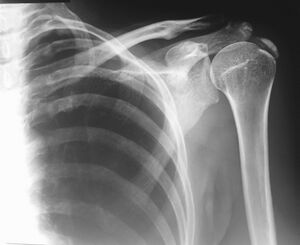
[[File:Dense calcification of the supraspinatus.jpeg|thumb|Calcification of the supraspinatus]] | [[File:Dense calcification of the supraspinatus.jpeg|thumb|Calcification of the supraspinatus]] | ||
Calcific tendonitis refers to the calcification and tendon degeneration around the shoulders rotator cuff insertions. It usually results in shoulder pain with decreased range of motion. Diagnosis is made by shoulder x-rays, with visible signs of calcium deposits overlying the rotator cuff insertion.Treatment consists of NSAIDs, physical therapy, corticosteroid injections and ultrasound-guided needle lavage. Those who fail conservative treatment may choose to have arthroscopic decompression of the calcium deposits.<ref>Orthobullets [https://www.orthobullets.com/shoulder-and-elbow/3042/calcific-tendonitis Calcific Tendonitis]Available:https://www.orthobullets.com/shoulder-and-elbow/3042/calcific-tendonitis (accessed 12.1.2023)</ref> | Calcific tendonitis refers to the calcification and tendon degeneration around the shoulders rotator cuff insertions. It usually results in shoulder pain with decreased range of motion. Diagnosis is made by shoulder x-rays, with visible signs of calcium deposits overlying the rotator cuff insertion.Treatment consists of NSAIDs, physical therapy, corticosteroid injections and ultrasound-guided needle lavage. Those who fail conservative treatment may choose to have arthroscopic decompression of the calcium deposits.<ref name=":1">Orthobullets [https://www.orthobullets.com/shoulder-and-elbow/3042/calcific-tendonitis Calcific Tendonitis]Available:https://www.orthobullets.com/shoulder-and-elbow/3042/calcific-tendonitis (accessed 12.1.2023)</ref> | ||
== Epidemiology == | == Epidemiology == | ||
| Line 75: | Line 75: | ||
== Medical Management == | == Medical Management == | ||
Nonoperative | |||
# NSAIDs, physical therapy, stretching & strengthening, steroid injections | |||
# Extracorporeal shock-wave therapy as an adjunct treatment. Most useful in refractory calcific tendonitis in the formative and resting phases | |||
# Ultrasound-guided needle lavage vs. needle barbotage (needle to break up calcium deposit) | |||
Operative: surgical decompression of calcium deposit.<ref name=":1" /> | |||
== <div id="Shockwave">Physical Therapy Management</div> == | == <div id="Shockwave">Physical Therapy Management</div> == | ||
Revision as of 08:06, 12 January 2023
Original Editors Mary Harris, Tom Lawlor, Patrick Bales, Misty Hillin, Rick Wetherald as part of the Texas Evidence Based Practice Project.
Top Contributors - Mary Harris, Thomas Lawlor, Karina Leahy, Rick Wetherald, Patrick Bales, Admin, Lucinda hampton, Fasuba Ayobami, Kim Jackson, Tony Lowe, Anthony Mertens, Wanda van Niekerk, 127.0.0.1, Naomi O'Reilly and WikiSysop
Introduction[edit | edit source]
Calcific tendonitis refers to the calcification and tendon degeneration around the shoulders rotator cuff insertions. It usually results in shoulder pain with decreased range of motion. Diagnosis is made by shoulder x-rays, with visible signs of calcium deposits overlying the rotator cuff insertion.Treatment consists of NSAIDs, physical therapy, corticosteroid injections and ultrasound-guided needle lavage. Those who fail conservative treatment may choose to have arthroscopic decompression of the calcium deposits.[1]
Epidemiology[edit | edit source]
Usually occurs in middle-aged patients between the ages of 30 and 60, with a slight preference for females.[2]
Pathogenesis[edit | edit source]
The exact pathogenesis of calcific tendinitis is unclear. Theories include:
- An association with endocrine disorders of thyroid and estrogen metabolism.
- Extracellular matrix vesicles are the origin of the pathologic calcification.
- Calcification occurs in the setting of tendon degeneration and necrosis, but finding now refute this.[3]
Localisation[edit | edit source]
- Supraspinatus tendon (80% of cases): critical zone - Most Common
- Infraspinatus tendon (15% of cases): lower 1/3
- Subscapularis tendon (5%of cases): pre-insertional fibers[4]
Characteristics/Clinical Presentation[edit | edit source]
The chief patient complaints to expect in calcific tendinopathy are:
- Night pain, causing loss of sleep.[5], [6], [7], [8].
- Constant dull ache[8].
- Pain increases considerably with AROM[8].
- Decrease in ROM, or complaint of stiffness [9], [7], [8].
- Radiating pain up into the suboccipital region, or down into the fingers[5], [6], [8].
The condition goes through 4 stage, see table below.
| Stages[8] | |
|---|---|
| Stage Name | Presentation |
| Chronic (Silent) Phase |
|
|
Acute Painful Phase |
|
|
Mechanical Phase |
|
Differential Diagnosis[edit | edit source]
- Incidental calcification: found in 2.5-20% of 'normal' healthy shoulders.
- Degenerative calcification: found tendons with tear history; generally smaller; slightly older individuals
- Loose bodies: associated chondral defect; associated secondary osteoarthritis[2]
Outcome Measures[edit | edit source]
Medical Management[edit | edit source]
Nonoperative
- NSAIDs, physical therapy, stretching & strengthening, steroid injections
- Extracorporeal shock-wave therapy as an adjunct treatment. Most useful in refractory calcific tendonitis in the formative and resting phases
- Ultrasound-guided needle lavage vs. needle barbotage (needle to break up calcium deposit)
Operative: surgical decompression of calcium deposit.[1]
Physical Therapy Management[edit | edit source]
There is evidence supporting the use of extracorporeal shock wave therapy (ESWT) as a potentially effective treatment of calcific tendinopathy. The modality administers high frequency sound waves to the affected area with the intent of breaking up the calcification. Researchers claim that this will cause the body to activate or increase the body’s calcium resorption system, removing the deposit. Depending on the frequency used, the treatment can be painful, but research shows the modality to be most effective at the highest frequency the patient can tolerate.ESWT is a potential alternative to surgery with good mid-term effectiveness and minimal side effects. [11] But ECSW is not free from complications, that included transient bone marrow edema and even reported cases of humeral head necrosis.[12][13]
Most authors report short term symptomatic improvement[14], but long term positive outcomes (past one year) have not been definitively demonstrated in research. [15]
Radial shock wave therapy (RSWT) is another modality that has been used in the treatment of calcific tendinopathy. RSWT is similar to ESWT in that it does not require puncture of the skin for treatment application. While RSWT has been shown to decrease pain and demonstrated at least partial deposit resorption in all subjects, long term positive outcomes (past 6 months) have not been demonstrated. [10]
Shock wave therapy increases shoulder function, reduces pain, and is effective in dissolving calcifications.[16] These results were maintained over the following 6 months.
Both ultrasound and pulsed electromagnetic field therapy resulted in improvement compared to placebo in pain in calcific tendinitis. [17]
Patients presenting with previously diagnosed calcific tendinopathy may have had medical treatment prior to PT. Limited research exists showing good short and long-term outcomes using an impairment based approach following medical treatment (aspiration or excision). These PT treatments were similar to treatment for adhesive capsulitis or rotator cuff impingment, including PROM/AAROM/AROM, capsule stretching and isometric activation of the affected rotator cuff musculature. Grade II-IV glenohumeral anterior-posterior and caudal glides should also be used when applicable restrictions are found.[8]
References[edit | edit source]
- ↑ 1.0 1.1 Orthobullets Calcific TendonitisAvailable:https://www.orthobullets.com/shoulder-and-elbow/3042/calcific-tendonitis (accessed 12.1.2023)
- ↑ 2.0 2.1 Radiopedia Calcific Tendinitis Available: https://radiopaedia.org/articles/calcific-tendinitis?lang=gb(accessed 12.1.2023)
- ↑ Siegal DS, Wu JS, Newman JS, Del Cura JL, Hochman MG. Calcific tendinitis: a pictorial review. Canadian Association of Radiologists Journal. 2009 Dec;60(5):263-72. Available:https://journals.sagepub.com/doi/10.1016/j.carj.2009.06.008 (accessed 12.1.2023)
- ↑ Serafini G, Sconfienza L, Lacelli F, Silvestri E, Aliprandi A, Sardanelli F. Rotator cuff calcific tendonitis: short-term and 10-year outcomes after two-needle us-guided percutaneous treatment--nonrandomized controlled trial. Radiology [serial online]. July 2009;252(1):157-164. Available from: CINAHL Plus with Full Text, Ipswich, MA. Accessed September 20, 2011.
- ↑ 5.0 5.1 Ebenbichler G R. et. al. Ultrasound therapy for calcific tendinitis of the shoulder. New England Journal of Medicine. 1999; Vol 340 (20): 1533-1538.
- ↑ 6.0 6.1 Gimblett P, Saville J, Ebrall P. A conservative management protocol for calcific tendinitis of the shoulder. Journal Of Manipulative And Physiological Therapeutics [serial online]. November 1999;22(9):622-627.
- ↑ 7.0 7.1 Alexander L D., et. al. Exposure to Low Amounts of Ultrasound Energy Does Not Improve Soft Tissue Shoulder Pathology: A Systematic Review. Physical Therapy. 2010; vol 90 (1): 14-25.
- ↑ 8.0 8.1 8.2 8.3 8.4 8.5 8.6 Wainner R, Hasz M. Management of acute calcific tendinitis of the shoulder. Journal Of Orthopaedic & Sports Physical Therapy [serial online]. March 1998;27(3):231-237. ( LOE 4 )
- ↑ Fusaro I, et. al. Functional results in calcific tendinitis of the shoulder treated with rehabilitation after ultrasonic-guided approach. Musculoskeletal Surgery. 2011 (95): S31–S36.
- ↑ 10.0 10.1 Cacchio A, Paoloni M, Spacca G, et al. Effectiveness of radial shock-wave therapy for calcific tendinitis of the shoulder: single-blind, randomized clinical study. Physical Therapy [serial online]. May 2006;86(5):672-682.( LOE 1b )
- ↑ Lee SY1, Cheng B, Grimmer-Somers K. The midterm effectiveness of extracorporeal shockwave therapy in the management of chronic calcific shoulder tendinitis. ( LOE 2a )
- ↑ Humeral head osteonecrosis after extracorporeal shock-wave treatment for rotator cuff tendinopathy. A case report. Liu HM, Chao CM, Hsieh JY, Jiang CC J Bone Joint Surg Am. 2006 Jun; 88(6):1353-6. ( LOE 4 )
- ↑ Osteonecrosis of the humeral head after extracorporeal shock-wave lithotripsy. Durst HB, Blatter G, Kuster MS J Bone Joint Surg Br. 2002 Jul; 84(5):744-6. ( LOE 4 )
- ↑ Arthroscopy surgery versus shock wave therapy for chronic calcifying tendinitis of the shoulder. Rebuzzi E, Coletti N, Schiavetti S, Giusto F J Orthop Traumatol. 2008 Dec; 9(4):179-85. ( LOE 1a )
- ↑ Harniman, E, Carette, S, Kennedy, C, Beaton, D. Extracorporeal shock wave therapy for calcific and non-calcific tendonitis of the rotator cuff: a systematic review. Journal of Hand Therapy, April 2004; 17(2), 132-151. ( LOE 1a )
- ↑ Ioppolo F, Tattoli M, Di Sante L, Venditto T, Tognolo L, Delicata M, Rizzo RS, Di Tanna G, Santilli V. Clinical improvement and resorption of calcifications in calcific tendinitis of the shoulder after shock wave therapy at 6 months' follow-up: a systematic review and meta-analysis. ( LOE 1a )
- ↑ Green S1, Buchbinder R, Hetrick S.2003 Physiotherapy interventions for shoulder pain. ( LOE 1a )


