Carpal Tunnel Syndrome
Original Editor - Kristin Sartore
Top Contributors - Jaroslaw Pospiech, Kristin Sartore, Admin, Laura Ritchie, Sien Bossant, Lucinda hampton, Kim Jackson, Rachael Lowe, Anas Mohamed, Mathieu Vanderroost, Sai Kripa, Camille Dewaele, Kai A. Sigel, Wanda van Niekerk, Erica Alexander, Chelsea Mclene, Blessed Denzel Vhudzijena and Stuart Wildman
Search Strategy[edit | edit source]
First we went looking in the library for useful information in anatomy books and physiotherapy books. Here we found relevant information. To add more scientific information we searched journal articles on the Internet using keywords we found in the library books. We also used some websites. We made sure that the websites we found all had references to journal articles or were written by actual doctors or physiotherapists. Pubmed was a very useful scientific database during our search.
For every individual item, our keyword existed of carpal tunnel syndrome or CTS and the name of the item we were searching information about. We only used articles of which the full text was available.
Which keywords did you use? Give some examples of successful combinations of keywords Epidemiology carpal tunnel syndrome, carpal tunnel syndrome medical, CTS, Carpal tunnel syndrome test,…
Definition/Description[edit | edit source]
Carpal tunnel syndrome (CTS) is a condition in the wrist. It is caused by compression, traction, pinching, squeezing or irritation of the median nerve, while it runs through the transverse carpal ligament. 1, 2, 3, 4
Clinically Revelant Anatomy[edit | edit source]
The carpal tunnel (CT) is situated on the palmar side of the wrist. The structures that form the tunnel are the scaphoid, lunate and triquetrium bones and the flexor retinaculum. The three bones form an arch. On top of the arch, running from the pisiform bone and the hamulus of the hamate bone to the scaphoid and the trapezium bone. 2, 4
The eight carpal bones are oriented in two rows between the ulna and radius and the metacarpal bones. These form the connection between the forearm and the hand. The first row exists from lateral to medial of the scaphoid, lunate, triquetrium and pisiform bones. The second row exists from lateral to medial of the trapezium, trapezoid, capitates and hamate bones. The hamate bone has an extra bone called the hamulus. An easy way to recall the orientation of the carpal bones is the sentence: Simply Learn The Parts That The Carpus Has. 4
Through the tunnel run the tendons of the flexor digitorum profundus, flexor digitorum superficialis and flexor pollicis longus muscles and the median nerve. The nerve runs in the middle of the tunnel. [gross] The median nerve runs from the forearm to the palm side of the hand. It controls sensations to the palm side of the thumb and fingers, except for the little finger. It also allows the fingers and thumb to move.3
The CT is very rigid and narrow, so a little swelling of the tendons causes a lot of compression on the median nerve.3
Table 1 4 describes the origin, insertion and function of the three muscles running through the CT. They are all innervated by the median nerve. The flexor digitorum profundus and the flexor pollicis longus muscles are also innervated by the ulnar nerve.
| Muscles | Origin | Insertion | function |
| flexor digitorum superficialis | - Medial epicondyle of the humeral head - Ulnar head - Coronoid process - Radial head - Anterior border of the radius |
Bodies of the middle phalanx of the second to the fifth finger | - Flexion of the middle and proximal phalanx of the second to the fifth finger - Flexion of the hand |
| flexor digitorum profundus | - Medial and anterior surface of proximal ¾ of ulna - Interosseous membrane |
Anterior base of distal phalanges of the second to the fifth finger | - Flexion of the distal phalanx of the second to the fifth finger - Hand flexion |
| flexor pollicis longus | - Anterior surface of the radius - Interosseous membrane |
Palmar base of distal phalanx of the thumb | - Flexion of the distal phalanx of the thumb |
Clinically Relevant Anatomy
[edit | edit source]
The anatomy of the carpal tunnel is such that you have 8 small wrist bones called carpals that make up 3 sides of the tunnel and a thick transverse ligament that makes up the fourth side. The carpal tunnel is a narrow passageway between the capal bones and the transverse ligament and is located on the palm side of your wrist. The flexor tendons of the wrist as well as the median nerve travel through this tunnel.
Mechanism of Injury / Pathological Process
[edit | edit source]
Carpal Tunnel Syndrome (CTS) is a cause of functional impairment and chronic wrist pain of the hand. It results from compression of the median nerve as it passes through the carpal tunnel. An increase in synovial fluid pressure and tendon tension/inflmmation can cause compression of the median nerve in the carpal tunnel. Excessive repetitive movements of the arms, wrists or hands from activities such as painting or typing can aggravate the carpal tunnel bringing out the symptoms of carpal tunnel syndrome.
The compression of the median nerve may results from numerous factors, several of which can easily be remembered by using the mnemonic PRAGMATIC: Pregnancy secondary to fluid retension, Renal dysfunction, Acromegaly, Gout and pseudogout, Myxedema or mass, Amyotrophy, Trauma, Infection, and Collagen disorders.[1]
Clinical Presentation[edit | edit source]
The clinical features of this syndrome include intermittent pain and paresthesias in median nerve distribution of the hand, muscle weakness, and night pain. Usually, people with CTS first notice a numbness or "falling asleep" sensation in their thumb, index and middle finger at night. As the symptoms progress, people with CTS may complain of burning pain and numbness along the median nerve distribution (radial three and a half digits on the palmar side; index, middle and ring finger on dorsal surface of the hand) up into the center of their forearm.
| [2] |
Median nerve conduction study and EMG study are two diagnostic test that can be performed to diagnosis CTS.
Tinel’s sign and Phalen’s test are two special test that can be performed in the clinic to help diagnose. Wainner et al developed a clinical prediction rule to help test for the presence of CTS. The rule consist of 5 predictor variables: Age greater than 45, patient reports shaking hands relieves symptoms, wrist ratio index >.67, reduced median sensory field of the first digit, and Symptom Severity Scale score >1.9.[3]
| [4] | [5] |
Outcome Measures
[edit | edit source]
Outcome Measures Symptom Severity Scale is a self-administered questionnarie for the assessment of severity of symptoms and functional status in paitents who have carpal tunnel syndrome. A study by Levine et al demonstrated that the instrument is reproducible, internally consistent, valid, and responsive to clinical change.[6]
DASH - The disabilities of the arm, shoulder and hand (DASH) questionnaire is a self-administered region-specific outcome instrument developed as a measure of self-rated upper-extremity disability and symptoms. The DASH consists mainly of a 30-item disability/symptom scale, scored 0 (no disability) to 100. The DASH can detect and differentiate small and large changes in disability over time after surgery in patients with upper extremity musculoskeletal disorders.[7]
Medical Management[edit | edit source]
Non-Surgical management
[edit | edit source]
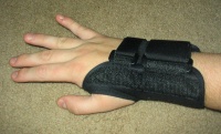
Non-surgical managment includes use of splints, activity modification, patient education, diuretics, and NSAIDs. A number of nonsurgical interventions benefit CTS in the short term, but there is sparse evidence on the midterm and long-term effectiveness of these interventions[8].
Surgical Management
[edit | edit source]
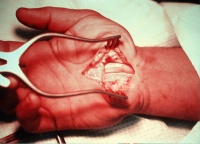
Surgical treatment seems to be more effective than splinting or anti-inflammatory drugs plus hand therapy in the midterm and long term to treat CTS[8]. However, there is no unequivocal evidence that suggests one surgical treatment is more effective than the other[8].
Differential Diagnosis
[edit | edit source]
Differential diagnosis for CTS includes cervical radiculopathy, thoracic outlet syndrome, pronator syndrome, wrist joint arthritis, tendonitis, and fibrositis.[1]
Key Evidence[edit | edit source]
Bionka M. Huisstede, Peter Hoogvliet, Manon S. Randsdorp, Suzanne Glerum, Marienke van Middelkoop and Bart W. Koes. Carpal tunnel syndrome. Part I: effectiveness of nonsurgical treatments – a systematic review. Arch Phys Med Rehabil. 2010 Jul;91(7):981-1004.
Bionka M. Huisstede, Manon S. Randsdorp, J. Henk Coert, Suzanne Glerum, Marienke van Middelkoop and Bart W. Koes. Carpal tunnel syndrome. Part II: effectiveness of surgical treatments–a systematic review. Arch Phys Med Rehabil. 2010 Jul;91(7):1005-24.
Resources [edit | edit source]
Dutton, M. Orthopaedic examination, evaluation, and intervention. New York: McGraw Hill; 2004.
Flynn TW, Cleland JA, Whitman JM. Users' guide to the musculoskeletal examination. Fundamentals for the evidence-based clinician. United States:Evidence in Motion; 2008.
Case Studies[edit | edit source]
add links to case studies here (case studies should be added on new pages using the case study template)
Carpal Tunnel Syndrome case study with MSK Ultrasound and US guided injection can be seen here http://theultrasoundsite.co.uk/carpal-tunnel-syndrome/
Recent Related Research (from Pubmed)[edit | edit source]
Failed to load RSS feed from http://www.ncbi.nlm.nih.gov/entrez/eutils/erss.cgi?rss_guid=1VePQbT36PByW9UeoZTRookH3hVMrxN0QI2FuBloyMUTmNx9UI|charset=UTF-8|short|max=10: Error parsing XML for RSS
References[edit | edit source]
References will automatically be added here, see adding references tutorial.
- ↑ 1.0 1.1 Dutton, M. Orthopaedic examination, evaluation, and intervention. New York: McGraw Hill; 2004
- ↑ Physical Therapy Nation. Tinel's Test for Median Nerve. Available from: http://www.youtube.com/watch?v=2wNGyEPdv_M [last accessed 14/12/13]
- ↑ Flynn TW, Cleland JA, Whitman JM. Users' guide to the musculoskeletal examination. Fundamentals for the evidence-based clinician. United States:Evidence in Motion; 2008.
- ↑ 3D Muscle Peep. 3D CGI medical video carpal tunnel syndrome . Available from: http://www.youtube.com/watch?v=u5dWTGYQ6PU [last accessed 22/02/13]
- ↑ Work Safe BC. Carpal Tunnel Syndrome. Available from: http://www.youtube.com/watch?v=J11EIfiHMYw [last accessed 22/02/13]
- ↑ Journal of Bone and Joint Surgery
- ↑ BMC Musculoskeletal Disorders
- ↑ 8.0 8.1 8.2 Bionka M. Huisstede, Peter Hoogvliet, Manon S. Randsdorp, Suzanne Glerum, Marienke van Middelkoop and Bart W. Koes. Carpal tunnel syndrome. Part I: effectiveness of nonsurgical treatments – a systematic review. Arch Phys Med Rehabil. 2010 Jul;91(7):981-1004.


