Cervical Stenosis: Difference between revisions
No edit summary |
No edit summary |
||
| Line 8: | Line 8: | ||
== Clinically Relevant Anatomy == | == Clinically Relevant Anatomy == | ||
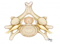
The cervical vertebrae are the smallest of the vertebrae, in comparison with the other spinal vertebrae. Their purpose is to contain and protect the spinal cord, support the skull, and enable diverse head movements. In general, the seven vertebrae of the vertebral spine are characterized by a large and triangular | The cervical vertebrae are the smallest of the vertebrae, in comparison with the other spinal vertebrae. Their purpose is to contain and protect the spinal cord, support the skull, and enable diverse head movements. In general, the seven vertebrae of the vertebral spine are characterized by a large and triangular vertebral foramen and small foramina in the transverse processes (except C7) that allow vertebral arteries, veins and nerves to pass through.<ref name="Moore">Moore KL, et al. Essential clinical anatomy. 4th ed. Baltimore, MD: Lippincott Williams and Wilkins, 2011.</ref> <br> | ||
{{#ev:youtube|vi7NuCGKzoY|300}} | |||
The ligaments of the cervical vertebrae guide the intra-articular movements in the most optimal directions in order to avoid cartilage damage and muscle hypertonicity. They also prevent excessive movements, which could otherwise lead to serious injuries. The [[Ligamentum flavum|ligamentum flavum]] is a broad, fibrous ligament that connects the laminae of adjacent vertebral arches. The elastic nature of the ligament helps to maintain the natural curvature of the spinal column and protects the intervertebral discs.<ref name="Moore" /><br> | The ligaments of the cervical vertebrae guide the intra-articular movements in the most optimal directions in order to avoid cartilage damage and muscle hypertonicity. They also prevent excessive movements, which could otherwise lead to serious injuries. The [[Ligamentum flavum|ligamentum flavum]] is a broad, fibrous ligament that connects the laminae of adjacent vertebral arches. The elastic nature of the ligament helps to maintain the natural curvature of the spinal column and protects the intervertebral discs.<ref name="Moore" /><br> | ||
| Line 32: | Line 34: | ||
<br> | <br> | ||
{| width="100%" cellspacing="1" cellpadding="1 | {| width="100%" cellspacing="1" cellpadding="1" | ||
|- | |- | ||
| [[Image: | | [[Image:Normal-cervical.jpg|thumb|center|200px|Normal cervical vertebrae]] | ||
| [[Image: | | [[Image:Cervica-Stenosis.jpg|thumb|center|200px|Cervical stenosis]] | ||
|} | |} | ||
| Line 42: | Line 44: | ||
== Characteristics/Clinical Presentation == | == Characteristics/Clinical Presentation == | ||
Cervical stenosis does not necessarily cause symptoms, but if symptoms are present they will mainly be caused by [[Cervical Radiculopathy|cervical radiculopathy]] or [[Cervical Myelopathy|cervical myelopathy]]. <br> | Cervical stenosis does not necessarily cause symptoms, but if symptoms are present they will mainly be caused by associated [[Cervical Radiculopathy|cervical radiculopathy]] or [[Cervical Myelopathy|cervical myelopathy]]. <br> | ||
Potential symptoms may include:<ref name="1">North American Spine Society Public Education Series. Cervical stenosis and myelopathy. http://www.spine.org/Documents/cervical_stenosis_2006.pdf (Accessed 22 November 2011).</ref><ref name="2">Williams SK, et al. Concomitant cervical and lumbar stenosis: Strategies for treatment and outcomes. Semin Spine Surg 2007;19(3):165-176.</ref><ref name="3">Countee RW, et al. Congenital stenosis of the cervical spine: Diagnosis and management. J Natl Med Assoc 1979;71(3):257-264.</ref> | |||
*Pain in neck or arms | *Pain in neck or arms | ||
| Line 53: | Line 55: | ||
*Frequent falling | *Frequent falling | ||
*The need to use a cane or walker | *The need to use a cane or walker | ||
*Urinary urgency which may | *Urinary urgency which may progress to bladder and bowel incontinence | ||
*Diminished proprioception | *Diminished proprioception | ||
The progression of the symptoms may also vary: | The progression of the symptoms may also vary: | ||
| Line 68: | Line 68: | ||
== Differential Diagnosis == | == Differential Diagnosis == | ||
*Acute cervical [[Disc Herniation|disc herniation]] | |||
*Cervical vertebral compression fracture | |||
*Cervical spine facet syndrome | |||
*[[Cervical Osteoarthritis|Osteoarthritis]] of intervertebral joints in cervical spine | |||
== Diagnostic Procedures == | == Diagnostic Procedures == | ||
| Line 74: | Line 77: | ||
Physical examination: <ref name="1" /><ref name="2" /><ref name="3" /><ref name="4">Santhosh A, et al. Spinal stenosis: history and physical examination. Phys Med Rehabil Clin N Am 2003;14.</ref><br> | Physical examination: <ref name="1" /><ref name="2" /><ref name="3" /><ref name="4">Santhosh A, et al. Spinal stenosis: history and physical examination. Phys Med Rehabil Clin N Am 2003;14.</ref><br> | ||
*Increased reflexes in the knee and ankle | *Hyper-reflexia: Increased reflexes in the knee and ankle | ||
*Changes in | *Changes in gait, such as clumsiness or loss of balance | ||
*Loss of sensitivity in the | *Loss of sensitivity in the hands or feet | ||
*Rapid foot beating that is triggered by turning the ankle upward | *Rapid foot beating that is triggered by turning the ankle upward | ||
*Babinski’s sign | *[[PLANTAR RESPONSE|Babinski’s sign]] | ||
*Hoffman’s sign | *Hoffman’s sign | ||
X- rays of the cervical spine | '''X-rays''' of the cervical spine do not provide enough information to confirm cervical stenosis, but can be used to rule out other conditions. Cervical stenosis can occur at one level or multiple levels of the spine, therefore an '''MRI''' is useful for looking at several levels at one time. A detailed MRI image may also be useful to show the tight spinal canal and pinching of the spinal cord. A '''CT scan''' can provide information about the bony invasion of the canal and can be combined with myelography. <ref name="1" /><ref name="2" /><br> | ||
{{#ev:youtube|9n09uGsCEkA|300}} | |||
== Outcome Measures == | == Outcome Measures == | ||
*[[Neck Disability Index]] | |||
*[[Neck Pain and Disability Scale]] | |||
See [[Outcome Measures|Outcome Measures Database]] for more | See [[Outcome Measures|Outcome Measures Database]] for more | ||
| Line 93: | Line 101: | ||
== Medical Management <br> == | == Medical Management <br> == | ||
For patients presenting with increasing weakness, pain or instability with walking, [[Surgical and Post‐Operative Management of Cervical Spine Stenosis|surgical management]] of cervical spine stenosis may be considered.<ref name="5">Fassett DR, et al. Asymptomatic cervical stenosis: To operate or not? Semin Spine Surg 2007;19(1):47-50.</ref><ref name="6">Kadanka Z, et al. Approaches to spondylotic cervical myelopathy conservative versus surgical results in a 3-year follow-up study. SPINE 2002;27(20):2205-2211.</ref><br> | |||
of the spine, which is called fusion, | [[Surgical and Post‐Operative Management of Cervical Spine Stenosis|Surgical options]] include anterior decompression and fusion, in which the disc and bone material that are causing spinal cord compression are removed from the anterior aspect and the spine is stabilized. The stabilizing of the spine, which is called fusion, involves placing an implant between the two cervical segments to support the spine and compensate for the bone and the disc that has been removed. <ref name="1" /><ref name="2" /><ref name="3" /><ref name="7">Boni M, et al. The cervical stenosis syndrome with a review of 83 patients treated by operation. Int Orthop 1982;6(3):185-195.</ref><ref name="8">Caron TH, et al. Combined (tandem) lumbar and cervical stenosis. Semin Spine Surg 2007;19(3):44-46.</ref><ref name="9">Engle CA, et al. Cervical stenosis in the athlete. Oper Tech Orthop 1995;5(3):218-222.</ref> | ||
Another surgical option is laminectomy. This is a procedure where the bone and ligaments that are pressing against the spinal cord are | Another surgical option is laminectomy. This is a procedure where the bone and ligaments that are pressing against the spinal cord are removed. In this treatment the surgeon might add also a fusion to stabilize the spine. <ref name="1" /><ref name="2" /><ref name="3" /><ref name="7" /><ref name="8" /><ref name="9" /> | ||
After the surgery, the patient has to remain in the hospital for several days. A postoperative rehabilitation program may be | After the surgery, the patient has to remain in the hospital for several days. A postoperative rehabilitation program may be provided, so that the patient can return to his activities and his typical daily function. <br> | ||
== Physical Therapy Management <br> == | == Physical Therapy Management <br> == | ||
Nonoperative treatments, such as physical therapy management, are aimed at reducing pain and increasing the function. Nonoperative treatments do not change the narrowing of the spinal canal, but | Nonoperative treatments, such as physical therapy management, are aimed at reducing pain and increasing the patient's function. Nonoperative treatments do not change the narrowing of the spinal canal, but can provide the patient of a long-lasting pain control and improved function without surgery. A rehabilitation program may require 3 or more months of supervised treatment. <ref name="1" /><br> | ||
A physical therapy or exercise program consists of the following exercises: <ref name="1" /><ref name="9" /> | A physical therapy or exercise program consists of the following exercises: <ref name="1" /><ref name="9" /> | ||
*Stretching exercises | *Stretching exercises: These exercises are aimed at restoring the flexibility of the muscles of the neck, trunk, arms and legs. | ||
*Cardiovascular exercises for arms and legs | *Cardiovascular exercises for arms and legs: This will improve blood circulation and enhance the patient's cardiovascular endurance. | ||
*Specific strengthening exercises for the arm, trunk and leg muscles. | *Specific strengthening exercises for the arm, trunk and leg muscles. | ||
*Training of activity of daily living (ADL). | *Postural re-education | ||
*Scapular stabilization | |||
*Cervical and thoracic joint manipulation | |||
*Training of activity of daily living (ADL) and functional movements. | |||
== Recent Related Research (from [http://www.ncbi.nlm.nih.gov/pubmed/ Pubmed]) == | == Recent Related Research (from [http://www.ncbi.nlm.nih.gov/pubmed/ Pubmed]) == | ||
Revision as of 04:23, 29 January 2014
Original Editors - Demol Yves as part of the Vrije Universiteit Brussel Evidence-based Practice Project
Definition/Description[edit | edit source]
Cervical stenosis is defined by the narrowing of the vertebral canal, which may result in a pinch of the spinal cord and/or the nerve roots. Because of this, the function of the spinal cord or the nerve may be affected, which may cause symptoms associated with cervical radiculopathy or cervical myelopathy.
Clinically Relevant Anatomy[edit | edit source]
The cervical vertebrae are the smallest of the vertebrae, in comparison with the other spinal vertebrae. Their purpose is to contain and protect the spinal cord, support the skull, and enable diverse head movements. In general, the seven vertebrae of the vertebral spine are characterized by a large and triangular vertebral foramen and small foramina in the transverse processes (except C7) that allow vertebral arteries, veins and nerves to pass through.[1]
The ligaments of the cervical vertebrae guide the intra-articular movements in the most optimal directions in order to avoid cartilage damage and muscle hypertonicity. They also prevent excessive movements, which could otherwise lead to serious injuries. The ligamentum flavum is a broad, fibrous ligament that connects the laminae of adjacent vertebral arches. The elastic nature of the ligament helps to maintain the natural curvature of the spinal column and protects the intervertebral discs.[1]
The following muscles act on the cervical spine and help to maintain its balance and stability:
- Longus capitis
- Longus colli
- Spinalis cervicis
- Semispinalis capitis
There is also a group of muscles that acts to ensure that the head can move in all directions:
Epidemiology /Etiology[edit | edit source]
Cervical stenosis typically has an insidious onset. The condition is characterized by a narrowing of the spinal canal, nerve root canal, or foramen. Pathological changes to a range of tissues in the region could be at fault. Examples include soft tissue damage (such as disc protrusion or fibrotic scars), boney tissue damage (such as osteophyte formation or spondylolisthesis), or impaired postural mechanics. Narrowing of the canal causes compression of the spinal cord and nerves at the effected level, leading to neurological symptoms as the condition progresses.[2]
Characteristics/Clinical Presentation[edit | edit source]
Cervical stenosis does not necessarily cause symptoms, but if symptoms are present they will mainly be caused by associated cervical radiculopathy or cervical myelopathy.
Potential symptoms may include:Cite error: Invalid <ref> tag; name cannot be a simple integer. Use a descriptive titleCite error: Invalid <ref> tag; name cannot be a simple integer. Use a descriptive titleCite error: Invalid <ref> tag; name cannot be a simple integer. Use a descriptive title
- Pain in neck or arms
- Arm and leg dysfunction
- Weakness, stiffness or clumsiness in the hands
- Leg weakness
- Difficulty walking
- Frequent falling
- The need to use a cane or walker
- Urinary urgency which may progress to bladder and bowel incontinence
- Diminished proprioception
The progression of the symptoms may also vary:
- A slow and steady decline
- Progression to a certain point and stabilizing
- Rapidly declining
Differential Diagnosis[edit | edit source]
- Acute cervical disc herniation
- Cervical vertebral compression fracture
- Cervical spine facet syndrome
- Osteoarthritis of intervertebral joints in cervical spine
Diagnostic Procedures[edit | edit source]
Physical examination: Cite error: Invalid <ref> tag; name cannot be a simple integer. Use a descriptive titleCite error: Invalid <ref> tag; name cannot be a simple integer. Use a descriptive titleCite error: Invalid <ref> tag; name cannot be a simple integer. Use a descriptive titleCite error: Invalid <ref> tag; name cannot be a simple integer. Use a descriptive title
- Hyper-reflexia: Increased reflexes in the knee and ankle
- Changes in gait, such as clumsiness or loss of balance
- Loss of sensitivity in the hands or feet
- Rapid foot beating that is triggered by turning the ankle upward
- Babinski’s sign
- Hoffman’s sign
X-rays of the cervical spine do not provide enough information to confirm cervical stenosis, but can be used to rule out other conditions. Cervical stenosis can occur at one level or multiple levels of the spine, therefore an MRI is useful for looking at several levels at one time. A detailed MRI image may also be useful to show the tight spinal canal and pinching of the spinal cord. A CT scan can provide information about the bony invasion of the canal and can be combined with myelography. Cite error: Invalid <ref> tag; name cannot be a simple integer. Use a descriptive titleCite error: Invalid <ref> tag; name cannot be a simple integer. Use a descriptive title
Outcome Measures[edit | edit source]
See Outcome Measures Database for more
Examination[edit | edit source]
add text here related to physical examination and assessment
Medical Management
[edit | edit source]
For patients presenting with increasing weakness, pain or instability with walking, surgical management of cervical spine stenosis may be considered.Cite error: Invalid <ref> tag; name cannot be a simple integer. Use a descriptive titleCite error: Invalid <ref> tag; name cannot be a simple integer. Use a descriptive title
Surgical options include anterior decompression and fusion, in which the disc and bone material that are causing spinal cord compression are removed from the anterior aspect and the spine is stabilized. The stabilizing of the spine, which is called fusion, involves placing an implant between the two cervical segments to support the spine and compensate for the bone and the disc that has been removed. Cite error: Invalid <ref> tag; name cannot be a simple integer. Use a descriptive titleCite error: Invalid <ref> tag; name cannot be a simple integer. Use a descriptive titleCite error: Invalid <ref> tag; name cannot be a simple integer. Use a descriptive titleCite error: Invalid <ref> tag; name cannot be a simple integer. Use a descriptive titleCite error: Invalid <ref> tag; name cannot be a simple integer. Use a descriptive titleCite error: Invalid <ref> tag; name cannot be a simple integer. Use a descriptive title
Another surgical option is laminectomy. This is a procedure where the bone and ligaments that are pressing against the spinal cord are removed. In this treatment the surgeon might add also a fusion to stabilize the spine. Cite error: Invalid <ref> tag; name cannot be a simple integer. Use a descriptive titleCite error: Invalid <ref> tag; name cannot be a simple integer. Use a descriptive titleCite error: Invalid <ref> tag; name cannot be a simple integer. Use a descriptive titleCite error: Invalid <ref> tag; name cannot be a simple integer. Use a descriptive titleCite error: Invalid <ref> tag; name cannot be a simple integer. Use a descriptive titleCite error: Invalid <ref> tag; name cannot be a simple integer. Use a descriptive title
After the surgery, the patient has to remain in the hospital for several days. A postoperative rehabilitation program may be provided, so that the patient can return to his activities and his typical daily function.
Physical Therapy Management
[edit | edit source]
Nonoperative treatments, such as physical therapy management, are aimed at reducing pain and increasing the patient's function. Nonoperative treatments do not change the narrowing of the spinal canal, but can provide the patient of a long-lasting pain control and improved function without surgery. A rehabilitation program may require 3 or more months of supervised treatment. Cite error: Invalid <ref> tag; name cannot be a simple integer. Use a descriptive title
A physical therapy or exercise program consists of the following exercises: Cite error: Invalid <ref> tag; name cannot be a simple integer. Use a descriptive titleCite error: Invalid <ref> tag; name cannot be a simple integer. Use a descriptive title
- Stretching exercises: These exercises are aimed at restoring the flexibility of the muscles of the neck, trunk, arms and legs.
- Cardiovascular exercises for arms and legs: This will improve blood circulation and enhance the patient's cardiovascular endurance.
- Specific strengthening exercises for the arm, trunk and leg muscles.
- Postural re-education
- Scapular stabilization
- Cervical and thoracic joint manipulation
- Training of activity of daily living (ADL) and functional movements.
Recent Related Research (from Pubmed)[edit | edit source]
References[edit | edit source]


