Frontal Lobe Brain Injury: Difference between revisions
Wendy Walker (talk | contribs) No edit summary |
Wendy Walker (talk | contribs) No edit summary |
||
| Line 52: | Line 52: | ||
=== Supplementary Motor Cortex === | === Supplementary Motor Cortex === | ||
Input: Thalamus, cingulate gyrus, sensory & prefrontal cortex | '''Input:''' Thalamus, cingulate gyrus, sensory & prefrontal cortex | ||
Output: Premotor cortex, primary motor cortex | '''Output:''' Premotor cortex, primary motor cortex | ||
Function: Intentional preparation for movement, procedural memory | '''Function:''' Intentional preparation for movement, procedural memory | ||
=== Prefrontal Cortex === | === Prefrontal Cortex === | ||
| Line 62: | Line 62: | ||
'''Function:''' working memory, executive functions including the ability to plan & implement (& monitor/evaluate) a series of goal-directed actions. | '''Function:''' working memory, executive functions including the ability to plan & implement (& monitor/evaluate) a series of goal-directed actions. | ||
=== Frontal Eye Fields === | === Frontal Eye Fields === | ||
Input: Parietal cortex, temporal cortex | '''Input:''' Parietal cortex, temporal cortex | ||
Output: Caudate, superior colliculus, paramedian pontine reticular formation | '''Output:''' Caudate, superior colliculus, paramedian pontine reticular formation | ||
Function: | Function: | ||
== Pathology/Injury<br> == | == Pathology/Injury<br> == | ||
Revision as of 23:13, 22 June 2016
Original Editor - Wendy Walker
Lead Editors - Wendy Walker, Lucinda hampton, Aminat Abolade, Kim Jackson, WikiSysop and Claire Knott
Introduction[edit | edit source]
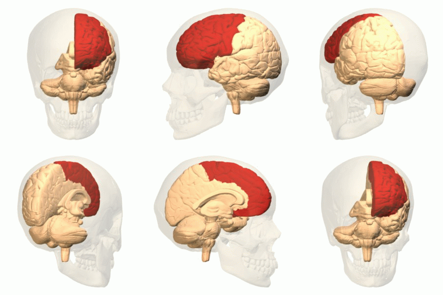
In evolutionary terms the frontal cortex has been the most recent to evolve, and the frontal lobes are the area of brain which has developed greatly in humans, differentiating us from other mammals. These lobes integrate the other brain areas, and are particularly responsible for higher level thinking and cognitive skills such as planning, evaluating likely outcomes, multitasking, performing risk assessment and the niceties of social interaction; it is the area of brain which deals in abstract concepts.
Anatomy
[edit | edit source]
The Frontal Lobes account for approximately one third of human brain mass.
They lie at the front of the brain, anterior to the parietal lobes and superior to the temporal lobe.
Each frontal lobe (left and right) is generally considered to have several distinct divisions:
- Motor cortex
- Premotor cortex
- Supplementary motor cortex
- Prefrontal cortex
- Orbital cortex AKA frontal eye fields
- Broca's Area
Vascular Supply[edit | edit source]
Medial frontal lobe = anterior cerebral artery
Deep & lateral regions = superior division of middle cerebral artery
Function
[edit | edit source]
Author Mesulam suggests that the frontal lobe is the part of the brain which modifies and imposes constraints on reflexive behaviours[1], and this control develops as the infant brain grows[2] and the frontal lobes become larger and more active.
- Motor cortex = voluntary movement
- Premotor cortex = storage of motor patterns & voluntary activities
- Prefrontal cortex = ability to concentrate; inhibition of reflexive behaviours; personality & emotional traits; abstract thinking
- Broca's Area = Motor control of speech
Primary Motor Cortex[edit | edit source]
Input: Basal gangliam thalamus, premotor cortex (in the frontal lobe), sensory cortex (in the parietal lobe)
Output: Motor fibres to spinal cord and brainstem, travelling in the corticospinal tract
Function: Movement
Pre motor Cortex[edit | edit source]
Input: Basal ganglia, thalamus, sensory cortex
Output: Primary motor cortex
Function: storage of motor programs, sensorimotor integration, facilitation of controlled, smooth movements
Supplementary Motor Cortex[edit | edit source]
Input: Thalamus, cingulate gyrus, sensory & prefrontal cortex
Output: Premotor cortex, primary motor cortex
Function: Intentional preparation for movement, procedural memory
Prefrontal Cortex[edit | edit source]
Function: working memory, executive functions including the ability to plan & implement (& monitor/evaluate) a series of goal-directed actions.
Frontal Eye Fields[edit | edit source]
Input: Parietal cortex, temporal cortex
Output: Caudate, superior colliculus, paramedian pontine reticular formation
Function:
Pathology/Injury
[edit | edit source]
It has been found that in traumatic brain injury contusions typically occur on the poles and the inferior aspects of the frontal lobes[3].
Resources[edit | edit source]
Recent Related Research (from Pubmed)[edit | edit source]
Extension:RSS -- Error: Not a valid URL: Feed goes here!!|charset=UTF-8|short|max=10
References[edit | edit source]
References will automatically be added here, see adding references tutorial.
- ↑ Mesulam MM. DT Stuss and RT Knight. The Human Frontal Lobes: Transcending the Default Mode through Continent Encoding. Principles of Frontal Lobe Function. Oxford: 2002. 8-30
- ↑ Luciana, ed. by Charles A. Nelson. Handbook of developmental cognitive neuroscience. Monica (2001). Cambridge, Mass. [u.a.]: MIT Press.
- ↑ Flint AC, Manley GT, Gean AD, et al. Post-operative expansion of hemorrhagic contusions after unilateral decompressive hemicraniectomy in severe traumatic brain injury. J Neurotrauma. Mar 17 2008


