Gemellus Superior: Difference between revisions
Abbey Wright (talk | contribs) (added content) |
Abbey Wright (talk | contribs) (content added) |
||
| Line 5: | Line 5: | ||
</div> | </div> | ||
== Description == | == Description == | ||
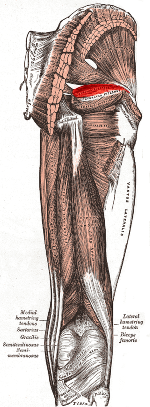
Gemellus superior works with [[Gemellus Inferior|gemellus inferior]] and [[Obturator Internus|obturator internus]] | Gemellus superior is a small muscle in the posterio-latereal portion of the [[hip]]. It works with [[Gemellus Inferior|gemellus inferior]] and [[Obturator Internus|obturator internus]] to externally rotate and extend the hip<ref>Palastanga, NIgel; Soames, Roger (November 2011). ''Physiotherapy Essentials : Anatomy and Human Movement : Structure and Function'' (6th ed.). London, GBR: Elsevier Health Sciences. p. 235</ref>. [[File:Gemellus superior.png|thumb]] | ||
=== Origin === | === Origin === | ||
Gemellus superior originates from the outer (gluteal) surface of the spine of the [[Pelvis|ischium]]<ref name=":0">Palastanga, NIgel; Soames, Roger (November 2011). ''Physiotherapy Essentials : Anatomy and Human Movement : Structure and Function'' (6th ed.). London, GBR: Elsevier Health Sciences. p. 237.</ref> | |||
=== Insertion === | === Insertion === | ||
It has a blended insertion with the upper part of the tendon of the Obturator internus.<ref name=":0" /> | |||
=== Nerve === | === Nerve === | ||
Innervated by the muscular branches of the sacral plexus | |||
=== Artery === | === Artery === | ||
Revision as of 15:54, 23 January 2020
Original Editor -
Top Contributors - Abbey Wright
Description[edit | edit source]
Gemellus superior is a small muscle in the posterio-latereal portion of the hip. It works with gemellus inferior and obturator internus to externally rotate and extend the hip[1].
Origin[edit | edit source]
Gemellus superior originates from the outer (gluteal) surface of the spine of the ischium[2]
Insertion[edit | edit source]
It has a blended insertion with the upper part of the tendon of the Obturator internus.[2]
Nerve[edit | edit source]
Innervated by the muscular branches of the sacral plexus
Artery[edit | edit source]
Function[edit | edit source]
Clinical relevance[edit | edit source]
Assessment[edit | edit source]
Treatment[edit | edit source]
Resources[edit | edit source]
- ↑ Palastanga, NIgel; Soames, Roger (November 2011). Physiotherapy Essentials : Anatomy and Human Movement : Structure and Function (6th ed.). London, GBR: Elsevier Health Sciences. p. 235
- ↑ 2.0 2.1 Palastanga, NIgel; Soames, Roger (November 2011). Physiotherapy Essentials : Anatomy and Human Movement : Structure and Function (6th ed.). London, GBR: Elsevier Health Sciences. p. 237.


