Gluteus Medius
Original Editor - Alex Palmer,
Top Contributors - Alex Palmer, George Prudden, Kim Jackson, Ahmed Nasr, Joao Costa, Joanne Garvey, Candace Goh, Nupur Smit Shah, WikiSysop, Pinar Kisacik, Rachael Lowe, Evan Thomas, Kai A. Sigel and Vidya Acharya;
Anatomy[edit | edit source]
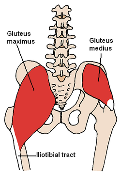
The gluteus medius is one of three gluteal muscles (minimus, medius and maximus). It is a superficial, fan shaped and broad muscle that lies in the posterolateral aspect of the pelvis, inferior to the iliac crest.[1] The gluteus medius has a broad origin on the external (gluteal) ilium and its tendon inserts into the lateral aspect of the greater trochanter.[2] The muscle is overlapped by the gluteus maximus and covered with a strong layer of fascia.[1]
Origin: External (gluteal) surface of ilium between anterior and posterior gluteal lines.[1] Reaches from iliac crest superiorly and as far as the sciatic notch inferiorly.[1] Superficial to gluteus maximus.[2]
Insertion: Lateral surface of greater trochanter.[2] A bursa seperates the tendon from the greater trochanter.[3]
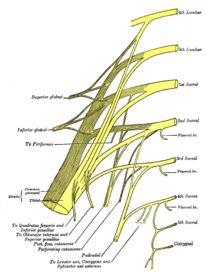
Nerve: The Superior Gluteal Nerve (SGN) supplies the gluteus medius. The SGN originates at the sacral plexus at levels L4, L5 & S1.[4] The SGN divides into several branches, supplying both the gluteus medius and minimus as it passes horizontally between them both, prior to terminating at the tensor facsia latae.[4]
Blood Supply: Superior gluteal artery and superior gluteal vein.[2]
Palpation[edit | edit source]
Find the centre of the iliac crest (directly above the greater trochanter of the femur) and palpate inferiorly two fingers breadth to find the bulk of the muscle.[1] To isolate in function, stand alternatively on one limb and then the other, feeling the muscle contract as you weight-bear through that limb.[1] The muscles will contract alternatively during walking, switching on during stance phase to stabilise the pelvis.
Function[edit | edit source]
The gluteus medius and minimus are strong abductors and medial rotators of the hip joint.[2] A contraction of the anterior fibers results in flexion and inward rotation and a contraction of the posterior fibers results in extension and external rotation.[5] Altogether they play an important role in the stabilization of the pelvis. Abnormality of this muscle can cause Trendelenburg’s sign.
Evidence Based Research
[edit | edit source]
Gowda AL, Mease SJ, Donatelli R, Zelicof S. Gluteus medius strengthening and the use of the Donatelli Drop Leg Test in the athlete. Physical Therapy in Sport 2014; 15(1) 15-19.
References[edit | edit source]
- ↑ 1.0 1.1 1.2 1.3 1.4 1.5 Palastanga N, Field D, Soames R. Anatomy and Human Movement, Structure and Function. 4th ed. Edinburgh: Butterworth Heinemann; 2002.
- ↑ 2.0 2.1 2.2 2.3 2.4 Drake RL, Vogl AW, Mitchell, AWM. Gray's Anatomy for Students. 2nd ed. Philadelphia: Churchill Livingstone; 2010.
- ↑ Diop M, Parratte B, Tatu L, Vuillier F, Faure A, Monnier G. Anatomical bases of superior gluteal nerve entrapment syndrome in the piriformis foramen. Surg Radiol Anat 2002; 24: 155-9.
- ↑ 4.0 4.1 Kenny P, O’Brien CP, Synnott K, Walsh MG. Damage to the superior gluteal nerve after two different approaches to the hip. J Bone Joint Surg Br 1999; 81: 979-81.
- ↑ Gowda AL, Mease SJ, Donatelli R, Zelicof S. Gluteus medius strengthening and the use of the Donatelli Drop Leg Test in the athlete. Physical Therapy in Sport 2014; 15(1) 15-19.


