Hemothorax: Difference between revisions
Nina Myburg (talk | contribs) m (Spelling and grammar) |
Kim Jackson (talk | contribs) (Formatted headings) |
||
| Line 1: | Line 1: | ||
<div class="editorbox"> '''Original Editor '''- [[Claire Knott]] '''Top Contributors''' - {{Special:Contributors/{{FULLPAGENAME}}}}</div> | <div class="editorbox"> '''Original Editor '''- [[Claire Knott]] '''Top Contributors''' - {{Special:Contributors/{{FULLPAGENAME}}}}</div> | ||
== Introduction == | |||
The term hemothorax can be defined as the entry of pleural fluid and blood into the pleural cavity. It needs to be pleural fluid with a hematocrit of 25% - 50% of the patient’s blood to be diagnosed as a hemothorax.<ref name=":0">Patrini D, Panagiotopoulos N, Pararajasingham J, Gvinianidze L, Iqbal Y, Lawrence DR. [https://www.ncbi.nlm.nih.gov/pmc/articles/PMC4387396/pdf/jtd-07-03-520.pdf Etiology and management of spontaneous haemothorax]. Journal of thoracic disease. 2015 Mar;7(3):520.</ref> | |||
The term hemothorax can be defined as the entry of pleural fluid and blood into the pleural cavity. It needs to be pleural fluid with a hematocrit of 25% - 50% of the patient’s blood to be diagnosed as a hemothorax.<ref name=":0">Patrini D, Panagiotopoulos N, Pararajasingham J, Gvinianidze L, Iqbal Y, Lawrence DR. [https://www.ncbi.nlm.nih.gov/pmc/articles/PMC4387396/pdf/jtd-07-03-520.pdf Etiology and management of spontaneous haemothorax]. Journal of thoracic disease. 2015 Mar;7(3):520.</ref> | |||
== Mechanism of Injury / Pathological Process == | == Mechanism of Injury / Pathological Process == | ||
There are two layers of pleura. One of which covers the lung surface (visceral pleura) and the other the inside of the chest wall (parietal pleura). These layers of pleura adhere to each other to keep the lung from collapsing, even with the expiration of air from the lung. If air or fluid enters the pleural cavity in between these layers of pleura, it causes the lung to collapse due to its elastic recoil. If it is only air entering the pleural cavity it causes a [[pneumothorax]]. If it is fluid or blood entering the pleural cavity it could cause a pleural effusion or hemothorax. A hemothorax could be caused by trauma, as a symptom of certain conditions or iatrogenic causes (through procedures like central line insertion or pleural biopsies).<ref name=":0" /> | There are two layers of pleura. One of which covers the lung surface (visceral pleura) and the other the inside of the chest wall (parietal pleura). These layers of pleura adhere to each other to keep the lung from collapsing, even with the expiration of air from the lung. If air or fluid enters the pleural cavity in between these layers of pleura, it causes the lung to collapse due to its elastic recoil. If it is only air entering the pleural cavity it causes a [[pneumothorax]]. If it is fluid or blood entering the pleural cavity it could cause a pleural effusion or hemothorax. A hemothorax could be caused by trauma, as a symptom of certain conditions or iatrogenic causes (through procedures like central line insertion or pleural biopsies).<ref name=":0" /> | ||
| Line 22: | Line 20: | ||
* CT scan | * CT scan | ||
* MRI scan | * MRI scan | ||
== Medical Management == | == Medical Management == | ||
Draining the blood as quickly as possible usually through a chest tube. Surgery could also be an option, depending on the clinical picture. | Draining the blood as quickly as possible usually through a chest tube. Surgery could also be an option, depending on the clinical picture. | ||
== Physiotherapy | == Physiotherapy Management == | ||
There are no published data regarding the physiotherapy management of patients with pneumothorax or hemothorax. | There are no published data regarding the physiotherapy management of patients with pneumothorax or hemothorax. | ||
Revision as of 03:29, 7 April 2019
Introduction[edit | edit source]
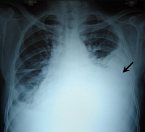
The term hemothorax can be defined as the entry of pleural fluid and blood into the pleural cavity. It needs to be pleural fluid with a hematocrit of 25% - 50% of the patient’s blood to be diagnosed as a hemothorax.[1]
Mechanism of Injury / Pathological Process[edit | edit source]
There are two layers of pleura. One of which covers the lung surface (visceral pleura) and the other the inside of the chest wall (parietal pleura). These layers of pleura adhere to each other to keep the lung from collapsing, even with the expiration of air from the lung. If air or fluid enters the pleural cavity in between these layers of pleura, it causes the lung to collapse due to its elastic recoil. If it is only air entering the pleural cavity it causes a pneumothorax. If it is fluid or blood entering the pleural cavity it could cause a pleural effusion or hemothorax. A hemothorax could be caused by trauma, as a symptom of certain conditions or iatrogenic causes (through procedures like central line insertion or pleural biopsies).[1]
Clinical Presentation[edit | edit source]
- Chest Pain
- Dyspnea
- Fever
- Tachycardia
- Reduced breath sounds on the affected side
- Pallor
- Cold Sweats
Diagnostic Procedures[edit | edit source]
- Chest X-ray
- Ultrasound
- CT scan
- MRI scan
Medical Management[edit | edit source]
Draining the blood as quickly as possible usually through a chest tube. Surgery could also be an option, depending on the clinical picture.
Physiotherapy Management[edit | edit source]
There are no published data regarding the physiotherapy management of patients with pneumothorax or hemothorax.
The following can be regarded as recommendations for management of patients with hemothorax:
- The patient's clinical picture should lead the physiotherapist in deciding what treatment is suitable.
- If the patient has a chest tube and intercostal drain in, the treatment might be different from when the patient had surgery.
- Help to improve ventilation, oxygenation and to re-inflate atelactic lung areas. This could be done by deep breathing exercise techniques.
- Help to improve the patient's exercise tolerance and mobility. This could be done by assisting with mobilisation or general strengthening exercises.
- Help to maintain airway clearance. This could be done by showing the patient assisted coughing techniques to help clear any secretions.
Differential Diagnosis[edit | edit source]
Through imaging, the diagnosis of a pneumothorax needs to be cancelled out. The hematocrit of the fluid from the pleural cavity could also be tested to see if it could be diagnosed as a pleural effusion or a hemothorax.
Resources[edit | edit source]
References[edit | edit source]
- ↑ 1.0 1.1 Patrini D, Panagiotopoulos N, Pararajasingham J, Gvinianidze L, Iqbal Y, Lawrence DR. Etiology and management of spontaneous haemothorax. Journal of thoracic disease. 2015 Mar;7(3):520.


