Kidney
Original Editor - User Name
Top Contributors - Manisha Shrestha and Lucinda hampton
Introduction[edit | edit source]
There are two kidneys present in the human body. Kidneys are bean-shaped, approximately fist-size internal organs located on either side of the spine in the retroperitoneal space between the parietal peritoneum and the posterior abdominal wall, well protected by muscle, fat, and ribs. The kidneys are well vascularized, receiving about 25 percent of the cardiac output at rest. The main function of the kidney is to excrete a variety of waste products produced by metabolism into the urine.
Gross Anatomy of Kidney[edit | edit source]
External Anatomy[edit | edit source]
- The left kidney is located at about the T12 to L3 vertebrae, whereas the right is lower due to slight displacement by the liver. The upper portions of the kidneys are somewhat protected by the eleventh and twelfth ribs.
- The right kidney sits just below the diaphragm and posterior to the liver. The left kidney sits below the diaphragm and posterior to the spleen.
- Each kidney weighs about 125–175 g in males and 115–155 g in females. They are about 11–14 cm in length, 6 cm wide, and 4 cm thick, and are directly covered by a fibrous capsule composed of dense, irregular connective tissue that helps to hold their shape and protect them.
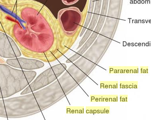
- Each kidney, with its adrenal gland on top is surrounded by shock-absorbing two layers of fat: the perirenal fat present between renal fascia and renal capsule and pararenal fat superior to the renal fascia.
- The renal fascia and, to a lesser extent, the overlying peritoneum serves to firmly anchor the kidneys to the posterior abdominal wall in a retroperitoneal position.
- So basically, the external layers of the kidney are a renal capsule in close proximity followed by perirenal fat pad, renal fascia, and then pararenal fat pad.
Internal Anatomy[edit | edit source]
- A frontal section through the kidney reveals an outer region called the renal cortex and an inner region called the medulla.
- The renal columns are connective tissue extensions that radiate downward from the cortex through the medulla to separate the most characteristic features of the medulla, the renal pyramids and renal papillae.
- The papillae are bundles of collecting ducts that transport urine made by nephrons to the calyces of the kidney for excretion.
- The renal columns also serve to divide the kidney into 6–8 lobes and provide a supportive framework for vessels that enter and exit the cortex.
- The pyramids and renal columns taken together constitute the kidney lobes.
Hilum
Microscopic Anatomy of Kidney[edit | edit source]
Blood supply of Kidney[edit | edit source]
Nerve supply of kidney[edit | edit source]
Function of Kidney[edit | edit source]
- It is a major excretory organ for elimination of metabolic wastes from the body.[4]
- The overall renal function of the system filters approximately 200 liters of fluid a day from renal blood flow which allows for toxins, metabolic waste products, and excess ion to be excreted while keeping essential substances in the blood.
- It regulates plasma osmolarity by modulating the amount of water, solutes, and electrolytes in the blood.
- It ensures long term acid-base balance and also produces erythropoietin which stimulates the production of red blood cell.
- It also produces renin for blood pressure regulation and carries out the conversion of vitamin D to its active form.[5]
Resources[edit | edit source]
References[edit | edit source]
- ↑ Anatomic Wisdom. The External Gross Anatomy of the Kidney. Available from: https://www.youtube.com/watch?v=OmMzMBSsaU4. [Lasted accessed: 2021-4-30]
- ↑ Kidneys and Urinary System LO2 - Internal Gross Anatomy. Available from: https://www.youtube.com/watch?v=25CO_f7o_c4. [lasted accessed: 2021-4-30]
- ↑ Microscopic structure of kidney-Nephron. Available from: https://www.youtube.com/watch?v=OoIZ1N-haL8. [Lasted accessed: 2021-4-30]
- ↑ Finco DR. Kidney function. In Clinical biochemistry of domestic animals 1997 Jan 1 (pp. 441-484). Academic Press.
- ↑ Ogobuiro I, Tuma F. Physiology, renal. StatPearls [Internet]. 2019 Feb 10.


