Lumbricals of the Hand: Difference between revisions
No edit summary |
No edit summary |
||
| Line 46: | Line 46: | ||
== Clinical relevance == | == Clinical relevance == | ||
'''Lumbrical-plus finger:''' when the tendons of the flexor digitorum profundus detach distal to the origin points of the lumbricals an interesting phenomenon occurs: when trying to close the fist, the fingers strangely extend instead. | |||
After detachment of the distal tendons, the lumbricals now serve as the new insertion surface of the flexor digitorum profundus. This means, even though the person consciously activates the flexor muscle, they actually move the lumbricals instead. And since the flexor digitorum profundus and the lumbricals are antagonists in the proximal interphalangeal and distal interphalangeal joints, the intended fist closure paradoxically leads to an extension of the fingers. This oddity is clinically referred to as the lumbrical-plus finger and can occur after injuries or amputations.<ref name=":1" /> | After detachment of the distal tendons, the lumbricals now serve as the new insertion surface of the flexor digitorum profundus. This means, even though the person consciously activates the flexor muscle, they actually move the lumbricals instead. And since the flexor digitorum profundus and the lumbricals are antagonists in the proximal interphalangeal and distal interphalangeal joints, the intended fist closure paradoxically leads to an extension of the fingers. This oddity is clinically referred to as the lumbrical-plus finger and can occur after injuries or amputations.<ref name=":1" /> | ||
Revision as of 14:41, 21 October 2020
Original Editor - Denys Nahornyi
Top Contributors - Denys Nahornyi, Shaimaa Eldib, Kim Jackson and Kirenga Bamurange Liliane
Description[edit | edit source]
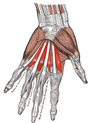
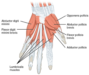
The lumbricals are deep muscles of the hand that flex the metacarpophalangeal joints and extend the interphalangeal joints. It has four, small, worm-like muscles on each hand. These muscles are unusual in that they do not attach to bone. Instead, they attach proximally to the tendons of flexor digitorum profundus and distally to the extensor expansions.[1]
Structure[edit | edit source]
The lumbricals are four, small, worm-like muscles on each hand. These muscles are unusual in that they do not attach to bone. Instead, they attach proximally to the tendons of flexor digitorum profundus and distally to the extensor expansions.[1]
| number of head | Origin | Insertion |
|---|---|---|
| First | It originates from the radial side of the most radial tendon of the flexor digitorum profundus (corresponding to the index finger). | It passes posteriorly along the radial side of the index finger to insert on the extensor expansion near the metacarpophalangeal joint. |
| Second | It originates from the radial side of the second most radial tendon of the flexor digitorum profundus (which corresponds to the middle finger). | It passes posteriorly along the radial side of the middle finger and inserts on the extensor expansion near the metacarpophalangeal joint. |
| Third | One head originates on the radial side of the flexor digitorum profundus tendon corresponding to the ring finger, while the other originates on the ulnar side of the tendon for the middle finger. | The muscle passes posteriorly along the radial side of the ring finger to insert on its extensor expansion. |
| Fourth | One head originates on the radial side of the flexor digitorum profundus tendon corresponding to the little finger, while the other originates on the ulnar side of the tendon for the ring finger. | The muscle passes posteriorly along the radial side of the little finger to insert on its extensor expansion. |
Nerve[edit | edit source]
The first and second lumbricals (the most radial two) are innervated by the median nerve. The third and fourth lumbricals (most ulnar two) are innervated by the ulnar nerve.
This is the usual innervation of the lumbricals (occurring in 60% of individuals). However 1:3 (median:ulnar - 20% of individuals) and 3:1 (median:ulnar - 20% of individuals) also exist. The lumbrical innervation always follows the innervation pattern of the associated muscle unit of flexor digitorum profundus (i.e. if the muscle units supplying the tendon to the middle finger are innervated by the median nerve, the second lumbrical will also be innervated by the median nerve).[2]
Artery[edit | edit source]
Four separate sources supply blood to these muscles: the superficial palmar arch, the common palmar digital artery, the deep palmar arch, and the dorsal digital artery.
Function[edit | edit source]
The fact that the lumbrical muscles of the hand originate from tendons and insert into the extensor expansions, instead of bony structures, makes both of their attachment points quite mobile. That means the muscles are capable of two different actions. These are flexion at the metacarpophalangeal joints and extension in both the proximal and distal interphalangeal joints . The reason for the opposite actions is that the tendons cross the metacarpophalangeal joints on the palmar side but distally insert at the dorsal side of the finger. These combined movements play a role in complex movement of the hand (e.g. for holding a pen), and contribute to the general dexterity of the hand.
In addition, it is found that the lumbrical muscles of the hand contain many muscle spindles and have a large fiber length, which indicates that they likely play a role in proprioception.[3]
Clinical relevance[edit | edit source]
Lumbrical-plus finger: when the tendons of the flexor digitorum profundus detach distal to the origin points of the lumbricals an interesting phenomenon occurs: when trying to close the fist, the fingers strangely extend instead.
After detachment of the distal tendons, the lumbricals now serve as the new insertion surface of the flexor digitorum profundus. This means, even though the person consciously activates the flexor muscle, they actually move the lumbricals instead. And since the flexor digitorum profundus and the lumbricals are antagonists in the proximal interphalangeal and distal interphalangeal joints, the intended fist closure paradoxically leads to an extension of the fingers. This oddity is clinically referred to as the lumbrical-plus finger and can occur after injuries or amputations.[3]
Resources[edit | edit source]
- ↑ 1.0 1.1 Gosling JA, Harris PF, Humpherson JR, Whitmore I, Willan PL. Human Anatomy: Color Atlas and Textbook. phot. by A.L. Bentley (5th ed.). Philadelphia: Mosby, 2008.
- ↑ Last's Anatomy - Regional and Applied, 10th ed. Chummy S. Sinnatamby, pg. 64 and pg. 82.
- ↑ 3.0 3.1 Kenhub. Available from:https://www.kenhub.com/en/library/anatomy/lumbrical-muscles-of-the-hand (accessed 21 october 2020)


