Lumbricals of the Hand
Original Editor - Denys Nahornyi
Top Contributors - Denys Nahornyi, Shaimaa Eldib, Kim Jackson and Kirenga Bamurange Liliane
Description[edit | edit source]
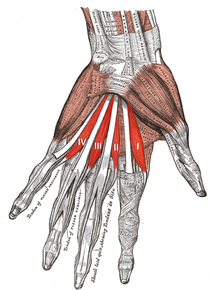
The lumbricals are intrinsic muscles of the hand that flex the metacarpophalangeal joints and extend the interphalangeal joints. The lumbricals are four, small, worm-like muscles on each hand. These muscles are unusual in that they do not attach to bone. Instead, they attach proximally to the tendons of flexor digitorum profundus and distally to the extensor expansions.
Structure[edit | edit source]
The lumbricals are four, small, worm-like muscles on each hand. These muscles are unusual in that they do not attach to bone. Instead, they attach proximally to the tendons of flexor digitorum profundus and distally to the extensor expansions.
| # | Origin | Insertion |
|---|---|---|
| First | It originates from the radial side of the most radial tendon of the flexor digitorum profundus (corresponding to the index finger). | It passes posteriorly along the radial side of the index finger to insert on the extensor expansion near the metacarpophalangeal joint. |
| Second | It originates from the radial side of the second most radial tendon of the flexor digitorum profundus (which corresponds to the middle finger). | It passes posteriorly along the radial side of the middle finger and inserts on the extensor expansion near the metacarpophalangeal joint. |
| Third | One head originates on the radial side of the flexor digitorum profundus tendon corresponding to the ring finger, while the other originates on the ulnar side of the tendon for the middle finger. | The muscle passes posteriorly along the radial side of the ring finger to insert on its extensor expansion. |
| Fourth | One head originates on the radial side of the flexor digitorum profundus tendon corresponding to the little finger, while the other originates on the ulnar side of the tendon for the ring finger. | The muscle passes posteriorly along the radial side of the little finger to insert on its extensor expansion. |


