Meningitis
Original Editors - Iris Partin
Lead Editors - Your name will be added here if you are a lead editor on this page. Read more.
Definition/Description[edit | edit source]
Meningitis is an infectious disease of the central nervous system that causes inflammation of the meningeal membranes. All three meninges may become involved, dura matter, arachnoid, and pia matter.[1][2] The pia matter and arachnoid layers become inflamed and opaque. The first two layers of the cortex and the spinal cord become inflamed as well.[1] As a result of the inflammation many complications may occur. There is an increased risk of infarctions and cortical veins may develop thromboses. In addition, blockage of the flow of CSF may occur secondary to scar tissue. This blockage is most common at the base of the brain. CSF blockage may result in subarachnoid cysts or hydrocephaly resulting in a headache, considered a cardinal sign.[1] The disease may present as acute (over a period of hours or days), subacute (more than two weeks), or chronic (more than one month).[2]
Aseptic and Bacterial meningitis are the most common forms of acute meningitis. Aseptic meningitis is usually a result of fungi, viruses, parasites, bacteria, or in some cases a noninfectious inflammation. This form of meningitis is usually self-limited. Bacterial meningitis is a much more serious illness and if goes untreated is fatal. Progression is very rapid and is distinguished by purulent CSF.[2] Below is a picture of the meninges which are affected by meningitis.
Prevalence[edit | edit source]
The prevalence of meningitis has greatly decreased over the last fifteen years due to the development of vaccines.[1] The second most common bacteria that causes acute bacterial meningitis, meningocicci, is present in the nasopharynx of approximately 5% of the population. Close contact or respiratory droplets may spread the bacteria. Of the population that has the bacteria, only a small fraction develops meningitis and the most prevalent age range is from birth to one year.[2] As individuals move through the late adolescence development stage, they experience a second period of increased vulnerability. In adulthood, bacterial meningitis usually is limited to individuals who have conditions that inhibit the immune system.[1] Bacterial meningitis is usually associated with this form of meningitis; it appears more frequently in populations that are in close living quarters such as college dormitories, military barracks, and boarding schools.[2]
Characteristics/Clinical Presentation[edit | edit source]
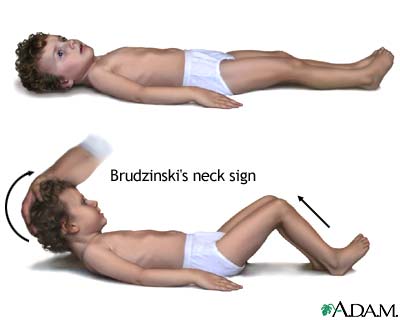
Headache, fever, vomiting, and rigidity of the neck are the most common symptoms that present with the onset of meningitis.[1][2][4] Pain in the posterior thigh or lumbar region may also be noted.[1] A rash on the skin, petechial in nature, appears on approximately 50-60% of individuals with bacterial meningitis.[4] Meningitis causes inflammation of the meningeal membranes, as a result nerve roots may endure tension as they pass through the inflamed membranes. Passive ROM of the neck into flexion will become painful and limited. Extension and rotation may be painful as well, but to the extent of flexion. In some cases passive neck flexion may produce flexion of the hips or knee, this is known as Brudzinki’s sign and is usually only seen in the most severe cases. Another sign that is usually seen in the most severe cases is the Kernig’s sign, which is defined as passive extension of the knee while the hip is flexed that produces restriction.[2]
In cases when meningitis is not treated immediately (especially bacterial meningitis), the parenchyma within the brain may be involved. As a result individuals may present with lethargy, vomiting, seizures, papilledema, confusion, coma, focal deficits, and cranial nerve palsies.[1][2]
Brudzinski's Sign[5]Image:Kernig's_sign.jpg [6]
Associated Co-morbidities[edit | edit source]
Predisposing conditions of meningitis include sinusitis, mastoiditis, and otitis. These conditions may require specialized treatment.[1] Damage or removal of the spleen increases the risk of pneumococcal disease which may lead to acute bacterial meningitis. Conditions in which an individuals immune system may become compromised increase the risk and severity of meningitis, such as HIV.[1][2] Other conditions that may predispose one for meningitis include alcoholism, prior neurosurgery, cancer, head trauma, parameningeal infection, and anatomical defects of the meningies.[4]
Medications[edit | edit source]
Quick introduction of antibiotics is crucial when acute meningitis is suspected to prevent progression of the disease and increase the chance of recovery.[1] If a patient presents with the cardinal signs and symptoms of meningitis antibiotics are immediately started after blood cultures are drawn. If the patient is not severely ill and the diagnosis of meningitis is questionable introduction of antibiotics is held until results from CSF stains are determined.[2] Once CSF gram stains are determined targeted antimicrobial treatment may begin. Duration of antimicrobial treatment is determined by CSF sterilization.[1] Organism specific antibiotics used to treat acute bacterial meningitis may be found in Table 1. The table lists the common organisms that result in acute meningitis along with the age appropriate antibiotics.[2]
Table 1: Common antibiotics used to treat specific acute bacterial meningitis. Adapted from The Merck Manual of Diagnosis and Therapy.[2]
| Organism | Age Group | Antibiotic |
| Unknown | Infants less than 1 month | Ampicillin
Cefotaxime Gentamicin
|
| Children over 1 month of age and Adults | Ampicillin
Cefotaxime Vancomycin
| |
| Gram-positive organisms (unidentified) | Children and Adults | Ceftriaxone
Vancomycin Ampicillin
|
| Gram-negative bacilli (unidentified) | Children and Adults | Cefazidime
Gentamicin
|
| Haemophilus influenza
type b
|
Children and Adults | Ceftriaxone |
| Meningococci | Children and Adults | Penicillin G plus ceftriaxone |
| Streptococci | Children and Adults | Vancomcin
Nafcillin
|
| Listeria sp | Children and Adults | Ampicillin
Gentamicin Trimethoprim-sulfamethoxaxzole
|
| Enteric gram-negative bacteria
(Escherichia coli, Proteus sp, Klebsiella sp)
|
Children and Adults |
Ceftriaxone Gentamicin
|
| Pseudomonas | Children and Adults | Ceftazidime
Cefepime
|
| Staphyococci | Children and Adults | Vancomycin
Nafcillin
|
In addition to antibiotics, medications to control and relieve the symptoms of meningitis are introduced as well. Alterations of CSF flow may be controlled using steroids. Inflammation of the subarachnoid space that is linked to mortality and morbidity is reduced with the use of dexamethasone. Antiseizure medication is used to control seizure activity. Viral meningitis is not as severe as bacterial and most patients recover within one to two weeks. Symptoms, nausea and headache, are controlled using the proper medications.[1]
Diagnostic Tests/Lab Tests/Lab Values[edit | edit source]
The gold standard for diagnosing meningitis is a lumbar puncture.[1] CSF obtained from a lumbar puncture is used to culture cells, glucose level, protein, cell count and differential, and to begin a Gram stain. The viruses that cause meningitis are easily isolated in the CSF. Gram stains are positive 80% of the time in bacterial meningitis, inexpensive, fast, and are 90% accurate. Lumbar punctures are performed immediately in patients with suspected meningitis to diagnosis and identify the causative organism. Once the organism is identified antimicrobial pharmacotherapy may begin immediately. CSF results may also be used to monitor progress of viral meningitis.[1][2]
While lumbar puncture is the gold standard, other diagnostic tests and lab tests are performed when meningitis is suspected. In situations when the patient presents with papilledema, seizures, focal deficits, or deterioration in consciousness an MRI or CT scan are performed to rule out a brain abscess or infarction. A brain abscess or infarction must be ruled out prior to the lumbar puncture to the risk of cerebral herniation. A combination of laboratory tests may be needed in addition to the lumbar puncture when the gram culture and stains are negative, but the CSF results suggest bacterial meningitis. Further lab tests are needed to examine glucose, protein, and white blood cell levels as well as close monitoring of pressure. [1][2]
Causes[edit | edit source]
Meningitis may result from a variety of causes. Bacterial meningitis is a result of infection from an organism. Neonates receive antibodies via placenta for bacteria such as Listeria monocytogenes, Escherichia coli, and group B streptococcus. With age, these antibodies decline resulting in increased susceptibility especially in ages 1 to 2 for meningococcus, pneumococcus, and Haemophilus influenzae type b (Hib). Geriatric and the adult population are more affected by Neisseria meningitis and Streptococcus pneumoniae. Pneumococci infections are especially common in adults who suffer from alcoholism, CSF leaks, chronic otitis, mastoiditis, sinusitis, sickle cell disease, pneumococcal pneumonia, and asplenia. Other organisms that may result in bacterial meningitis may be found above in Table 1. These organisms are most commonly found in the upper respiratory tract in the mucosal layers.[2][7]
Meningitis is most commonly a result of a viral infection. Enteroviruses (echovirus and coxsackievirus) and herpes simplex virus are the most common viruses representing 40% of the cases of meningitis in individuals 30-60 years old and 20% of all individuals with meningitis, respectively. Individuals in late adolescence and early adulthood who develop meningitis usually came in contact with the Epstein-Barr virus (EBV). Other causes of viral meningitis include intracranial tumor rupture, mumps, systemic lupus erythematosus (SLE), radiopaque agents, lead poisoning, itrathecal drug use, and NSAIDs especially as a result of exposure during surgery.[2][7]
Systemic Involvement[edit | edit source]
Nervous
• Inflammation of subarachnid space
• Spread of inflammation to parenchyma
• Focal ischemic lesions
• Hydrocephaly
• Impaired consciousness
o Stages include irritability, confusion, drowsiness, stupor, and coma
• Hemiparesis
• Seizures
• Cranial nerve palsy
• Hypothalamic dysfunction in children
Vascular
• Inflammation of small subarachnoid vessels (especially veins)
• Thrombotic obstruction of vessels
Musculoskeletal
• Opisthotonic posture
o See image below
• Infectious spread to joints
Sensory
• Impaired hearing
• Loss of vision
Metabolic
• Dehydration
• Hyponatremia
Gastrointestinal
• Vomiting
Integumentary
• Petechial rash of skin (usually associated with bacterial meningitis)
Opisthotonic PosturingOpisthotonic Posturing [8]
Medical Management (current best evidence)[edit | edit source]
In children and infants treatment usually consist of six or more days of inpatient antimicrobial therapy followed by close follow-up outpatient management. Duration of inpatient treatment is dependant upon absence of fever for at least 24 to 48 hours, no focal findings, ability to take fluids by mouth, no seizure activity, no significant neurologic dysfunction, and improvement or stabilization of condition.[1]
Currently there are vaccines available that are highly effective and safe for some serogroups of N. meningitides, Haemophilus influenzae type b (Hib), and many types of Streptococcus pneumoniae. It is recommended that the meningococcal conjugate vaccine be given between the ages of 11-18 due to the increased prevalence during adolescence. The Advisory Committee on Immunization Practices (ACIP) suggests the vaccines be given at the earliest. The ACIP highly recommends that prior to living in dormitories college freshmen should be vaccinated.[9]
The vaccine for Streptococcus pneumoniae is known and the pneumococcal polysaccharide vaccine (PPSV) and is recommended for individuals between the ages 19-64 with asthma or who smoke and individuals older than 65 and at least 2 years of age with certain medical problems. Another form of the vaccine to prevent infection of pneumococcal is approved and routinely given to children younger than the age of 2.[9]
- See Resources for more information on the prevention of meningococcal disease.
Physical Therapy Management (current best evidence)[edit | edit source]
add text here
Alternative/Holistic Management (current best evidence)[edit | edit source]
add text here
Case Reports[edit | edit source]
add links to case studies here (case studies should be added on new pages using the case study template)
Resources
[edit | edit source]
add appropriate resources here
Recent Related Research (from Pubmed)[edit | edit source]
see tutorial on Adding PubMed Feed
Extension:RSS -- Error: Not a valid URL: Feed goes here!!|charset=UTF-8|short|max=10
References[edit | edit source]
see adding references tutorial.
- ↑ 1.00 1.01 1.02 1.03 1.04 1.05 1.06 1.07 1.08 1.09 1.10 1.11 1.12 1.13 1.14 1.15 1.16 1. Goodman C, Fuller K. Pathology: Implications for the Physical Therapist. 3rd ed. St. Louis, Missouri: Saunders Elsevier, 2009.
- ↑ 2.00 2.01 2.02 2.03 2.04 2.05 2.06 2.07 2.08 2.09 2.10 2.11 2.12 2.13 2.14 2.15 2.16 2. Beers MH, et. al. eds. The Merck Manual of Diagnosis and Therapy. 18th ed. Whitehouse Station, NJ: Merck Research Laboratories; 2006
- ↑ National Library of Medicine. Medline Plus, Encyclopedia: Meninges of the brain. http://www.nlm.nih.gov/medlineplus/ency/imagepages/19080.htm (accessed 2 March 2010).
- ↑ 4.0 4.1 4.2 4.3 Aminoff M, Greenberg D, Simon R. Clinical Neurology. 6th ed. New York, NY: Lange Medical Books/McGraw-Hill, 2005.
- ↑ Neisseri Meningitidis. Brudzinski’s sign. http://bioweb.uwlax.edu/bio203/s2008/bingen_sama/neck.jpg (accessed 6 April 2010)
- ↑ National Library of Medicine. Kernig’s sign. http://www.nlm.nih.gov/medlineplus/ency/images/ency/fullsize/19077.jpg (accessed 6 April 2010)
- ↑ 7.0 7.1 7.2 Goodman C, Fuller K. Pathology: Implications for the Physical Therapist. 3rd ed. St. Louis, Missouri: Saunders Elsevier, 2009
- ↑ Roll Back Malaria. Children and Malaria. http://www.rollbackmalaria.org/cmc_upload/0/000/015/367/RBMInfosheet_6.htm (accessed 6 April 2010)
- ↑ 9.0 9.1 Centers for Disease Control and Prevention. Meningitis Question and Answer. http://www.cdc.gov/meningitis/about/faq.html (accessed 6 April 2010)

