Obliquus Capitis Inferior: Difference between revisions
Evan Thomas (talk | contribs) No edit summary |
Kim Jackson (talk | contribs) m (Text replacement - "Category:Cervical_Anatomy" to "Category:Cervical Spine - Anatomy") |
||
| (19 intermediate revisions by 3 users not shown) | |||
| Line 3: | Line 3: | ||
'''Lead Editors''' - {{Special:Contributors/{{FULLPAGENAME}}}} | '''Lead Editors''' - {{Special:Contributors/{{FULLPAGENAME}}}} | ||
</div> | </div> | ||
== Description == | == Description == | ||
Obliquus Capitis Inferior (also known as the Inferior Oblique) is a small muscle that runs posteriorly and inferomedially from C1 to C2. It is situated under the deep cervical vein and comprises the inferior boarder of the suboccipital triangle. It is the only suboccipital muscle that does not attach to the skull. | Obliquus Capitis Inferior (also known as the Inferior Oblique) is a small muscle that runs posteriorly and inferomedially from C1 to C2. It is situated under the deep cervical vein and comprises the inferior boarder of the suboccipital triangle.<ref name="Grants">Agur AMR, Dalley AF (2012). Grant's Atlas of Anatomy (13th ed). Philadelphia, PA: Lippincott Williams & Wilkins.</ref> It is the only suboccipital muscle that does not attach to the skull.<ref name="T&S">Travell JG, Simons DG, Simons LS (1998). Travell and Simons' Myofascial Pain and Dysfunction: The Trigger Point Manual, Volume 1: Upper Half of Body (2nd ed). Baltimore, MD: Williams & Wilkins.</ref> | ||
{| cellpadding="2" border="0;" | {| cellpadding="2" border="0;" | ||
| Line 17: | Line 16: | ||
== Origin == | == Origin == | ||
Base of | Base of spinous process and adjoining lamina of the axis.<ref name="AE">http://www.anatomyexpert.com/structure_detail/5213/</ref> | ||
== Insertion == | == Insertion == | ||
Along the inferior aspect of the tip of the transverse process of the atlas. | Along the inferior aspect of the tip of the transverse process of the atlas.<ref name="AE" /> | ||
== Nerve Supply == | == Nerve Supply == | ||
Suboccipital nerve or dorsal ramus of cervical spinal nerve (C1). | Suboccipital nerve or dorsal ramus of cervical spinal nerve (C1).<ref name="AE" /> | ||
== Blood Supply == | == Blood Supply == | ||
Vertebral artery and the deep descending branch of the occipital artery. | Vertebral artery and the deep descending branch of the occipital artery.<ref name="AE" /> | ||
== Action == | == Action == | ||
Ipsilateral rotation of the atlantoaxial joint. | Ipsilateral rotation of the atlantoaxial joint.<ref name="Grants" /> | ||
== | == Trigger Point Referral Pattern<ref name="T&S" /> == | ||
[[Image:OCI RCPM TrP Referral.png|center|600x348px|OCI_post_view]]<div class="researchbox"> | |||
<div class="researchbox"> | |||
</div> | </div> | ||
== References == | == References == | ||
<references /> | |||
[[Category:Cervical Spine - Anatomy]] [[Category:Muscles]] | |||
Latest revision as of 14:37, 16 August 2019
Original Editor - Evan Thomas
Lead Editors - Evan Thomas, WikiSysop, Tarina van der Stockt and Kim Jackson
Description[edit | edit source]
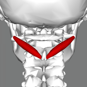
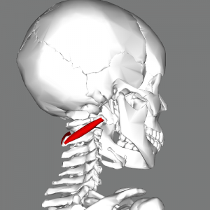
Obliquus Capitis Inferior (also known as the Inferior Oblique) is a small muscle that runs posteriorly and inferomedially from C1 to C2. It is situated under the deep cervical vein and comprises the inferior boarder of the suboccipital triangle.[1] It is the only suboccipital muscle that does not attach to the skull.[2]
Origin[edit | edit source]
Base of spinous process and adjoining lamina of the axis.[3]
Insertion[edit | edit source]
Along the inferior aspect of the tip of the transverse process of the atlas.[3]
Nerve Supply[edit | edit source]
Suboccipital nerve or dorsal ramus of cervical spinal nerve (C1).[3]
Blood Supply[edit | edit source]
Vertebral artery and the deep descending branch of the occipital artery.[3]
Action[edit | edit source]
Ipsilateral rotation of the atlantoaxial joint.[1]
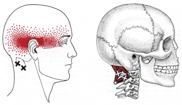
Trigger Point Referral Pattern[2][edit | edit source]
References[edit | edit source]
- ↑ 1.0 1.1 Agur AMR, Dalley AF (2012). Grant's Atlas of Anatomy (13th ed). Philadelphia, PA: Lippincott Williams & Wilkins.
- ↑ 2.0 2.1 Travell JG, Simons DG, Simons LS (1998). Travell and Simons' Myofascial Pain and Dysfunction: The Trigger Point Manual, Volume 1: Upper Half of Body (2nd ed). Baltimore, MD: Williams & Wilkins.
- ↑ 3.0 3.1 3.2 3.3 http://www.anatomyexpert.com/structure_detail/5213/


