Osteosarcoma
Original Editors -Jody Swimmer from Bellarmine University's Pathophysiology of Complex Patient Problems project.
Lead Editors - Your name will be added here if you are a lead editor on this page. Read more.
Definition/Description[edit | edit source]
Osteosarcoma is also known as osteogenic sarcoma. Osteosarcoma is an extremely malignant primary cancer of long bones. Evidence of malignant osteoid bone and/or cartilage formation with destructive lesions and sclerosis is characteristic of the disease.1 Differentiating osteosarcoma from other tumors is defined by the production of extensive, incompletely mineralized matrix that is seen with histological staining.2
http://www.healthofchildren.com/images/gech_0001_0004_0_img0237.jpg
http://t0.gstatic.com/images?q=tbn:ANd9GcS7CHkoETuNqWluHna8FPvTUORXHV1zaxtxroUWKI5sLtH9lW4HGQ
Prevalence[edit | edit source]
Osteosarcoma accounts for 15% to 20% of all primary bone tumors and is the second most malignant condition of bone. It occurs most frequently in male adolescents and young adults under the age of 30, peaking in frequency during the adolescent growth spurt with another smaller peak in adults over the age of 50.1 Review of data from the Surveillance, Epidemiology and End Results program of the NCI resulted in an estimate of 4.4 per million new cases of osteosarcoma each year in people aged newborn to 24 years.3 “The U.S. Census Bureau estimates that there will be 110 million people in this age range in 2010, resulting in an incidence of roughly 450 cases per year in children and young adults less than 25 years old. Osteosarcoma accounts for approximately 5% of childhood tumors. In children and adolescents, more than 50% of these tumors arise from the bones around the knee.”3
childrensspecialists.com
Characteristics/Clinical Presentation[edit | edit source]
The patient most often presents with pain prior to soft tissue swelling and an enlarging bone mass. This is due to the stretching of the periosteum which usually causes pain before the tumor is detected. Pain could also result from the weakening of the bone and the development of minute stress fractures. The majority of patients with osteosarcoma present with localized pain at the primary tumor site.3
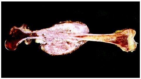
The most commonly affected bones are the metaphyseal region of long bones such as the distal femur, proximal tibia, and proximal humerus although osteosarcoma can arise in any bone in the body”.4 Systemic symptoms such as weight loss, pallor and fever are uncommon. The patient may be a tall adolescent, with males presenting more often than females.
Distal locations of the tumor have a more favorable prognosis than proximal sites. “Axial skeleton primary tumors are associated with the greatest risk of progression and death, primarily related to the inability to achieve a complete surgical resection. Pelvic osteosarcomas make up 7%-9% of all osteosarcomas; survival rates for patients with pelvic primary tumors are 20% to 47%.”3
Associated Co-morbidities[edit | edit source]
Patients with Rothmund-Thomson Syndrome (RTS) have an increased risk of developing osteosarcoma when compared with the general population. They also develop osteosarcoma at an earlier age. RTS is also called poikiloderma congenital, which is a rare autosomal recessive condition attributed to mutations of the RECQLF helicase gene on 8q24.3 This condition is characterized with skin issues such as atrophy, telangiectasias, pigmentation change, thinning or sparse hair, cataracts, small stature and skeletal anomalies.
Other familial osteosarcoma syndromes which predispose an individual to osteosarcoma include: retinoblastoma, Li-Fraumeni syndrome, Werner syndrome, Blooms syndrome, Paget’s disease, Fibrodysplasia, Enchondromatosis (Ollier disease), hereditary multiple exostoses.5
“Another condition which may predispose the patient to the development of osteosarcoma is radiotherapy. For a sarcoma to be considered a post radiation tumor, the following conditions must be met: (1) A history of previous radiation, (2) Development of the sarcoma in a bone that was in the field of radiation. (3) A latent period between radiation and the development of osteosarcoma….most osteosarcomas arise between 5 and 10 years after radiation, (4) Histologic difference from the previous tumor, which may have been irradiated. Most sarcomas arising in the field of radiation are high-grad osteosarcomas.” 6
Among 220 children who were given Ra IV as therapy for tuberculosis, many developed osteosarcoma.7
Medications[edit | edit source]
- Chemotherapy – Combination therapy with gemcitabine and docetaxel in refractory bone sarcomas
- Immunotherapy - Interferon-alpha
- Emerging therapies: 4
- Immunostimulant: muramyl-tripeptide phosphatidyl-ethanolamine (MTP-PE) – macrophage inhibitor. Recent addition of liposomal MTP-PE in combination with adjuvant chemotherapy resulted in a statistically significant increase in overall survival versus standard combination chemotherapy.
- T-cell responses by vaccination with the anti-idiotypic antibody mimicking CD55, a complement regulatory protein expressed by many solid tumors including osteosarcoma. The use of dendritic cell vaccines to enhance cytotoxic T-cell activation is being evaluated in xenograft models.
- Small molecule therapy with inhibition of the Src kinase pathway involved in osteoclast activity. The orally available Src tyrosine kinase inhibitor AZD0530 is currently being investigated in a phase II clinical trial in osteosarcoma with pulmonary recurrence post-metastasectomy.
Diagnostic Tests/Lab Tests/Lab Values[edit | edit source]
- Radiographs should be taken in any patient with prolonged and unexplained bone pain.
- Magnetic resonance imaging (MRI) - evaluates bone lesions, determine their exact location and proximity to neurovascular structures, delineate how far the tumor extends and whether systemic disease is present.
- Chest CT Scan to detect pulmonary metastasis
- Nuclear Imaging to aid in the initial staging/metastatic evaluation and response to therapy
- Technetium-99 bone scans
- Positron Emission Tomography (PET) Scan
- Bone Biopsy – Biopsy is the gold standard for diagnosis of bone sarcoma.
- Blood Tests – Blood tests are not conclusive, but they may be helpful once a diagnosis is made to help determine the staging of cancer. High levels of alkaline phosphatase and lactate dehydrogenase (LDH) may suggest that the osteosarcoma is more advanced.8
- http://t1.gstatic.com/images?q=tbn:ANd9GcT7oDH6sIKJ0W6d3ZfUI4NGdlYgpthr3kG2CnwbLe3G5tzXZcUD
Etiology/Causes[edit | edit source]
The pathogenesis and etiology of osteosarcoma remains obscure. Several chemical agents such as beryllium, viruses such as FBJ, (which contain the src-oncogene), and radiation have shown to be inducers of osteosarcoma. Paget’s disease, electrical burn and trauma may also contribute to the pathogenesis of osteosarcoma. Numerous genes have been shown recently to be associated with osteosarcoma.8
There is a suggestion of genetic predisposition to osteosarcoma, with the strongest genetic predisposition found in patients with hereditary retinoblastoma.9
Systemic Involvement[edit | edit source]
add text here
Medical Management (current best evidence)[edit | edit source]
add text here
Physical Therapy Management (current best evidence)[edit | edit source]
add text here
Alternative/Holistic Management (current best evidence)[edit | edit source]
add text here
Differential Diagnosis[edit | edit source]
add text here
Case Reports/ Case Studies[edit | edit source]
add links to case studies here (case studies should be added on new pages using the case study template)
Resources
[edit | edit source]
add appropriate resources here
Recent Related Research (from Pubmed)[edit | edit source]
see tutorial on Adding PubMed Feed
Extension:RSS -- Error: Not a valid URL: Feed goes here!!|charset=UTF-8|short|max=10
References[edit | edit source]
see adding references tutorial.

