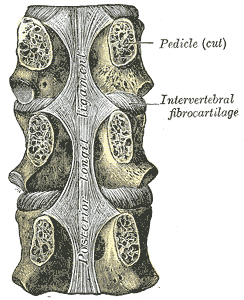
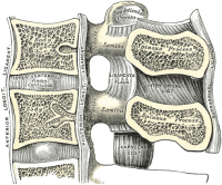
Posterior longitudinal ligament
Original Editor - Rachael Lowe
Top Contributors - Rachael Lowe, Adam Vallely Farrell, Kim Jackson, Evan Thomas, WikiSysop and Lucinda hampton
Description[edit | edit source]
Forming the anterior wall of the vertebral canal, this strong ligament spans from the body of the Axis (C2) to the posterior surface of the sacrum. Like its anterior counterpart the Anterior longitudinal ligament, its deep fibres are intersegmental while the more superficial fibres can span up to four vertebral levels.
In the cervical and thoracic regions it has a uniform width over the bodies and discs, but in the lumbar region the ligament is widest at the levels of the intervertebral discs where it is firmly anchored to the Annulus fibrosus, cartilage of the Vertebral end plates, and the margins of the vertebrae.
Superiorly it blends with the Tectorial membrane.
Attachments[edit | edit source]
Arising at the superior margin of one vertebra they span to the inferior margin of the vertebra that they attach to.Forming the anterior wall of the vertebral canal, this strong ligament arises from the body of the axis (C2) body and travels downward and posterior to the vertebral bodies (attached loosely) and intervertebral discs (firmly attaching to the posterior annulus), attaching to the back of the sacrum. It narrows as it travels downward and also has a serrated edge[1].
Like its anterior counterpart, the anterior longitudinal ligament, its deep fibers are intersegmental, while the more superficial fibers can span up to four vertebral levels. In the cervical and thoracic regions it has a uniform width over the bodies and discs, but in the lumbar region the ligament is widest at the levels of the intervertebral discs where it is firmly anchored to the annulus fibrosus, cartilage of the vertebral end plates, and the margins of the vertebrae. Its cranial counterpart is the tectorial membrane[2][3].
It is broader above than below, and thicker in the thoracic than in the cervical and lumbar regions. The ligament is more narrow at the vertebral bodies and wider at the intervertebral disc space which is more pronounced than the anterior longitudinal ligament. This is significant in understanding certain pathological conditions of the spine such as the typical location for a spinal disc herniation.
In the situation of the intervertebral fibrocartilages and contiguous margins of the vertebrae, where the ligament is more intimately adherent, it is broad, and in the thoracic and lumbar regions presents a series of dentations with intervening concave margins; but it is narrow and thick over the centers of the bodies, from which it is separated by the basivertebral veins.
This ligament is composed of smooth, shining, longitudinal fibers, denser and more compact than those of the anterior ligament, and consists of superficial layers occupying the interval between three or four vertebræ, and deeper layers which extend between adjacent vertebrae.
Iconsists of two layers. The superficial layer is a continuation of the tectorial membrane at the body of axis while the deep layer is a continuation of the cruciform ligament of the atlas. The posterior longitudinal ligament runs in the spinal canal attaching to the vertebral bodies and vertebral discs and tightens with cervical flexion.
Function[edit | edit source]
Limits flexion of the vertebral column and reinforces the intervertebral disc[2].
Clinical Relevance[edit | edit source]
Assessment[edit | edit source]
Treatment[edit | edit source]
References[edit | edit source]
- ↑ https://radiopaedia.org/articles/posterior-longitudinal-ligament
- ↑ 2.0 2.1 http://www.anatomyexpert.com/app/structure/15110/79/
- ↑ Nigel Palastanga; Roger W. Soames (2012). Churchill Livingstone, ed. Anatomy and Human Movement: Structure and Function.


