Rubrospinal Tract
Original Editor - Kate Sampson
Lead Editors - Kate Sampson, Lucinda hampton, Evan Thomas, Wendy Walker, WikiSysop and Kim Jackson
Description[edit | edit source]
The Rubrospinal tract is a descending pathway which originates in the Red Nucleus and descends to the spinal cord.[1]
Part of the extrapyramidal system.
Anatomy[edit | edit source]
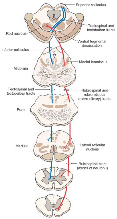 Origin[edit | edit source]

- The Red Nucleus of the midbrain tegmentum[1]
Course / Path[edit | edit source]
- Fibres pass ventromedially and cross the ventral tegmental decussation [1]
- Fibres descend to the spinal cord where they lie ventrolateral to and intertwined with the corticospinal tract [1]
Function[edit | edit source]
The most important function of the rubrospinal tract is the control of muscle tone in flexor muscle groups. It is tempting to speculate that this system is particularly important in the term newborn because of the impressive flexor tone in the limbs.[2]Its functions are believed to be:
- Modulation of Flexor Muscle Tone: plays an important role in the modulation of motor tone, causing flexors to contract and extensors to relax and vice versa. This function is associated with the rubrospinal tract because the red nucleus receives input fibers from reticular formation. Under the influence of these connections, the rubrospinal tract inhibits the activity of extensors and facilitates the activity of flexor muscles.
- Modulation of Reflex Activity: facilitates the reflex activity due to the connections of the red nucleus with reticular formation. It plays an important role in reflex activity that involve flexors eg withdrawing hand after touching a hot object, flexion of limbs when something hits the flexor surface, etc.
- Inhibition of Anti-gravity Muscles: iinhibits the contraction of anti-gravity muscles. Important in the prevention of decerebrate posture (abnormal posture in which all the body parts are extended and stretched due to unregulated activity of extensors or antigravity muscles)[3].
Pathology[edit | edit source]
Within the literature, selective lesions have not been reported of solely the red nucleus or rubrospinal tract. Lesions within the region of the red nucleus can result in movement disorders and tremor, but these effects may be more associated with damage to fibers which are associated with the cerebellar and basal ganglia systems.[4]
In primate studies, it has been suggested that the rubrospinal tract is responsible for fractionation of movement.[5]. Fractionation is the ability to isolate movement to one joint independent of another.[6] Therefore, it could be hypothesised that if the rubrospinal tract was affected, this could have an impact on the fine tuning and fractionation of movement.
However, even though it has been shown within animal studies that the rubrospinal tract can assist with functional recovery, it is unclear as to how this can be generalised to humans.[7]
References[edit | edit source]
- ↑ 1.0 1.1 1.2 1.3 Crossman AR, Neary D. Neuroanatomy: An Illustrated Colour Text. Third Edition. London: Elsevier, 2004
- ↑ JJ V. Volpe’s Neurology of the Newborn.2018 Available: https://www.sciencedirect.com/topics/neuroscience/rubrospinal-tract(accessed 25.4.2022)
- ↑ Brain made simple Rubrospinal tract Available:https://brainmadesimple.com/rubrospinal-tract/ (accessed 25.4.2022)
- ↑ What-When-How. The Upper Motor Neurons (Motor Systems) Part 3. http://what-when-how.com/neuroscience/the-upper-motor-neurons-motor-systems-part-3/ (accessed on 1/4/2016)
- ↑ Belhaj-Saif A, Cheney PD. Plasticity in the distribution of the red nucleus output to forearm muscles after unilateral lesions of the pyramidal tract. J Neurophysiol 2000; 83: 3147–53
- ↑ Scheets PL, Sahrmann SA, Norton BJ. Use of movement system diagnoses in the management of patients with neuromuscular conditions: a multiple-patient case report. Physical therapy. 2007 May 15.
- ↑ Onodera S, Hicks TP. Carbocyanine dye usage in demarcating boundaries of the aged human red nucleus. PloS one 2010; 5: e14430.

