Rupture of the Triceps Brachii muscle: Difference between revisions
No edit summary |
No edit summary |
||
| (20 intermediate revisions by 4 users not shown) | |||
| Line 1: | Line 1: | ||
<div class="editorbox"> '''Original Editor '''- [[User:Marlies Verbruggen |Marlies Verbruggen ]] '''Top Contributors''' - {{Special:Contributors/{{FULLPAGENAME}}}}</div> | |||
( | ===Introduction === | ||
[[File:Triceps .png|right|frameless]] | |||
The triceps brachii is a large, thick [[muscle]] on the dorsal part of the upper arm. It often appears as the shape of a horseshoe on the posterior aspect of the arm. The main function of the triceps is the extension of the [[Elbow|elbow joint .]]<ref>Singh RK, Pooley J. [https://bjsm.bmj.com/content/36/6/467 Complete rupture of the triceps brachii muscle.] British journal of sports medicine. 2002 Dec 1;36(6):467-9.Available: https://bjsm.bmj.com/content/36/6/467<nowiki/>(accessed 30.12.2021)</ref> | |||
Triceps Ruptures are rare injuries to the elbow extensor mechanism that most commonly occurs as a result of a sudden forceful elbow contraction in weightlifters or older males with underlying systemic illness<ref name=":1">Othrobullets [https://www.orthobullets.com/shoulder-and-elbow/3071/triceps-rupture Triceps Rupture] Available: https://www.orthobullets.com/shoulder-and-elbow/3071/triceps-rupture<nowiki/>(accessed 30.12.2021)</ref>. | |||
==Etiology == | |||
Triceps | [[File:Tri2.jpg|alt=|right|frameless]] | ||
== | Triceps tendon tear is a relatively rare injury and the rupture of the distal triceps is the most uncommon rupture in the upper extremity (less than 1% of all the upper extremity tendon injuries)<ref name="Black">Blackmore S.M. et al, Management of distal biceps and triceps ruptures. Journal of hand therapy, 2006; 19 : 154-169. Level B</ref><ref name="Rin">Rineer C.A. et al., Elbow tendinopathy and tendon ruptures : Epicondylitis, biceps and triceps ruptures. Journal of hand surgery, 2009 ; 34 A : 566 – 576 Level B</ref>. | ||
[[File:Tri2.jpg| | |||
Triceps tendon tear is a relatively rare injury and the rupture of the distal triceps is the most uncommon rupture in the upper extremity | |||
== | * Rupture is often associated with pre-existing systemic conditions or drug treatments, including the local or systemic of steroids or systematic endocrine disorders, renal failure, anabolic steroid use, local steroid injection. | ||
* Patients tend to be men who practice sports and are from 30 to 50 years of age. | |||
* Injury is commonly caused by falls on an outstretched hand, direct trauma on the elbow, lifting against resistance<ref>Sollender JL, Rayan GM, Barden GA. Triceps tendon rupture in weight lifters. Journal of shoulder and elbow surgery. 1998 Mar 1;7(2):151-3.</ref> or sometimes it occurs after a surgical procedure where the triceps was reattached. eg some case reports confirm triceps ruptures after following total elbow arthroplasty. | |||
==Clinical Presentation== | |||
Clinical signs and symptoms vary depend upon: | |||
* The type of the lesions (tendinous, tendon avulsion or inside the muscle belly), | * The type of the lesions (tendinous, tendon avulsion or inside the muscle belly), | ||
* The degree of the extension (partial or total). | * The degree of the extension (partial or total). | ||
* The time (acute or chronic). | * The time (acute or chronic). | ||
In general, the triceps lesions are characterised by | In general, the triceps lesions are characterised by | ||
=== | * Pain or tenderness and a palpable defect in the tendon that can be seen proximal to the olecranon<ref name=":0">Celli A. Triceps tendon rupture: the knowledge acquired from the anatomy to the surgical repair. Musculoskeletal surgery. 2015 Sep 1;99(1):57-66.</ref><ref name="Rin2">Rineer C.A. et al., Elbow tendinopathy and tendon ruptures : Epicondylitis, biceps and triceps ruptures. Journal of hand surgery, 2009 ; 34 A : 566 – 576 Level B</ref>. | ||
* Patient describes often an unexpected “pop” or giving way, with subsequent pain and weakness in the extremity. | |||
* When the patient is unable to extend his arm against gravity, you can expect a complete triceps rupture. A palpable defect is not always present, but sometimes can be found in the posterior arm. | |||
* Swelling and echymosis (bruising) are other nonspecific findings. <ref name="Black2">Blackmore S.M. et al, Management of distal biceps and triceps ruptures. Journal of hand therapy, 2006; 19 : 154-169. Level B</ref> | |||
==Examination== | |||
Inspection | |||
* | * pain, swelling, and ecchymosis over the posterior aspect of the elbow | ||
* may have palpable defect | |||
Motion | |||
* inability to extend elbow against resistance | |||
* not always present -- some patients are able to extend elbow against resistance if intact lateral expansion or compensating anconeus muscle | |||
Provocative tests | |||
Modified Thompson squeeze test: patient lies prone with the elbow at the end of the table and forearm hanging down, triceps muscle is firmly squeezed, inability to extend the elbow against gravity suggests complete disruption of triceps proper and lateral expansion.<ref name=":1" /> | |||
==== | ==Imaging Studies== | ||
Magnetic resonance imaging is widely accepted as the gold standard to evaluate the size and extension of the tear: triceps lesions often occur at the tendon insertion and result in either partial or total tears. | |||
== Complications == | |||
< | # Elbow stiffness/weakness | ||
# Ulnar nerve injury | |||
# Failure of repair<ref name=":1" /> | |||
== Differential Diagnosis == | |||
* Fracture<ref name="Black" /> | |||
* Joint dislocation<ref name="Black" /> | |||
<br> | * Intramuscular tear<ref name="Black" />,Weakness due to the neurological radial nerve problems and Triceps tendinitis but it is an uncommon condition reported in literature<ref name=":0" /> <ref>Kapandji IA (1970) The elbow. In: The physiology of the joints, vol 1. Churchill Livingstone, London, pp 78–121</ref> <br> | ||
==Treatment == | |||
'''Non-operative''': splint immobilization | |||
Indications | |||
* partial tears and able to extend against gravity | |||
* low demand patients in poor health | |||
[[ | Techniques: immobilize elbow in 30 degrees of flexion for 4 weeks. Immobilization is followed by range of motion and [[Strength Training|strengthening exercises]]. Six months after the injury, the full strength and ROM should be achieved.<ref>Morrey BF. Morrey BF, Sanchez-Sotelo J. Functional evaluation of the elbow. The Elbow and Its Disorders. 2009.</ref> | ||
'''Operative:''' primary surgical repair | |||
Indications | |||
* acute complete tears | |||
* partial tears (>50%) with significant weakness | |||
Technique: delayed reconstruction may need tendon graft | |||
==Physical Therapy Mangement== | |||
The treatment after triceps surgery contains several steps : | |||
=== '''Protected phase''' === | |||
1.Splinting following triceps repair: | |||
After the repair of the triceps due to surgery, the elbow is immobilized in a long arm [[splint]] initially, thereby the elbow is placed in 30 – 45° elbow flexion with the forearm in neutral position and the wrist is often supported. There is a huge variability in recommendations for management after the operation. But the postoperative position is decided by the surgeon, based on tension and quality of the tendon repair, other injuries and the medical history of the patient. This position is also unique to each patient. <ref name="Black" /><ref>Blackmore SM, Jander RM, Culp RW. Management of distal biceps and triceps ruptures. Journal of Hand Therapy. 2006 Apr 1;19(2):154-69.</ref> | |||
[[File:Echte_hinged_splint.jpg|alt=|right|frameless|279x279px]] | |||
A hinged splint can be used after triceps repair if early controlled motion is desired. This splint blocks elbow flexion on the one hand, on the other hand it allows dynamic/gravity assisted elbow extension. The goal is a progressive elbow flexion by weekly advancing the range of motion block. So this splint allows passive elbow flexion and active elbow flexion through a range which is defined. When the patient is not exercising , the splint is locked in one position. In this phase patient education is also very important. The patient must be aware of the prohibition for active elbow extension, because this could lead to avulsion or rupture of the repaired tendon. For example : pushing himself out of a chair may not be performed by his operated arm.<ref name="Black" /><ref>(1) MacInnes SJ, Crawford LA, Shahane SA. Disorders of the biceps and triceps tendons at the elbow. Orthopaedics and Trauma 2016 August 2016;30(4):346-354</ref><br> <br>When the patient has difficulties with relaxing the repaired muscle or when he is untrustworthy during the performances of the exercises out of a protective splint, in this cases we use a dynamic traction splint.<ref name="Black" /> | |||
2. Therapy program | |||
<br> | In this phase we can start with early controlled motion (ECM), whether the patient uses a static, hinged or dynamic splint. <br>When the patient has a static splint it’s an absolute must that the patient is reliable. We can prevent this problem by using a hinged splint, because then there are blocks placed to limit end range and it also assists dynamic motion.<ref name="Black" /><br>There is not really a consensus in literature about the optimum time frame to begin early controlled motion. In this article the ECM starts from day 10 to day 14 postoperatively. This has some advantages such as: the reduction of pain and oedema. As you can see on the photos below, the ECM program contains four exercises namely<ref name="Black" /> : | ||
[[File:ECM_triceps.jpg|alt=|right|frameless]] | |||
A) full passive elbow extension<br>B) active and/or passive elbow flexion to 30 °<br>C) the use of a template splint, to block degree of active flexion<br>D) passive forearm rotation with the elbow held in extension<br> <br>The motion performance during the [[Adherence to Home Exercise Programs|home program]] can be assisted by the gravity. Increased flexion is allowed with each successive week. The full active elbow and forearm range motion is allowed at week 6. After the operation the patient has often edema in the hand. We can explain this by the fact that the hand is in a dependant position for much of the day caused by the immobilization of the elbow in extended position. Therefore it’s highly recommended to start immediately after the surgery with some exercises like: hand pumping, elevation above the heart level, the use of compressive glove or wrap. <ref name="Black" /> | |||
Additional therapy interventions include ''':''' thermal agents; therapist assisted motion within range limits; edema control; pain management; scar mobilization | |||
If it is necessary : isometric strengthening for the hand and shoulder rotation, abduction and adduction.<ref name="Black" />It’s very important to be carefully with an elbow flexion combined with end range of shoulder elevation because this will stress the triceps repair. Full-time immobilization is used for three to four weeks, if the patient follows not the ECM program because more protection for the repair is necessary .<ref name="Black" /> | |||
=== '''Progressive motion phase''' === | |||
Six weeks after the operation , the patient begins with active contraction of the triceps. Elbow extension against gravity is definitely emphasized to encourage active motor recruitment rather than using the gravity to assist the extension.<br>There are some techniques to restore end range of active motion such as <ref name="Black" />: | |||
< | <nowiki>*</nowiki> Place and hold exercises at available end range<br>* PNF = Proprioceptive neuromuscular facilitation patterns of exercise<br>* Neuromuscular electrical stimulation | ||
It’s possible that treatment is demanded for capsular tightness or joint contractures. This treatment contains<ref name="Black" /> : | |||
<br> | <nowiki>*</nowiki> [[Thermotherapy|Thermal agents]]<br>* Joint mobilization<br>* Sustained positioning<br>* Splints to increase range of motion | ||
'''C) Strengthening phase''': | |||
This phase begins 10-12 weeks postoperatively. It may take several months before the strength is returned. It’s highly important that the active [[Range of Motion|range of motion]] is equal to passive range of motion when starting this phase. Passive limitations can be treated as long as there are still improvements. The fact that this phase starts after 10 – 12 weeks guarantees that the tendon is adequately healed so that it can tolerate the stress exerted by the strengthening exercises. <ref name="Black" /> | |||
[[ | The strengthening starts with up to 50 % effort isometric contraction of the muscle tendon unit. The effort is determined by measuring the maximal voluntary contraction (MVC) of the not- operated side with a hand held dynamometer and having the patient exercise up to 50% of MVC. The contraction is tarted in midrange and later progressed to the end range of motion. If the patient doesn’t develop pain, the effort is increased to the maximum. <br>These are the exercises for the triceps strengthening, note position the forearm in supination, neutral and pronation to address all three heads of the triceps.<ref name="Black" /><br>The strengthening program is finally advanced to : | ||
[[File:Mobilisation_exercises.jpg|alt=|right|frameless]] | |||
Isotonic concentric exercises , using :<br>• Free weights eg [[Dumbbell Exercise|dumbbells]]<br>• Elastic bands<br>• PNF diagonal patterns<br>eventually advanced to eccentric muscle contraction | |||
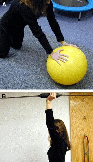
These two photos at R demonstrate exercises to complete the stabilisation and mobilization exercises.<br>A : Demonstration of weight-bearing and [[Scapular Dyskinesia|scapular control]] exercise to facilitate full limb reconditioning | |||
B: Demonstration of the Body Blade. Bodyblade uses vibration and the power of inertia to rapidly contract your muscles up to 270 times per minute, stimulate your nervous system, and transform your body.<ref>Body Blade Body blade Available: https://bodyblade.com/<nowiki/>(accessed 30.12.2021)</ref> | |||
= | If there’s a development of pain at the surgical site, the intensity of the strengthening program is immediately reduced. <br>Sixteen weeks after the operation , sports-specific and work -specific activities begin.<ref name="Black" /><ref>Monasterio M, Longsworth KA, Viegas S. Dynamic hinged orthosis following a surgical reattachment and therapy protocol of a distal triceps tendon avulsion. Journal of Hand Therapy. 2014 Oct 1;27(4):330-4.</ref><br> | ||
==References== | |||
<references /> | <references /> | ||
Latest revision as of 12:58, 2 January 2022
Introduction[edit | edit source]
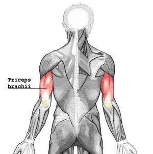
The triceps brachii is a large, thick muscle on the dorsal part of the upper arm. It often appears as the shape of a horseshoe on the posterior aspect of the arm. The main function of the triceps is the extension of the elbow joint .[1]
Triceps Ruptures are rare injuries to the elbow extensor mechanism that most commonly occurs as a result of a sudden forceful elbow contraction in weightlifters or older males with underlying systemic illness[2].
Etiology[edit | edit source]
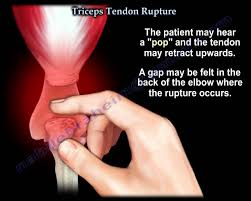
Triceps tendon tear is a relatively rare injury and the rupture of the distal triceps is the most uncommon rupture in the upper extremity (less than 1% of all the upper extremity tendon injuries)[3][4].
- Rupture is often associated with pre-existing systemic conditions or drug treatments, including the local or systemic of steroids or systematic endocrine disorders, renal failure, anabolic steroid use, local steroid injection.
- Patients tend to be men who practice sports and are from 30 to 50 years of age.
- Injury is commonly caused by falls on an outstretched hand, direct trauma on the elbow, lifting against resistance[5] or sometimes it occurs after a surgical procedure where the triceps was reattached. eg some case reports confirm triceps ruptures after following total elbow arthroplasty.
Clinical Presentation[edit | edit source]
Clinical signs and symptoms vary depend upon:
- The type of the lesions (tendinous, tendon avulsion or inside the muscle belly),
- The degree of the extension (partial or total).
- The time (acute or chronic).
In general, the triceps lesions are characterised by
- Pain or tenderness and a palpable defect in the tendon that can be seen proximal to the olecranon[6][7].
- Patient describes often an unexpected “pop” or giving way, with subsequent pain and weakness in the extremity.
- When the patient is unable to extend his arm against gravity, you can expect a complete triceps rupture. A palpable defect is not always present, but sometimes can be found in the posterior arm.
- Swelling and echymosis (bruising) are other nonspecific findings. [8]
Examination[edit | edit source]
Inspection
- pain, swelling, and ecchymosis over the posterior aspect of the elbow
- may have palpable defect
Motion
- inability to extend elbow against resistance
- not always present -- some patients are able to extend elbow against resistance if intact lateral expansion or compensating anconeus muscle
Provocative tests
Modified Thompson squeeze test: patient lies prone with the elbow at the end of the table and forearm hanging down, triceps muscle is firmly squeezed, inability to extend the elbow against gravity suggests complete disruption of triceps proper and lateral expansion.[2]
Imaging Studies[edit | edit source]
Magnetic resonance imaging is widely accepted as the gold standard to evaluate the size and extension of the tear: triceps lesions often occur at the tendon insertion and result in either partial or total tears.
Complications[edit | edit source]
- Elbow stiffness/weakness
- Ulnar nerve injury
- Failure of repair[2]
Differential Diagnosis[edit | edit source]
- Fracture[3]
- Joint dislocation[3]
- Intramuscular tear[3],Weakness due to the neurological radial nerve problems and Triceps tendinitis but it is an uncommon condition reported in literature[6] [9]
Treatment[edit | edit source]
Non-operative: splint immobilization
Indications
- partial tears and able to extend against gravity
- low demand patients in poor health
Techniques: immobilize elbow in 30 degrees of flexion for 4 weeks. Immobilization is followed by range of motion and strengthening exercises. Six months after the injury, the full strength and ROM should be achieved.[10]
Operative: primary surgical repair
Indications
- acute complete tears
- partial tears (>50%) with significant weakness
Technique: delayed reconstruction may need tendon graft
Physical Therapy Mangement[edit | edit source]
The treatment after triceps surgery contains several steps :
Protected phase [edit | edit source]
1.Splinting following triceps repair:
After the repair of the triceps due to surgery, the elbow is immobilized in a long arm splint initially, thereby the elbow is placed in 30 – 45° elbow flexion with the forearm in neutral position and the wrist is often supported. There is a huge variability in recommendations for management after the operation. But the postoperative position is decided by the surgeon, based on tension and quality of the tendon repair, other injuries and the medical history of the patient. This position is also unique to each patient. [3][11]
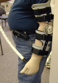
A hinged splint can be used after triceps repair if early controlled motion is desired. This splint blocks elbow flexion on the one hand, on the other hand it allows dynamic/gravity assisted elbow extension. The goal is a progressive elbow flexion by weekly advancing the range of motion block. So this splint allows passive elbow flexion and active elbow flexion through a range which is defined. When the patient is not exercising , the splint is locked in one position. In this phase patient education is also very important. The patient must be aware of the prohibition for active elbow extension, because this could lead to avulsion or rupture of the repaired tendon. For example : pushing himself out of a chair may not be performed by his operated arm.[3][12]
When the patient has difficulties with relaxing the repaired muscle or when he is untrustworthy during the performances of the exercises out of a protective splint, in this cases we use a dynamic traction splint.[3]
2. Therapy program
In this phase we can start with early controlled motion (ECM), whether the patient uses a static, hinged or dynamic splint.
When the patient has a static splint it’s an absolute must that the patient is reliable. We can prevent this problem by using a hinged splint, because then there are blocks placed to limit end range and it also assists dynamic motion.[3]
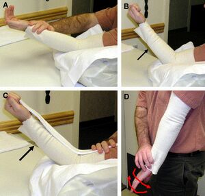
There is not really a consensus in literature about the optimum time frame to begin early controlled motion. In this article the ECM starts from day 10 to day 14 postoperatively. This has some advantages such as: the reduction of pain and oedema. As you can see on the photos below, the ECM program contains four exercises namely[3] :
A) full passive elbow extension
B) active and/or passive elbow flexion to 30 °
C) the use of a template splint, to block degree of active flexion
D) passive forearm rotation with the elbow held in extension
The motion performance during the home program can be assisted by the gravity. Increased flexion is allowed with each successive week. The full active elbow and forearm range motion is allowed at week 6. After the operation the patient has often edema in the hand. We can explain this by the fact that the hand is in a dependant position for much of the day caused by the immobilization of the elbow in extended position. Therefore it’s highly recommended to start immediately after the surgery with some exercises like: hand pumping, elevation above the heart level, the use of compressive glove or wrap. [3]
Additional therapy interventions include : thermal agents; therapist assisted motion within range limits; edema control; pain management; scar mobilization
If it is necessary : isometric strengthening for the hand and shoulder rotation, abduction and adduction.[3]It’s very important to be carefully with an elbow flexion combined with end range of shoulder elevation because this will stress the triceps repair. Full-time immobilization is used for three to four weeks, if the patient follows not the ECM program because more protection for the repair is necessary .[3]
Progressive motion phase[edit | edit source]
Six weeks after the operation , the patient begins with active contraction of the triceps. Elbow extension against gravity is definitely emphasized to encourage active motor recruitment rather than using the gravity to assist the extension.
There are some techniques to restore end range of active motion such as [3]:
* Place and hold exercises at available end range
* PNF = Proprioceptive neuromuscular facilitation patterns of exercise
* Neuromuscular electrical stimulation
It’s possible that treatment is demanded for capsular tightness or joint contractures. This treatment contains[3] :
* Thermal agents
* Joint mobilization
* Sustained positioning
* Splints to increase range of motion
C) Strengthening phase:
This phase begins 10-12 weeks postoperatively. It may take several months before the strength is returned. It’s highly important that the active range of motion is equal to passive range of motion when starting this phase. Passive limitations can be treated as long as there are still improvements. The fact that this phase starts after 10 – 12 weeks guarantees that the tendon is adequately healed so that it can tolerate the stress exerted by the strengthening exercises. [3]
The strengthening starts with up to 50 % effort isometric contraction of the muscle tendon unit. The effort is determined by measuring the maximal voluntary contraction (MVC) of the not- operated side with a hand held dynamometer and having the patient exercise up to 50% of MVC. The contraction is tarted in midrange and later progressed to the end range of motion. If the patient doesn’t develop pain, the effort is increased to the maximum.
These are the exercises for the triceps strengthening, note position the forearm in supination, neutral and pronation to address all three heads of the triceps.[3]
The strengthening program is finally advanced to :
Isotonic concentric exercises , using :
• Free weights eg dumbbells
• Elastic bands
• PNF diagonal patterns
eventually advanced to eccentric muscle contraction
These two photos at R demonstrate exercises to complete the stabilisation and mobilization exercises.
A : Demonstration of weight-bearing and scapular control exercise to facilitate full limb reconditioning
B: Demonstration of the Body Blade. Bodyblade uses vibration and the power of inertia to rapidly contract your muscles up to 270 times per minute, stimulate your nervous system, and transform your body.[13]
If there’s a development of pain at the surgical site, the intensity of the strengthening program is immediately reduced.
Sixteen weeks after the operation , sports-specific and work -specific activities begin.[3][14]
References[edit | edit source]
- ↑ Singh RK, Pooley J. Complete rupture of the triceps brachii muscle. British journal of sports medicine. 2002 Dec 1;36(6):467-9.Available: https://bjsm.bmj.com/content/36/6/467(accessed 30.12.2021)
- ↑ 2.0 2.1 2.2 Othrobullets Triceps Rupture Available: https://www.orthobullets.com/shoulder-and-elbow/3071/triceps-rupture(accessed 30.12.2021)
- ↑ 3.00 3.01 3.02 3.03 3.04 3.05 3.06 3.07 3.08 3.09 3.10 3.11 3.12 3.13 3.14 3.15 3.16 Blackmore S.M. et al, Management of distal biceps and triceps ruptures. Journal of hand therapy, 2006; 19 : 154-169. Level B
- ↑ Rineer C.A. et al., Elbow tendinopathy and tendon ruptures : Epicondylitis, biceps and triceps ruptures. Journal of hand surgery, 2009 ; 34 A : 566 – 576 Level B
- ↑ Sollender JL, Rayan GM, Barden GA. Triceps tendon rupture in weight lifters. Journal of shoulder and elbow surgery. 1998 Mar 1;7(2):151-3.
- ↑ 6.0 6.1 Celli A. Triceps tendon rupture: the knowledge acquired from the anatomy to the surgical repair. Musculoskeletal surgery. 2015 Sep 1;99(1):57-66.
- ↑ Rineer C.A. et al., Elbow tendinopathy and tendon ruptures : Epicondylitis, biceps and triceps ruptures. Journal of hand surgery, 2009 ; 34 A : 566 – 576 Level B
- ↑ Blackmore S.M. et al, Management of distal biceps and triceps ruptures. Journal of hand therapy, 2006; 19 : 154-169. Level B
- ↑ Kapandji IA (1970) The elbow. In: The physiology of the joints, vol 1. Churchill Livingstone, London, pp 78–121
- ↑ Morrey BF. Morrey BF, Sanchez-Sotelo J. Functional evaluation of the elbow. The Elbow and Its Disorders. 2009.
- ↑ Blackmore SM, Jander RM, Culp RW. Management of distal biceps and triceps ruptures. Journal of Hand Therapy. 2006 Apr 1;19(2):154-69.
- ↑ (1) MacInnes SJ, Crawford LA, Shahane SA. Disorders of the biceps and triceps tendons at the elbow. Orthopaedics and Trauma 2016 August 2016;30(4):346-354
- ↑ Body Blade Body blade Available: https://bodyblade.com/(accessed 30.12.2021)
- ↑ Monasterio M, Longsworth KA, Viegas S. Dynamic hinged orthosis following a surgical reattachment and therapy protocol of a distal triceps tendon avulsion. Journal of Hand Therapy. 2014 Oct 1;27(4):330-4.
