Sartorius: Difference between revisions
Rotimi Alao (talk | contribs) (Kk) |
Rotimi Alao (talk | contribs) (Jjj) |
||
| Line 28: | Line 28: | ||
The muscle may be absent in some people.<ref>Wysocki J, Krasuski P, Czubalski A. Vascularization of the sartorius muscle. Folia Morphol (Warsz). 1996;55(2):115-20.</ref> | The muscle may be absent in some people.<ref>Wysocki J, Krasuski P, Czubalski A. Vascularization of the sartorius muscle. Folia Morphol (Warsz). 1996;55(2):115-20.</ref> | ||
Revision as of 09:49, 12 March 2018
Description[edit | edit source]
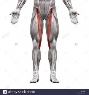
The sartorius muscle is a thin, long, superficial muscle in the anterior compartment of the thigh.[1] It runs down the length of the thigh, runs over 2 joints- hip and knee joints [2] and is the longest muscle in the human body.[1] An exceptional length of this muscle often exceeds 50cm.[3] In such long muscles not all muscle fibres run through the whole length of the muscle belly.[2] It is estimated that at the most 30–50% of fibres run from tendon to tendon.[2] The rest of them end intrafascicularly.[2] The length of a single fibre is estimated at 35–45 cm.[4]
Just like an S-shaped tape, the muscle belly twists around the anterior, and medial surface of the thigh.[2] It passes behind the medial condyle of the femur to end in a tendon.[1] The tendon, after taking an anterior curve joins with the tendon of the Gracilis and Semitendinosus in the pes anserinus before its final insertion.[1]

Etymology[edit | edit source]
Sartorius derives from the Latin word sartor, meaning tailor, [6] and it is occasionally referred to as the tailor's muscle.[1] This name was chosen in reference to the cross-legged position in which tailors once sat.[1]
Origin[edit | edit source]
Anterior superior iliac spine (1)(2)
Anterior superior iliac spine[1][2]
Insertion[edit | edit source]
The superomedial surface of the tibia[1]
Nerve supply[edit | edit source]
Femoral Nerve[1]
Variation[edit | edit source]
There are slight adaptive ethnic differences in width and the range of muscle belly and tendon of the sartorius muscle.[4]
The muscle may be absent in some people.[7]
- ↑ 1.0 1.1 1.2 1.3 1.4 1.5 1.6 1.7 1.8 Moore, Keith L.; Dalley, Arthur F.; Agur, A. M. R. (2013). Clinically Oriented Anatomy. Lippincott Williams & Wilkins. pp. 545–546. ISBN 9781451119459.
- ↑ 2.0 2.1 2.2 2.3 2.4 2.5 Dziedzic D, Bogacka U, Ciszek B. (2014) Anatomy of sartorius muscle. Folia Morphol (Warsz). 73(3):359-62. doi: 10.5603/FM.2014.0037.
- ↑ Clavert P, Cognet JM, Baley S, Stussi D, Prevost P, Babin SR, Simon P, Kahn JL (2008). Anatomical basis for distal sartorius muscle flap for reconstructive surgery below the knee. Anatomical study and case report. J Plastic, Reconstr Aesthetic Surg, 61: 50–54.
- ↑ 4.0 4.1 Klein Horsman M, Koopman H (2007) Morphological muscle and joint parameters for musculoskeletal modelling of the lower extremity. Clin Biomech, 22: 239–247.
- ↑ http://www.innerbody.com/image_musfov/musc20-new.html
- ↑ Mosby's Medical, Nursing & Allied Health Dictionary, Fourth Edition, Mosby-Year Book Inc., 1994, p. 1394
- ↑ Wysocki J, Krasuski P, Czubalski A. Vascularization of the sartorius muscle. Folia Morphol (Warsz). 1996;55(2):115-20.


