Cerebral Atrophy
Original Editor - Rahma Ahmed Ahmed Bahbah
Top Contributors - Rahma Ahmed Ahmed Bahbah and Lucinda hampton
Introduction[edit | edit source]

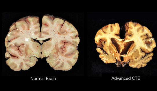
Brain atrophy relates to changes seen with brain aging in both the healthy and pathological brains. Brain atrophy is characterized by several degrees of cognitive decline that strongly correlate with morphological changes. These characteristic morphological changes include cortical thinning, white and gray matter volume loss, ventricular enlargement, and loss of gyrification, a a result of a myriad of subcellular and cellular aging processes. These morphological changes are part of healthy brain aging as yet we do not know how these observed changes relate to cognitive deficits. When accelerated aging occurs , as occurs in neurodegenerative diseases, these observed changes are a result of the presence of neurotoxic proteins that propagate through the brain. Research focusing on the mechanics of brain aging are needed.[1]
- Brain atrophy can also be observed in the pediatric age group, where it carries forward the small volume of the brain into middle age. It is important to note that some atrophic changes may be reversed during childhood.
- In the normal aging, brain atrophy tends to be accelerated by the presence of other risk factors such as high blood pressure, cardiac disease, diabetes mellitus, smoking practice, and regular alcohol intake. It's been observed that glycated hemoglobin (HbA1c) was the most significant risk factor for accelerating of brain atrophy, which is average blood sugar levels over a period of weeks/months.
Types[edit | edit source]
Cerebral atrophy is classified into two categories. According to:
- The affected brain area- global or focal.
- The volume loss distributed zone - central or cortical. In central atrophy, it's found that white matter volume loss is more than grey matter, and the opposite is seen in cortical atrophy.
There's also brain hemiatrophy in which volume loss involves one hemisphere.
Causes[edit | edit source]
There are many factors cause atrophy;[2]
- Aging
- Infections of central nervous system (CNS)
- Nutritional deficiency
- Metabolic and endocrine causes
- Traumatic causes
- Drug induced brain atrophy
- Radiation induced brain atrophy
- Increased intracranial pressure
- Perinatal and birth injury induced atrophy
- Neurodegenerative diseases causing brain atrophy
- Other causes
Clinical picture[edit | edit source]
There are various clinical features of cerebral atrophy like poor levels of intelligence especially in growing children. Memory loss is common among elderly individuals. Elderly patients with brain atrophy often experience acute confusional state.
Brain atrophy can result in loss of functional recovery following an infarct, which may also lead to death due to poor brain functioning.
Conditions[edit | edit source]
Brain atrophy does not always occur in isolation; unlike some other conditions such as leukoaraiosis and stroke are known to accompany brain atrophy.[2]
Some conditions that featured with cerebral atrophy are; Alzheimer's disease, Cerebral Palsy , Multiple sclerosis, Epilepsy, Pick's Disease, Dementia, ALS, Prion disease, Encephalitis, AIDS, Neurosyphilis.
Neuroimaging[edit | edit source]
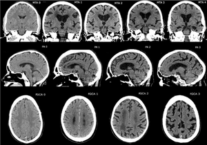
The features appear in imaging may include the following;
- Widening of cortical sulci
- Enlargement of ventricles
- Thinning of cortex
- Shrinking of hippocampus
Management[edit | edit source]
There’s no fix for brain atrophy however it can be a sign of one or more diseases. Management involves personalised treatment plans aimed at managing the symptoms of the underlying condition. Treatment may include a mix of:
- Medication.
- Physical and occupational therapy. See dementia and stroke physiotherapy
- Counseling.
- Speech therapy.
- Studies show good outcome in nutritional related brain atrophy especially that related to protein-energy and vitamin related malnutrition. The use of vitamin B12 has shown significant improvement from brain volume loss especially in children.[2]
- Surgery, in some cases eg. For ischemic stroke clot dissolving medication and/or endovascular therapy (EVT) (a minimally invasive surgical procedure to remove a blood clot from an artery -thrombectomy)[3],may benefit the client and result in less atrophy. [4].
Brain atrophy tends to be permanent and the damage can’t be reversed once it has occurred.
Brain Atrophy Types[edit | edit source]
Table 1 shows Brain atrophy types and their clinical implication.[2]
| Brain atrophy type | Common differential determinants | Clinical presentation | Current treatment options |
|---|---|---|---|
| Global brain atrophy | Advanced form degenerative atrophy
Infection such as encephalitis Malnutrition |
Headache
Tremors Loss of memory Convulsions Dizziness Vertigo |
-Treatment of offending cause (eg HIV)
-Mainly supportive -Nutritional supplements involving Vitamin B1, B12 and proteins |
| Focal brain atrophy | Head trauma
Localized space occupying lesion Stroke or ischemia |
Headache
Convulsions One side body weakness |
-Treatment of offending cause
-Mainly supportive involving anticonvulsant drugs. -Neurofeedback stimulation |
| Central brain atrophy | Previous hydrocephalus
Multiple sclerosis Malnutrition |
Headache
Tremors |
-Treatment of offending cause (eg hydrocephalus)
-Mainly supportive -Nutritional supplements involving Vitamin B1, B12 and proteins |
| Cortical brain atrophy | Degenerative age related atrophy
HIV encephalitis Hypoxic ischemic encephalopathy Malnutrition |
Headache
Tremors Loss of memory |
-Treatment of offending cause
-Mainly supportive care. -Nutritional supplements involving Vitamin B1, B12 and proteins |
| Brain hemiatrophy | Sturge Weber syndrome
Dyke-Davidoff Mason’s syndrome Tuberous sclerosis Stroke or ischemia |
Headache
One side body weakness Convulsions |
-Mainly supportive involving anticonvulsant drugs |
References[edit | edit source]
- ↑ Yana Blinkouskaya et al. Brain Shape Changes Associated With Cerebral Atrophy in Healthy Aging and Alzheimer’s Disease. Front. Mech. Eng., 19 July 2021 Sec. Micro- and Nanoelectromechanical Systems Available: https://www.frontiersin.org/articles/10.3389/fmech.2021.705653/full (accessed 11.3.24)
- ↑ 2.0 2.1 2.2 2.3 Sungura R, Onyambu C, Mpolya E, Sauli E, Vianney JM. The extended scope of neuroimaging and prospects in brain atrophy mitigation: a systematic review. Interdisciplinary Neurosurgery. 2021 Mar 1;23:100875.
- ↑ Ansari J, Triay R, Kandregula S, Adeeb N, Cuellar H, Sharma P. Endovascular intervention in acute ischemic stroke: history and evolution. Biomedicines. 2022 Feb 10;10(2):418. BibTeXEndNoteRefManRefWorks
- ↑ Luijten SP, Compagne KC, van Es AC, Roos YB, Majoie CB, van Oostenbrugge RJ, van Zwam WH, Dippel DW, Wolters FJ, van der Lugt A, Bos D. Brain atrophy and endovascular treatment effect in acute ischemic stroke: a secondary analysis of the MR CLEAN trial. International journal of stroke. 2022 Oct;17(8):881-8. BibTeXEndNoteRefManRefWorks


