Atlanto-axial joint: Difference between revisions
No edit summary |
Kim Jackson (talk | contribs) No edit summary |
||
| (23 intermediate revisions by 6 users not shown) | |||
| Line 6: | Line 6: | ||
== Description == | == Description == | ||
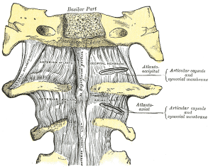
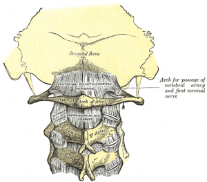
[[Image:Atlanto-occipital joint anterior.png|thumb|right|Atlanto-occipital joint (anterior)]][[Image:Atlanto-occipital joint posterior.png|thumb|right|Atlanto-occipital joint (posterior)]]The atlanto-axial joint is a joint | [[Image:Atlanto-occipital joint anterior.png|thumb|right|Atlanto-occipital joint (anterior)]][[Image:Atlanto-occipital joint posterior.png|thumb|right|Atlanto-occipital joint (posterior)]]The atlanto-axial joint is a joint between the first and second cervical vertebrae; the [[Atlas]] and [[Axis]]. It is a complex joint made up of three synovial joints and constitutes the most mobile articulation of the spine<ref>Magee, D. Orthopedic Physical Assessment. Elsevier</ref>. The middle (or median) joint is classified as a '''[[Joint Classification|pivot]]''' [[Joint Classification|joint]] and the lateral joints are '''[[Joint Classification|plane]]''' [[Joint Classification|articulations]]. <br> | ||
== Articulating Surfaces == | == Articulating Surfaces == | ||
There are two '''lateral atlanto-axial joints''' which are concave in an anterior-posterior direction, this allows rotation. | There are two '''lateral atlanto-axial joints''' which are concave in an anterior-posterior direction, this allows rotation. | ||
The '''median atlantoaxial joint''' is the articulation of the: . | The '''median atlantoaxial joint''' is the articulation of the: . | ||
*posterior surface of the anterior arch of atlas and the front of the odontoid process | *posterior surface of the anterior arch of atlas and the front of the odontoid process | ||
| Line 19: | Line 19: | ||
Between the articular processes of the two bones there is on either side an arthrodial or gliding joint. | Between the articular processes of the two bones there is on either side an arthrodial or gliding joint. | ||
== Capsule | == Capsule == | ||
The atlantoaxial articular capsules are thick and loose, and connect the margins of the lateral masses of the atlas with those of the posterior articular surfaces of the axis. | The atlantoaxial articular capsules are thick and loose, and connect the margins of the lateral masses of the atlas with those of the posterior articular surfaces of the axis. | ||
Each is strengthened at its posterior and medial part by an accessory ligament, which is attached below to the body of the axis near the base of the odontoid process, and above to the lateral mass of the atlas near the transverse ligament.<br> | Each is strengthened at its posterior and medial part by an accessory ligament, which is attached below to the body of the [[axis]] near the base of the [[Odontoid process|odontoid]] process, and above to the lateral mass of the [[atlas]] near the transverse ligament.<br> | ||
== Ligaments == | == Ligaments == | ||
| Line 32: | Line 32: | ||
*[[Anterior atlantoaxial ligament]] | *[[Anterior atlantoaxial ligament]] | ||
*[[Posterior atlantoaxial ligament]] | *[[Posterior atlantoaxial ligament]] | ||
*[[Transverse | *[[Transverse Ligament of the Atlas]] | ||
*[[Alar ligaments]] | |||
*[[Apical ligament]] | |||
*[[Tectorial membrane]] | |||
* | |||
* | |||
== Muscles == | == Muscles == | ||
| Line 45: | Line 41: | ||
== Motions Available == | == Motions Available == | ||
'''Rotation''' is the primary movement at this joint - 60% of cervical rotation (50°) comes from the atlanto-axial articulation. This is allowed by the pivot articulation between the odontoid process of the axis and the ring formed by the anterior arch and the transverse ligament of the atlas.<br> | |||
'''Flexion (10'''<span style="line-height: 1.5em;">°</span>''')'''<span style="line-height: 1.5em;"> and </span>'''Extension'''<span style="line-height: 1.5em;"> is limited by tectoral membrane.</span> | |||
<span style="line-height: 1.5em;">Side flexion is around 5°.</span> | |||
== Pathology == | |||
Because of its proximity to the [[Brainstem|brain stem]] and importance in stabilization, fracture or injury at this level can be catastrophic. Common trauma and pathologies include (but are not limited to): | |||
*The Dens: significant depression on the skull can push the dens into the brainstem, causing death. The dens itself is vulnerable to fracture due to trauma or ossification. | |||
*Transverse ligament: Should the transverse ligament of the atlas fail due to trauma or disease (such as [[Rheumatoid Arthritis|rheumatoid arthritis]]), the dens is no longer anchored and can travel up the cervical spine, causing paralysis. If it reaches the medulla death can result. | |||
*Alar ligaments: stress or trauma can stretch the weaker alar ligaments, causing an increase in range of motion of approximately 30%. | |||
*Posterior Atlanto-Occipital Membrane: genetic traits can sometimes result in ossification, turning the groove into an foramen. | |||
== References == | == References == | ||
<references /> | |||
[[Category:Cervical Spine - Anatomy]] | |||
[[Category:Cervical Spine - Joints]] | |||
[[Category:Cervical Spine]] | |||
Latest revision as of 17:48, 2 January 2021
Original Editor - Rachael Lowe
Lead Editors
Description[edit | edit source]
The atlanto-axial joint is a joint between the first and second cervical vertebrae; the Atlas and Axis. It is a complex joint made up of three synovial joints and constitutes the most mobile articulation of the spine[1]. The middle (or median) joint is classified as a pivot joint and the lateral joints are plane articulations.
Articulating Surfaces[edit | edit source]
There are two lateral atlanto-axial joints which are concave in an anterior-posterior direction, this allows rotation.
The median atlantoaxial joint is the articulation of the: .
- posterior surface of the anterior arch of atlas and the front of the odontoid process
- anterior surface of the transverse ligament and the back of the odontoid process
Between the articular processes of the two bones there is on either side an arthrodial or gliding joint.
Capsule[edit | edit source]
The atlantoaxial articular capsules are thick and loose, and connect the margins of the lateral masses of the atlas with those of the posterior articular surfaces of the axis.
Each is strengthened at its posterior and medial part by an accessory ligament, which is attached below to the body of the axis near the base of the odontoid process, and above to the lateral mass of the atlas near the transverse ligament.
Ligaments[edit | edit source]
The ligaments connecting these bones are:
- Articular capsules
- Anterior atlantoaxial ligament
- Posterior atlantoaxial ligament
- Transverse Ligament of the Atlas
- Alar ligaments
- Apical ligament
- Tectorial membrane
Muscles[edit | edit source]
Motions Available[edit | edit source]
Rotation is the primary movement at this joint - 60% of cervical rotation (50°) comes from the atlanto-axial articulation. This is allowed by the pivot articulation between the odontoid process of the axis and the ring formed by the anterior arch and the transverse ligament of the atlas.
Flexion (10°) and Extension is limited by tectoral membrane.
Side flexion is around 5°.
Pathology[edit | edit source]
Because of its proximity to the brain stem and importance in stabilization, fracture or injury at this level can be catastrophic. Common trauma and pathologies include (but are not limited to):
- The Dens: significant depression on the skull can push the dens into the brainstem, causing death. The dens itself is vulnerable to fracture due to trauma or ossification.
- Transverse ligament: Should the transverse ligament of the atlas fail due to trauma or disease (such as rheumatoid arthritis), the dens is no longer anchored and can travel up the cervical spine, causing paralysis. If it reaches the medulla death can result.
- Alar ligaments: stress or trauma can stretch the weaker alar ligaments, causing an increase in range of motion of approximately 30%.
- Posterior Atlanto-Occipital Membrane: genetic traits can sometimes result in ossification, turning the groove into an foramen.
References[edit | edit source]
- ↑ Magee, D. Orthopedic Physical Assessment. Elsevier


