
Rheumatoid Arthritis
Introduction
Rheumatoid arthritis (RA) is a “chronic inflammatory autoimmune systemic disease that usually presents with joint inflammation leading to pain, fatigue, and impaired physical functioning and work productivity, all of which negatively impact health-related quality of life.”[1]
RA commonly affects the hands, wrists, shoulders, elbows, knees, ankles and feet.[2] As it is a systemic condition, it can significantly affect a number of body systems, including the cardiovascular and respiratory systems.[2][3][4] Without adequate treatment, RA can cause long-term disability, pain and premature death.[5][6] While pharmacological management is the mainstay treatment for RA,[5] rehabilitation professionals can play a role in the non-pharmacological management of this condition.[2]
Epidemiology
RA affects around 1% of the global population,[3] and up to 3% of older persons.[5] It is 2-3 times more likely to occur in females.[6] However, with improving medical management, the severity, mortality and associated comorbidities appear to be decreasing.[7]
RA can develop at any age. The literature has traditionally described a peak age of onset between 30 and 60 years,[8][9] distinguishing younger-onset RA from elderly-onset RA (i.e. after 60 years).[8][10] However, the peak age of onset has increased,[11] with recent data showing the highest prevalence in people aged 75-79 years.[12]
Risk Factors
RA is a multifactorial disease that has been linked to various genetic, environmental and other factors.[3] The HLA-DRB1 alleles are linked with an increased risk of developing RA.[13] In genetically predisposed individuals, certain environmental exposures, such as smoking, infections (e.g. periodontal disease,[3] Epstein-Barr virus, bacterial superantigens etc.[14]), and dietary agents[3] may contribute. Hormonal factors are also implicated: the effect of oestrogen on immune function is thought to play a role in the female predominance in RA, and other sex-related factors are probably involved as well.[14]
Clinical Characteristics
Common joint symptoms include symmetrical joint pain, with the wrists, fingers (MCP), knees, ankles, feet and upper cervical spine commonly affected. People typically experience morning stiffness lasting more than one hour, or stiffness after a period of inactivity, and affected joints may feel warm and tender. As the disease progresses, there is loss of or limitation in range of motion, and joint deformity may develop.[2][15]
Typical Joint Deformities
Cervical spine: The prevalence of atlantoaxial instability (AAI) in individuals with RA is reported as 40–85%. RA can cause laxity of the transverse ligament and hypermobility between C1 and C2, with a risk of subluxation and, in some cases, neurological involvement.[16]
Hand: Characteristic hand deformities include swan neck deformity, boutonnière deformity, ulnar drift, and thumb metacarpophalangeal flexion with interphalangeal hyperextension.[15]
-
 Swan neck deformity
 -
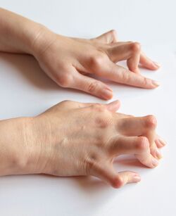 Boutonniere deformity


Lower limb: In the knee, genu valgus and Baker's cysts may develop, while in the ankle and foot, pronation, hallux valgus, and claw toes are common.[15]
-
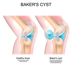 Baker cyst
 -
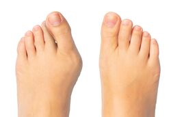 Hallux valgus
 -
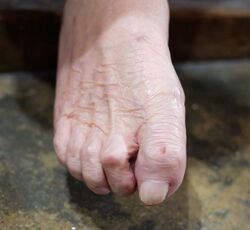 Claw toe


Other symptoms and extra-articular manifestations: Because RA is systemic, a range of extra-articular features may occur. These include pulmonary issues (e.g. pleuritis, pleural effusions, pulmonary fibrosis and interstitial lung disease) and cardiac issues (e.g. atherosclerosis, arterial stiffness, pericarditis and valvular disease). Other manifestations include Felty's syndrome (low white blood cell count, splenomegaly and RA[17]), dry eyes and mouth (Sjögren's syndrome), numbness, tingling or burning in the hands and feet, sleep difficulties, and rheumatic nodules, which occur more often in people with seropositive, erosive disease. People with RA may also experience fatigue, reduced cognitive function, sarcopenia, osteoporosis and vasculitis.[2][3][15][18]
Pathophysiology
There are two main subtypes of RA. In seropositive RA, autoantibodies such as rheumatoid factor (RF) and anti-citrullinated protein antibodies (ACPA) are present. These are found in around 50% of individuals with early RA and up to 80% of those with established RA, with ACPAs found in around 67% of people with RA.[18][19]
In seronegative RA, these autoantibodies are absent, but other types of autoantibodies may be present.[20] Seronegative disease is often considered less severe, although there is some controversy about this.[21] Volkov et al. note that many individuals with RF-negative or ACPA-negative RA do not develop “a chronic progressive disease”[19] and Carbonell-Bobadilla et al.[21] suggest there may be less joint damage than in seropositive RA, but a similar long-term cardiovascular risk.[18][19]
Basic Overview of Rheumatoid Arthritis Pathophysiology
The pathophysiology of RA is complex. The following describes the pathophysiology of seropositive RA, though it should be noted that patients can have different pathogenic pathways and cell lineages.[14][18][22][23]
A genetically predisposed individual is exposed to an environmental trigger, such as smoking or infection, which can cause disordered immunity. This disordered immunity may lead to the citrullination of some proteins (citrullination is when the amino acid arginine converts into citrulline[24]). Antigen-presenting cells recognise these modified proteins as foreign and transport them to lymph nodes and mucosal and lymphoid tissues, where they activate CD4+ T-helper cells. This stimulates B-cells to proliferate and differentiate into plasma cells, which produce autoantibodies (ACPAs and rheumatoid factor). ACPAs can be detected before joint symptoms develop in people with RA, and their production indicates a "break of immunological tolerance".[18]
The autoantibodies can then migrate to the joint synovium, and the symmetrical swelling in the small joints is the "external reflection of synovial membrane inflammation following immune activation".[18] Once in the synovium, autoantibodies start producing proinflammatory cytokines, such as interferon-gamma and interleukins. This causes macrophages, monocytes and synovial fibroblasts to release cytokines including tumour necrosis factor alpha (TNF alpha), interleukin 6 (IL-6) and IL-1. The synovial fibroblasts then begin to proliferate, which increases joint inflammation (i.e. synovial hyperplasia), and the proliferating synovial cells produce a pannus (a thickened synovial membrane made of destructive granulation tissue). This pannus causes the erosion of cartilage and bone, while activated synovial cells also produce proteases that break down cartilage. Inflammatory cytokines increase the activity of osteoclasts (cells that break down bone) and decrease the activity of osteoblasts (cells that repair or form new bone), which leads to articular bone loss and osteoporosis.[14][18][22][23]
ACPAs and RF form immune complexes that activate the complement system (an important part of the innate immune system[25]), which leads to chronic inflammation. Angiogenesis (the formation of new capillaries from existing vasculature[26]) occurs alongside this chronic inflammation, enabling more inflammatory cells to travel to the joints. This produces additional pro-inflammatory factors that exacerbate existing processes. As RA progresses, multiple joints on both sides of the body are affected, and inflammatory cytokines also spread to other parts of the body. This causes extra-articular symptoms such as vasculitis, rheumatoid nodules and protein breakdown in skeletal muscle. RA can wax and wane, with various exacerbations (flares) and periods of remission.[14][18][22][23][27]
This complex process is explained in detail in Rheumatoid arthritis: pathological mechanisms and modern pharmacologic therapies[18] and in the following videos:
Diagnosis
Diagnostic Tests
Several laboratory tests support the diagnosis of seropositive RA.[15] Around 80% of people with RA have a positive rheumatoid factor, although there is roughly a 5% false-positive rate. The anti-cyclic citrullinated peptide antibody (anti-CCP/ACPA) test is frequently used to diagnose and predict RA; it has a high specificity (>97%[18]), meaning a positive result strongly supports the diagnosis, but its sensitivity rates vary in the literature from 50% to 95%.[28][29][30]
Other tests include C-reactive protein (CRP), a general inflammatory marker, and erythrocyte sedimentation rate (ESR), a non-specific measure of inflammation that reflects the relative activity of the disease process. A complete blood count may also be performed. X-rays will not show early-onset RA,[31] but during the later stages they may reveal irregular joint surfaces, decreased joint space and abnormal joint alignment. Synovial fluid analysis may show an increased white blood cell count, increased collagenase and increased debris (proteins).[15]
Diagnostic Criteria
1987 criteria:
The 1987 American College of Rheumatology (ACR) classification criteria for RA are: morning stiffness in and around the joints lasting at least one hour; soft tissue swelling of three or more joint areas; swelling of the proximal interphalangeal, metacarpophalangeal or wrist joints; symmetric swelling; rheumatoid nodules; the presence of rheumatoid factor; and radiographic erosions and/or periarticular osteopenia in the hand and/or wrist joints. The first four criteria must have been present for at least six weeks, and under this system, a diagnosis of rheumatoid arthritis requires four or more of the above criteria.[32]
While these criteria “are well accepted as providing the benchmark for disease definition”,[31] there are limitations because they are designed to differentiate individuals with established RA from other rheumatological conditions. They don’t, therefore, help identify individuals with early RA.[31]
2010 criteria:
The 2010 ACR/EULAR (European League Against Rheumatism) diagnostic criteria for RA score four domains. A score of 6 or more is required for a person to be classified as having RA.[31]
| Domain | Criteria | Score |
|---|---|---|
| Joint involvement | 1 large joint | 0 |
| 2-10 large joints | 1 | |
| 1-3 small joints (with or without large joint involvement) | 2 | |
| 4-10 small joints (with or without large joint involvement) | 3 | |
| >10 joints (at least 1 small joint) | 5 | |
| Serology (at least one test result needed) | Negative rheumatoid factor and negative ACPA | 0 |
| Low-positive RF or low-positive ACPA | 2 | |
| High-positive RF or high-positive ACPA | 3 | |
| Acute-phase reactants (at least one test result needed) | Normal CRP and normal ESR | 0 |
| Abnormal CRP or abnormal ESR | 1 | |
| Duration of symptoms | Less than 6 weeks | 0 |
| 6 or more weeks | 1 |
Treatment for Rheumatoid Arthritis
There have been many developments in the treatment of RA in recent decades.[2] While the primary management is pharmacological, various non-pharmacological interventions also play a role, including patient education, exercise, physiotherapy and occupational therapy.[33]
Pharmacological Management
Pharmacological management aims to decrease joint inflammation and inhibit disease progression.[15]
Disease-Modifying Antirheumatic Drugs (DMARD)
“Early diagnosis and intervention with a disease-modifying antirheumatic drug (DMARD) remain the cornerstone of treatment to control inflammation, prevent joint and organ damage, and reduce the risk of death.”
Current guidelines recommend a treat-to-target approach: a target is set (remission or, where this is not achievable, low disease activity), disease activity is monitored regularly, and treatment is altered/escalated until the target is reached.[34][35][36] Low-dose methotrexate is the mainstay initial treatment for RA, and 25–40% of patients experience significant improvements with methotrexate alone; if the response is inadequate, other agents such as a JAK inhibitor or a biological agent may be introduced.[14] [37]
For a detailed discussion of DMARDs for RA, see: Current therapeutic options in the treatment of rheumatoid arthritis.[33] The following videos provide a simple summary of DMARDs for RA:
Other Medications
Non-steroidal anti-inflammatory drugs (NSAIDs), such as ibuprofen and naproxen, decrease pain and improve function by reducing acute inflammation; they may help with symptoms but do not change the disease course in RA.[15] Corticosteroids, such as low-dose prednisolone, have a number of side effects and, like NSAIDs, may reduce symptoms but do not change the disease course in RA.[15][40] They are often used short term as "bridging" therapy, such as while waiting for a DMARD to take effect, or during a flare.[41]
Non-pharmacological Management
“Despite the availability of effective medication, there is a substantial proportion of patients with persisting or recurring disease activity, with or without joint damage. Moreover, the evidence for an increased cardiovascular risk in patients with inflammatory joint diseases is growing. As a result, many patients are in need of additional, nonpharmacological treatment, including physical therapy.”[2]
While rehabilitation cannot influence the disease process of RA, it can help with function. NICE recommends that individuals with RA receive input from various members of the rehabilitation team, including physiotherapy, occupational therapy, podiatry and psychology.[42]
Exercise / Physical Activity
There is evidence that increasing physical activity levels / exercise can help with symptoms and reduce the impact of systemic manifestations of RA.[43] However, people with rheumatic and musculoskeletal diseases (RMDs) are, in general, less active than healthy controls,[44] typically engaging in less physical activity than is recommended in international guidelines, especially those aged <55 years.[45]
The following video will help you to learn more about exercise for rheumatoid arthritis. Key points from the video include:[46]
- all types of exercise produce similar benefits, so consider your patient’s preferences
- exercise must be individualised to suit the patient’s requirements (e.g. available time, interests, access to equipment, function, etc.)
- look at the current function and compare this to your patient's required function
- consider general physical activity levels alongside exercise plans
- include strength, range and cardiovascular components in an exercise plan
Peter et al.[2] produced a clinical practice guideline detailing the physiotherapy assessment and management of individuals with RA, which is available here: Clinical practice guideline for physical therapist management of people with rheumatoid arthritis.
The Strengthening And Stretching for Rheumatoid Arthritis of the Hand (SARAH) programme is considered a clinically effective and cost-effective adjunct treatment to improving hand function in individuals with RA.[47][48] More information on this programme is available here and in the following video:
Other Non-pharmacological Interventions
Patient education is relevant at all stages of the disease course. It should include information on RA and the importance of exercise and a healthy lifestyle, along with strategies to reduce disability within your specific scope of practice, such as distributing load and using assistive devices. Patients should be supported to be physically active while distributing energy across the day or week based on fatigue, and it is important to acknowledge and address barriers to exercise and physical activity; barriers can include a lack of knowledge, a lack of social support, pain, fatigue and the fear that exercise will damage the joints.[50][2][15] Joint protection involves avoiding positions of deformity, adopting the most stable positions and avoiding repetitive tasks. Energy conservation should take account of systemic complications, such as reduced cardiovascular function, and assistive devices may also be of benefit.[15]
Additional Resources
- Peter WF, Swart NM, Meerhoff GA, Vliet Vlieland TPM. Clinical practice guideline for physical therapist management of people with rheumatoid arthritis. Phys Ther. 2021 Aug 1;101(8):pzab127.
- Majnik J, Császár-Nagy N, Böcskei G, Bender T, Nagy G. Non-pharmacological treatment in difficult-to-treat rheumatoid arthritis. Front Med (Lausanne). 2022 Aug 29;9:991677. doi: 10.3389/fmed.2022.991677. PMID: 36106320; PMCID: PMC9465607.
- National Institute for Health and Care Excellence (NICE). Rheumatoid arthritis in adults: management: NICE guideline [NG100]. 2018 (updated 2020).
References
- ↑ Küçükdeveci AA, Turan BK, Arienti C, Negrini S. Overview of Cochrane Systematic Reviews of rehabilitation interventions for persons with rheumatoid arthritis: a mapping synthesis. Eur J Phys Rehabil Med. 2023 Apr;59(2):259-69.
- ↑ 2.0 2.1 2.2 2.3 2.4 2.5 2.6 2.7 2.8 Peter WF, Swart NM, Meerhoff GA, Vliet Vlieland TPM. Clinical practice guideline for physical therapist management of people with rheumatoid arthritis. Phys Ther. 2021 Aug 1;101(8):pzab127.
- ↑ 3.0 3.1 3.2 3.3 3.4 3.5 Radu AF, Bungau SG. Management of rheumatoid arthritis: an overview. Cells. 2021 Oct 23;10(11):2857.
- ↑ Metsios GS, Kitas GD. Physical activity, exercise and rheumatoid arthritis: Effectiveness, mechanisms and implementation. Best Pract Res Clin Rheumatol. 2018 Oct;32(5):669-82.
- ↑ 5.0 5.1 5.2 Turk MA, Liu Y, Pope JE. Non-pharmacological interventions in the treatment of rheumatoid arthritis: A systematic review and meta-analysis. Autoimmun Rev. 2023 Jun;22(6):103323.
- ↑ 6.0 6.1 GBD 2021 Rheumatoid Arthritis Collaborators. Global, regional, and national burden of rheumatoid arthritis, 1990-2020, and projections to 2050: a systematic analysis of the Global Burden of Disease Study 2021. Lancet Rheumatol. 2023 Sep 25;5(10):e594-e610.
- ↑ Finckh A, Gilbert B, Hodkinson B, Bae SC, Thomas R, Deane KD, et al. Global epidemiology of rheumatoid arthritis. Nat Rev Rheumatol. 2022 Oct;18(10):591-602.
- ↑ 8.0 8.1 Yazici Y, Paget SA. Elderly-onset rheumatoid arthritis. Rheum Dis Clin North Am. 2000 Aug;26(3):517-26.
- ↑ Bullock J, Rizvi SAA, Saleh AM, Ahmed SS, Do DP, Ansari RA, Ahmed J. Rheumatoid arthritis: A brief overview of the treatment. Med Princ Pract. 2018;27(6):501-7.
- ↑ Pavlov-Dolijanovic S, Bogojevic M, Nozica-Radulovic T, Radunovic G, Mujovic N. Elderly-onset rheumatoid arthritis: characteristics and treatment options. Medicina (Kaunas). 2023 Oct 23;59(10):1878.
- ↑ Slobodin G. Rheumatoid arthritis. In: Slobodin G, Shoenfeld Y, editors. Rheumatic Disease in Geriatrics. Springer, Cham, 2020.
- ↑ Su QY, Yang L, Qi XY, Wang MY, Cheng JW, Niu H, Zhang SX. Global, regional, and national burdens of rheumatoid arthritis among people aged 60 years and older from 1990 to 2021: a trend analysis for the Global Burden of Disease Study 2021. Front Public Health. 2025 May 26;13:1527680.
- ↑ Chauhan K, Jandu JS, Brent LH, et al. Rheumatoid Arthritis. [Updated 2023 May 25]. In: StatPearls [Internet]. Treasure Island (FL): StatPearls Publishing; 2024 Jan-. Available from: https://www.ncbi.nlm.nih.gov/books/NBK441999/
- ↑ 14.0 14.1 14.2 14.3 14.4 14.5 Gravallese EM, Firestein GS. Rheumatoid arthritis - common origins, divergent mechanisms. N Engl J Med. 2023 Feb 9;388(6):529-42.
- ↑ 15.00 15.01 15.02 15.03 15.04 15.05 15.06 15.07 15.08 15.09 15.10 Cunningham S. Rheumatoid Arthritis Course. Physiopedia Plus, 2024.
- ↑ Cunningham S. Upper cervical instability associated with rheumatoid arthritis: a case report. J Man Manip Ther. 2016 Jul;24(3):151-7.
- ↑ Arthritis Society Canada. Felty's syndrome. Available from: https://arthritis.ca/about-arthritis/arthritis-types-(a-z)/types/felty-s-syndrome (last accessed 22 May 2024).
- ↑ 18.00 18.01 18.02 18.03 18.04 18.05 18.06 18.07 18.08 18.09 Guo Q, Wang Y, Xu D, Nossent J, Pavlos NJ, Xu J. Rheumatoid arthritis: pathological mechanisms and modern pharmacologic therapies. Bone Res. 2018 Apr 27;6:15.
- ↑ 19.0 19.1 19.2 Volkov M, van Schie KA, van der Woude D. Autoantibodies and B cells: The ABC of rheumatoid arthritis pathophysiology. Immunol Rev. 2020 Mar;294(1):148-63.
- ↑ NEJM Group. What Is Rheumatoid Arthritis? | NEJM. Available from: https://www.youtube.com/watch?v=e_NZk8nFSPA (last accessed 23 May 2024).
- ↑ 21.0 21.1 Carbonell-Bobadilla N, Soto-Fajardo C, Amezcua-Guerra LM, Batres-Marroquín AB, Vargas T, et al. Patients with seronegative rheumatoid arthritis have a different phenotype than seropositive patients: A clinical and ultrasound study. Front Med (Lausanne). 2022 Aug 16;9:978351.
- ↑ 22.0 22.1 22.2 22.3 NEJM Group. What Is Rheumatoid Arthritis? | NEJM. Available from: http://www.youtube.com/watch?v=e_NZk8nFSPA [last accessed 23/05/2025]
- ↑ 23.0 23.1 23.2 23.3 Osmosis from Elsevier. Rheumatoid arthritis - causes, symptoms, diagnosis, treatment, pathology. Available from: https://www.youtube.com/watch?v=EB5zxdAQGzU [last accessed 23/05/2024]
- ↑ Gazitt T, Lood C, Elkon KB. Citrullination in rheumatoid arthritis-a process promoted by neutrophil lysis? Rambam Maimonides Med J. 2016 Oct 31;7(4):e0027.
- ↑ Holers VM, Banda NK. Complement in the initiation and evolution of rheumatoid arthritis. Front Immunol. 2018 May 28;9:1057.
- ↑ Elshabrawy HA, Chen Z, Volin MV, Ravella S, Virupannavar S, Shahrara S. The pathogenic role of angiogenesis in rheumatoid arthritis. Angiogenesis. 2015 Oct;18(4):433-48.
- ↑ Orange DE, Yao V, Sawicka K, Fak J, Frank MO, Parveen S, et al. RNA identification of PRIME cells predicting rheumatoid arthritis flares. N Engl J Med. 2020 Jul 16;383(3):218-28.
- ↑ Melbourne Pathology. Anti-CCP Insight – March 2019. Available from: https://www.mps.com.au/media/6479/mp_insight_anti-ccp_final-in-house-march-19.pdf (last accessed 8 June 2024).
- ↑ Cho J, Pyo JY, Fadriquela A, Uh Y, Lee JH. Comparison of the analytical and clinical performances of four anti-cyclic citrullinated peptide antibody assays for diagnosing rheumatoid arthritis. Clin Rheumatol. 2021 Feb;40(2):565-573.
- ↑ Zhu JN, Nie LY, Lu XY, Wu HX. Meta-analysis: compared with anti-CCP and rheumatoid factor, could anti-MCV be the next biomarker in the rheumatoid arthritis classification criteria? Clin Chem Lab Med. 2019 Oct 25;57(11):1668-79.
- ↑ 31.0 31.1 31.2 31.3 31.4 Aletaha D, Neogi T, Silman AJ, Funovits J, Felson DT, Bingham CO 3rd, et al. 2010 rheumatoid arthritis classification criteria: an American College of Rheumatology/European League Against Rheumatism collaborative initiative. Arthritis Rheum. 2010 Sep;62(9):2569-81.
- ↑ Arnett FC, Edworthy SM, Bloch DA, McShane DJ, Fries JF, Cooper NS, et al. The American Rheumatism Association 1987 revised criteria for the classification of rheumatoid arthritis. Arthritis Rheum. 1988 Mar;31(3):315-24.
- ↑ 33.0 33.1 Köhler BM, Günther J, Kaudewitz D, Lorenz HM. Current therapeutic options in the treatment of rheumatoid arthritis. J Clin Med. 2019 Jun 28;8(7):938.
- ↑ Smolen JS, Edwards CJ, Konzett V, Laskou F, Aletaha D, Caporali R, et al. EULAR recommendations for the management of rheumatoid arthritis with synthetic and biologic disease-modifying antirheumatic drugs: 2025 update. Ann Rheum Dis. 2026 Jun;85(6):991-1009.
- ↑ Gunkl-Tóth L, McInnes IB, Nagy G. Bridging the gap: combining treat-to-target and difficult-to-treat strategies in the management of rheumatoid arthritis. Nat Rev Rheumatol. 2026 May;22(5):319-327.
- ↑ Heckert SL, Maassen JM, Nevins I, Baudoin P, Steup-Beekman GM, Huizinga TWJ, et al. Long-term clinical outcomes in early rheumatoid arthritis that was treated-to-target in the BeSt and IMPROVED studies. Rheumatology (Oxford). 2025 Mar 1;64(3):1052-1059.
- ↑ Bonfiglioli KR. From concept to practice: real-world safety of JAK inhibitors in rheumatoid arthritis treatment. Front Med (Lausanne). 2026 Feb 13;13:1729135.
- ↑ Johns Hopkins Rheumatology. Rheumatoid Arthritis - Treatment | Johns Hopkins. Available from: https://www.youtube.com/watch?v=2kDbztlKXKM [last accessed 23/05/2024]
- ↑ Johns Hopkins Rheumatology. Treatment Options for Rheumatoid Arthritis | Johns Hopkins Rheumatology. Available from: https://www.youtube.com/watch?v=YSOhxIJCV-o [last accessed 23/05/2024]
- ↑ Johns Hopkins Arthritis Center. Rheumatoid arthritis treatment. Available from: https://www.hopkinsarthritis.org/arthritis-info/rheumatoid-arthritis/ra-treatment/ (last accessed 25 May 2024).
- ↑ Brinkmann GH, Sundlisæter NP, Nordberg LB, Sexton J, Ikdahl E, Aga AB, et al. Successful discontinuation of oral glucocorticoids after short-term bridging therapy in patients with newly diagnosed rheumatoid arthritis. Ann Rheum Dis. 2026 Jun;85(6):1073-1080.
- ↑ National Institute for Health and Care Excellence (NICE), Rheumatoid arthritis in adults: management: NICE guideline [NG100]. 2018 (updated 2020). Available from: https://www.nice.org.uk/guidance/ng100/ [accessed 24 May 2024].
- ↑ Metsios GS, Kitas GD. Physical activity, exercise and rheumatoid arthritis: Effectiveness, mechanisms and implementation. Best Pract Res Clin Rheumatol. 2018 Oct;32(5):669-82.
- ↑ Rausch Osthoff AK, Niedermann K, Braun J, Adams J, Brodin N, Dagfinrud H, et al. 2018 EULAR recommendations for physical activity in people with inflammatory arthritis and osteoarthritis. Ann Rheum Dis. 2018 Sep;77(9):1251-60.
- ↑ Lange E, Kucharski D, Svedlund S, Svensson K, Bertholds G, Gjertsson I, Mannerkorpi K. Effects of aerobic and resistance exercise in older adults with rheumatoid arthritis: A randomized controlled trial. Arthritis Care Res (Hoboken). 2019 Jan;71(1):61-70.
- ↑ 46.0 46.1 The Rheumatology Physio. Exercise and rheumatoid arthritis webinar. Available from: https://www.youtube.com/watch?v=QdtXacpfBg8 [last accessed 25 May 2024].
- ↑ NDORMS. SARAH Implementation. Available from: https://www.ndorms.ox.ac.uk/rrio/sarah-implementation (last accessed 24 May 2024).
- ↑ Lamb SE, Williamson EM, Heine PJ, Adams J, Dosanjh S, Dritsaki M, et al. Exercises to improve function of the rheumatoid hand (SARAH): a randomised controlled trial. Lancet. 2015 Jan 31;385(9966):421-9.
- ↑ British Society for Rheumatology. Hand exercises for people with rheumatoid arthritis. Available from: http://www.youtube.com/watch?v=q1pNhMkJHhg [last accessed 24/05/2024]
- ↑ Zangi HA, Ndosi M, Adams J, Andersen L, Bode C, Boström C, et al. EULAR recommendations for patient education for people with inflammatory arthritis. Ann Rheum Dis. 2015 Jun;74(6):954-62.