
Osteoarthritis
Top Contributors - Jess Bell and Wanda van Niekerk
Introduction
Osteoarthritis (OA) is a common chronic health condition. It can cause pain, decreased function, poor sleep, decreased mental health and reduced quality of life.[1][2] It is also associated with an increased risk of cardiovascular disease, diabetes, hypertension and mortality.[3][4] Conservative management strategies for osteoarthritis include education, exercise and weight management. This page provides an overview of osteoarthritis, including epidemiology, risk factors and pathology, before considering diagnosis and management trends.
Definition
The Osteoarthritis Research Society International (OARSI) defines osteoarthritis as: “a disorder involving movable joints characterized by cell stress and extracellular matrix degradation initiated by micro- and macro-injury that activates maladaptive repair responses including pro-inflammatory pathways of innate immunity. The disease manifests first as a molecular derangement (abnormal joint tissue metabolism) followed by anatomic, and/or physiologic derangements (characterized by cartilage degradation, bone remodeling, osteophyte formation, joint inflammation and loss of normal joint function), that can culminate in illness.”[5]
Key point: osteoarthritis has traditionally been described as a degenerative cartilage disease, but our understanding has evolved, and we now know that there is a breakdown of the cartilage, as well as structural changes across the whole joint.[6]
Epidemiology
According to the 2021 Global Burden of Disease Study, 595 million people had osteoarthritis in 2020 (i.e. 7.6% of the global population).[7] This number has increased by 132.2% since 1990. These figures are expected to continue to increase. By 2050, cases of knee osteoarthritis are projected to increase by 74.9%, cases of hip osteoarthritis are projected to increase by 78.6%, cases of hand osteoarthritis are projected to increase by 48.6%, and cases of other types of osteoarthritis are projected to increase by 95.1%.[7]
Increased prevalence has been linked to ageing populations and an increase in obesity and joint injuries.[8][9]
Risk Factors
Known risk factors for osteoarthritis include ageing, obesity, acute trauma, chronic overload, gender and hormone profile, metabolic syndrome, genetic predisposition[8] and the gut-joint axis.[10] However, osteoarthritis is not "the inevitable consequence of these factors [...and…] different risk factors may act together in the pathogenesis of osteoarthritis".[8]
Ageing is characterised by progressive tissue loss and decreased organ function, and it "represents the single greatest risk factor for OA."[8]
Obesity is considered "the most prevalent preventable risk factor for developing osteoarthritis".[11] Previously, obesity was considered a primary risk factor in knee osteoarthritis because of its impact on biomechanics, but it is now understood that it increases risk by altering metabolism and inflammation.[11] Obesity increases the risk of osteoarthritis in various joints, including the hand,[12] hip, knee, ankle and spine.[11] This effect is seen in both males and females, although it is greater in females.[11]
Acute trauma and joint injury are considered "potent" risk factors for osteoarthritis.[1]
Chronic overload: various occupational ergonomic risk factors for osteoarthritis have been proposed, including force exertion, demanding postures, repetitive movements, hand-arm vibration, kneeling/squatting, lifting and climbing.[13] These risk factors can increase the risk of developing knee or hip osteoarthritis compared to no exposure. However, because the quality of evidence is currently low, there is "limited evidence of harmfulness".
Another systematic review found that physically demanding jobs (e.g. construction work, floor and bricklaying, fishing, and farming) are associated with an increased risk of knee and hip osteoarthritis, and there may be a dose-response relationship.[1]
Gender and hormone profile: there are gender differences in osteoarthritis across all joints (the cervical spine is one potential exception),[1] with females more likely to develop osteoarthritis.[14]
Anatomic factors (joint deformities): the shape of a joint can impact the development of osteoarthritis[10] and certain variations in the shape of bones/joints have been associated with osteoarthritis of the hip and knee.[1]
Pathology of Osteoarthritis
Osteoarthritis is a "dynamic and complex process, involving inflammatory, mechanical, and metabolic factors that result in the inability of the articular surface to serve its function of absorbing and distributing the mechanical load through the joint that ultimately leads to joint destruction."[8]
Osteoarthritis is typically characterised by:[8][14]
- degradation/destruction of the articular cartilage
- surface irregularities
- osteophyte formation
- subchondral bone remodelling/thickening
- synovial inflammation
- inflammation of periarticular structures
However, the exact pathological mechanisms of osteoarthritis remain unknown. Changes across the whole joint are thought to result from an interplay between various tissues in the osteochondral complex (e.g. adipose tissue, synovial tissue, ligaments, tendons, muscles),[8] and there is increasing evidence that low-grade, chronic inflammation plays a part in osteoarthritis.[15]
It is a "heterogeneous disease that impacts all component tissues of the articular joint organ."[8]
Please watch the following video if you want to learn more about the pathology of osteoarthritis:
Clinical Features
The pathological changes and symptoms caused by osteoarthritis vary considerably in each person.[8] But typical signs and symptoms associated with osteoarthritis include decreased range of motion, stiffness, pain, deformity, crepitus, decreased mobility and functional limitations, reduced/loss of ability to engage in “valued activities”, such as walking, sports, and dancing, and bony tenderness.[8][14][17][18]
Diagnosis
Osteoarthritis can be confirmed radiographically on x-ray. Radiographic findings include:[8][14][17]
- joint space narrowing
- osteophytes
- subchondral sclerosis
- cyst formation
- abnormalities of bone contour
- ankylosis
However, x-rays can "underestimate the joint tissue involvement in OA, since they only visualize a component of the condition including cartilage loss that result in joint space narrowing and bony changes that result in subchondral sclerosis, cysts, and osteophyte formation. Once these changes are apparent on radiographs, the condition has significantly advanced."[10]
MRI is also used to evaluate and monitor osteoarthritis.[19] You can read more about the use of MRI in osteoarthritis here: MR-Imaging in osteoarthritis: Current standard of practice and future outlook.
Clinical vs Radiographic Osteoarthritis
Knee Osteoarthritis
Radiographic knee osteoarthritis requires structural changes on x-ray while clinical knee osteoarthritis is diagnosed based on a patient’s symptoms and the clinical examination.[20] Radiography "is disputed because structural findings appear relatively late in the course of the disease and symptoms are not always associated with the structural findings."[20]
There are different clinical classification criteria used to help identify individuals with osteoarthritis, including the European League Against Rheumatism (EULAR), the American College of Rheumatology (ACR), and the National Institute for Health and Care Excellence (NICE) clinical classification criteria.[21] For more information on these criteria, please see Comparison of three sets of clinical classification criteria for knee osteoarthritis: a cross-sectional study of 13,459 patients treated in primary care.[21]
The following clinical criteria for knee osteoarthritis are based on the NICE guidelines:[21][22]
- aged ≥ 45 years
- has movement-related joint pain
- has no knee morning stiffness or has knee morning stiffness that lasts ≤ 30 minutes
Hip Osteoarthritis
Hip osteoarthritis has traditionally been diagnosed based on radiographic features, like the Kellgren and Lawrence score, but many guidelines recommend against using radiography as a diagnostic tool.[23] While there is no validated diagnostic criteria for early hip osteoarthritis,[23] the American College of Rheumatology clinical classification criteria for hip osteoarthritis are as follows:[24][18]
- hip pain in combination with:
- <15° of hip internal rotation
- erythrocyte sedimentation rate (ESR) of ≤45 mm/hour OR ≤115° of hip flexion (a hip flexion measurement is used if ESR is not obtained)
Or
- hip pain and:
- aged >50 years
- ≥15° of hip internal rotation
- pain with internal rotation
- ≤60 minutes of morning stiffness
Spine Osteoarthritis
Specific clinical criteria for identifying spine osteoarthritis (e.g. pain, morning stiffness, painful/reduced range of motion) are not yet available, but there is a known link between these criteria and lumbar disc degeneration.[25]
Hand Osteoarthritis
The American College of Rheumatology criteria for osteoarthritis of the hand are as follows:[26]
- hand pain, aching or stiffness on most days of the previous month, plus three of the following four criteria:
- hard tissue enlargement of ≥ 2 of 10 selected hand joints (the 10 selected hand joints include bilateral second and third distal interphalangeal joints, second and third proximal interphalangeal joints, and first carpometacarpal joints)
- metacarpophalangeal joint swelling in ≤ 2 joints
- enlargement of the hard tissue of ≥ 2 of the distal interphalangeal joints
- deformity of ≥ 1 of 10 selected hand joints
Outcome Measures
There are a range of outcome measures for osteoarthritis. Please see here for more information.
Joint Deformities
Specific joint deformities to look out for include Heberden's nodes, Bouchard's nodes and genu varus/ valgus.[14]
-
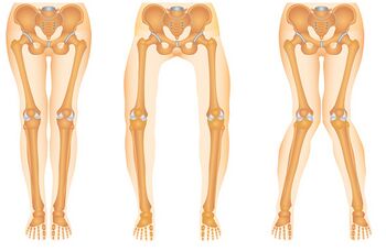 Figure 1. From left to right: knee with normal alignment, genu varus and genu valgus.
 -
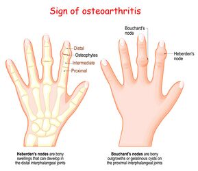 Figure 2. Heberden's and Bouchard's nodes.


Osteoarthritis Management
"Clinical guidelines for management of knee OA emphasise non-drug non-surgical strategies that focus on self-help and patient-driven options rather than clinician-delivered passive therapies."[27]
General management goals include maintaining/gaining range of motion, increasing muscular support, decreasing joint stress and managing pain.[14]
International guidelines consistently recommend education, structured exercise and weight management as core, first-line treatments for hip and knee osteoarthritis.[22][28][29]
Medical management may include medication, including anti-inflammatories,[22] weight management, intra-articular corticosteroid injections,[22] and surgical intervention (i.e. total joint replacement). Note that the American Academy of Orthopaedic Surgeons (AAOS) has "published moderate recommendations against using joint lavage and debridement as a treatment for symptomatic OA"[30]
Physiotherapy Management
"The clinical effect of PT [physical therapy] on pain and disability in hip or knee OA is substantial, while its associated costs are low."[9]
Key physiotherapy interventions for the hip and knee include patient education and exercise rehabilitation.[9]
Patient Education
Patient education topics might include information about osteoarthritis and the benefits of exercise and strengthening, a healthy lifestyle and weight management. These conversations may vary depending on your scope of practice and may require referral to another health professional. Education will also cover treatment options, as well as treatment benefits and risks to ensure informed consent. If arthroplasty (joint replacement) is planned, physiotherapists might provide information on rehabilitation timeframes, assistive devices, benefits of prehabilitation (i.e. maintaining strength and fitness pre-operatively), post-operative considerations, lifestyle restrictions and any precautions that may be necessary.[9][22]
The EULAR 2023 update identified information, education and self-management as the highest priority recommendation for implementation in hip and knee OA care.[29]
Exercise Rehabilitation
Exercise rehabilitation should focus on joint-specific and general exercise/physical activity. Exercise programmes should be individualised for each person's goals, requirements and preferences.
Current evidence on exercise for osteoarthritis has been re-examined in several recent reviews. The 2024 Cochrane update on knee OA found exercise probably produces small improvements in pain, physical function, and quality of life in the short-term.[31] A 2026 review also noted that effects tend to be small and short-lasting, based on very low certainty evidence.[32] For hip OA, a 2023 review concluded that while exercise therapy is effective, the effect is small and that future research should identify which patients benefit most and which types of exercise are most effective.[33] Yet despite these modest effect sizes, exercise remains a core, first-line treatment in current international guidelines for hip and knee OA.[22][28][29]
Programmes should be tailored to the individual, with attention to dose, progression and ongoing engagement. The following table provides a summary of Frequency, Intensity, Time, and Type (FITT) principles for hip and knee osteoarthritis, based on recent research. Most of the information is adapted from Van Doormaal et al. A clinical practice guideline for physical therapy in patients with hip or knee osteoarthritis.[9]
| FITT principle | Exercise recommendations |
|---|---|
| Frequency | Aim for at least 2 days per week of strengthening and 5 days of aerobic exercise
|
| Intensity | Muscle strength training:
Aerobic training: |
| Type | Aim for a combination of strength, aerobic and functional training
Strength exercises:
Aerobic exercise:
Functional training:
Can also include balance, coordination, neuromuscular and range of motion exercises if indicated |
| Time |
|
The following videos include some ideas for exercises. The first video focuses on knee osteoarthritis, and the second demonstrates hip exercises. The strengthening exercises used in the PEAK study for knee osteoarthritis are listed here.
Key Considerations for Exercise Rehabilitation
Key considerations for exercise rehabilitation include incorporating a warm-up and cool down, and increasing the intensity of training gradually (e.g. once per week) to the maximum level for the patient. If joint pain increases after a workout and persists for more than 2 hours, reduce the intensity of the next session. For patients who are untrained or limited by pain or mobility, start with short sessions of 10 minutes or less, and offer alternatives to exercises where needed.[9]
Other Rehabilitation Interventions
Other interventions might include manual therapy and assistive mobility devices. Manual therapy should only be considered alongside therapeutic exercise,[22] and assistive mobility devices, such as walking sticks, may be useful for individuals with lower limb osteoarthritis.[14]
The NICE guidelines recommend against the routine use of insoles, braces, tape, splints or supports, unless all of the following apply:[22]
- there is evidence of joint instability or abnormal biomechanical loading
- therapeutic exercise will not be effective or is not suitable without an aid or device being used
- the aid or device will likely improve function or movement
The NICE guidelines do not recommend acupuncture, transcutaneous electrical nerve stimulation (TENS), ultrasound, interferential therapy, laser therapy, pulsed short-wave therapy, or neuromuscular electrical stimulation (NMES) for individuals with osteoarthritis, due to a lack of benefits.[22]
References
- ↑ 1.0 1.1 1.2 1.3 1.4 Allen KD, Thoma LM, Golightly YM. Epidemiology of osteoarthritis. Osteoarthritis Cartilage. 2022 Feb;30(2):184-95.
- ↑ Osteoarthritis Research Society International (OARSI). Improving care for osteoarthritis: the forgotten chronic disease infographic. Available from: https://oarsi.org/sites/oarsi/files/docs/2022/oarsi_infographic_for_policymakers_2022_final.pdf (last accessed 13 May 2024).
- ↑ Constantino de Campos G, Mundi R, Whittington C, Toutounji MJ, Ngai W, Sheehan B. Osteoarthritis, mobility-related comorbidities and mortality: an overview of meta-analyses. Ther Adv Musculoskelet Dis. 2020 Dec 25;12:1759720X20981219.
- ↑ Osteoarthritis Research Society International (OARSI). Is osteoarthritis a series disease infographic. Available from: https://oarsi.org/sites/oarsi/files/images/2020/oarsi-20-final-oa-infographic-_revised_copyright.pdf (last accessed 13 May 2024).
- ↑ Osteoarthritis Research Society International (OARSI). Standardization of osteoarthritis definitions. Available from: https://oarsi.org/research/standardization-osteoarthritis-definitions (last accessed 13 May 2024).
- ↑ Coaccioli S, Sarzi-Puttini P, Zis P, Rinonapoli G, Varrassi G. Osteoarthritis: new insight on its pathophysiology. J Clin Med. 2022 Oct 12;11(20):6013.
- ↑ 7.0 7.1 GBD 2021 Osteoarthritis Collaborators. Global, regional, and national burden of osteoarthritis, 1990-2020 and projections to 2050: a systematic analysis for the Global Burden of Disease Study 2021. Lancet Rheumatol. 2023 Aug 21;5(9):e508-e522.
- ↑ 8.00 8.01 8.02 8.03 8.04 8.05 8.06 8.07 8.08 8.09 8.10 He Y, Li Z, Alexander PG, Ocasio-Nieves BD, Yocum L, Lin H, Tuan RS. Pathogenesis of osteoarthritis: risk factors, regulatory pathways in chondrocytes, and experimental models. Biology (Basel). 2020 Jul 29;9(8):194.
- ↑ 9.0 9.1 9.2 9.3 9.4 9.5 9.6 9.7 Van Doormaal MCM, Meerhoff GA, Vliet Vlieland TPM, Peter WF. A clinical practice guideline for physical therapy in patients with hip or knee osteoarthritis. Musculoskeletal Care. 2020 Dec;18(4):575-95.
- ↑ 10.0 10.1 10.2 Yunus MHM, Nordin A, Kamal H. Pathophysiological perspective of osteoarthritis. Medicina (Kaunas). 2020 Nov 16;56(11):614.
- ↑ 11.0 11.1 11.2 11.3 Batushansky A, Zhu S, Komaravolu RK, South S, Mehta-D'souza P, Griffin TM. Fundamentals of OA. An initiative of osteoarthritis and cartilage. Obesity and metabolic factors in OA. Osteoarthritis Cartilage. 2022 Apr;30(4):501-15.
- ↑ Plotz B, Bomfim F, Sohail MA, Samuels J. Current epidemiology and risk factors for the development of hand osteoarthritis. Curr Rheumatol Rep. 2021 Jul 3;23(8):61.
- ↑ Hulshof CTJ, Pega F, Neupane S, Colosio C, Daams JG, Kc P, et al. The effect of occupational exposure to ergonomic risk factors on osteoarthritis of hip or knee and selected other musculoskeletal diseases: A systematic review and meta-analysis from the WHO/ILO Joint Estimates of the Work-related Burden of Disease and Injury. Environ Int. 2021 May;150:106349.
- ↑ 14.0 14.1 14.2 14.3 14.4 14.5 14.6 Cunningham S. Osteoarthritis Course. Plus, 2024.
- ↑ De Roover A, Escribano-Núñez A, Monteagudo S, Lories R. Fundamentals of osteoarthritis: Inflammatory mediators in osteoarthritis. Osteoarthritis Cartilage. 2023 Oct;31(10):1303-11.
- ↑ Osmosis from Elsevier. Osteoarthritis - causes, symptoms, diagnosis, treatment & pathology. Available from: http://www.youtube.com/watch?v=sUOlmI-naFs [last accessed 13/05/2024]
- ↑ 17.0 17.1 Katz JN, Arant KR, Loeser RF. Diagnosis and Treatment of Hip and Knee Osteoarthritis: A Review. JAMA. 2021 Feb 9;325(6):568-78.
- ↑ 18.0 18.1 Schiphof D, Runhaar J, Waarsing JH, van Spil WE, van Middelkoop M, Bierma-Zeinstra SMA. The clinical and radiographic course of early knee and hip osteoarthritis over 10 years in CHECK (Cohort Hip and Cohort Knee). Osteoarthritis Cartilage. 2019 Oct;27(10):1491-1500.
- ↑ Ehmig J, Engel G, Lotz J, Lehmann W, Taheri S, Schilling AF, et al. MR-Imaging in osteoarthritis: Current standard of practice and future outlook. Diagnostics (Basel). 2023 Aug 3;13(15):2586.
- ↑ 20.0 20.1 Törnblom M, Bremander A, Aili K, Andersson MLE, Nilsdotter A, Haglund E. Development of radiographic knee osteoarthritis and the associations to radiographic changes and baseline variables in individuals with knee pain: a 2-year longitudinal study. BMJ Open. 2024 Mar 8;14(3):e081999.
- ↑ 21.0 21.1 21.2 Skou ST, Koes BW, Grønne DT, Young J, Roos EM. Comparison of three sets of clinical classification criteria for knee osteoarthritis: a cross-sectional study of 13,459 patients treated in primary care. Osteoarthritis Cartilage. 2020 Feb;28(2):167-72.
- ↑ 22.0 22.1 22.2 22.3 22.4 22.5 22.6 22.7 22.8 National Institute for Health and Care Excellence (NICE). Osteoarthritis in over 16s: diagnosis and management: NICE Guideline [NG226]. 2022. Available from https://www.nice.org.uk/guidance/ng226 [Accessed 14th May 2024].
- ↑ 23.0 23.1 Runhaar J, Özbulut Ö, Kloppenburg M, Boers M, Bijlsma JWJ, Bierma-Zeinstra SMA; CREDO expert group. Diagnostic criteria for early hip osteoarthritis: first steps, based on the CHECK study. Rheumatology (Oxford). 2021 Nov 3;60(11):5158-64.
- ↑ Altman R, Alarcón G, Appelrouth D, Bloch D, Borenstein D, Brandt K, et al. The American College of Rheumatology criteria for the classification and reporting of osteoarthritis of the hip. Arthritis Rheum. 1991 May;34(5):505-14.
- ↑ Van den Berg R, Chiarotto A, Enthoven WT, de Schepper E, Oei EHG, Koes BW, Bierma-Zeinstra SMA. Clinical and radiographic features of spinal osteoarthritis predict long-term persistence and severity of back pain in older adults. Ann Phys Rehabil Med. 2022 Jan;65(1):101427.
- ↑ Kloppenburg M, Stamm T, Watt I, Kainberger F, Cawston TE, Birrell FN, et al. Research in hand osteoarthritis: time for reappraisal and demand for new strategies. An opinion paper. Ann Rheum Dis. 2007 Sep;66(9):1157-61.
- ↑ 27.0 27.1 27.2 27.3 27.4 27.5 Hinman RS, Kimp AJ, Campbell PK, Russell T, Foster NE, Kasza J, et al. Technology versus tradition: a non-inferiority trial comparing video to face-to-face consultations with a physiotherapist for people with knee osteoarthritis. Protocol for the PEAK randomised controlled trial. BMC Musculoskelet Disord. 2020 Aug 7;21(1):522.
- ↑ 28.0 28.1 Bannuru RR, Osani MC, Vaysbrot EE, Arden NK, Bennell K, Bierma-Zeinstra SMA, et al. OARSI guidelines for the non-surgical management of knee, hip, and polyarticular osteoarthritis. Osteoarthritis Cartilage. 2019 Nov;27(11):1578-1589.
- ↑ 29.0 29.1 29.2 Moseng T, Vliet Vlieland TPM, Battista S, Beckwée D, Boyadzhieva V, Conaghan PG, et al. EULAR recommendations for the non-pharmacological core management of hip and knee osteoarthritis: 2023 update. Ann Rheum Dis. 2024 May 15;83(6):730-740.
- ↑ Gress K, Charipova K, An D, Hasoon J, Kaye AD, Paladini A, et al. Treatment recommendations for chronic knee osteoarthritis. Best Pract Res Clin Anaesthesiol. 2020 Sep;34(3):369-82.
- ↑ Lawford BJ, Hall M, Hinman RS, Van der Esch M, Harmer AR, Spiers L, Kimp A, Dell'Isola A, Bennell KL. Exercise for osteoarthritis of the knee. Cochrane Database Syst Rev. 2024 Dec 3;12(12):CD004376.
- ↑ Schleimer T, Teichert F, Henriksen M, Doeding R, Innocenti T, Brisby H, et al. Effectiveness of exercise therapy for osteoarthritis: an overview of systematic reviews and randomised controlled trials. RMD Open. 2026 Feb 17;12(1):e006275.
- ↑ Teirlinck CH, Verhagen AP, van Ravesteyn LM, Reijneveld-van de Vendel EAE, Runhaar J, van Middelkoop M, et al. Effect of exercise therapy in patients with hip osteoarthritis: A systematic review and cumulative meta-analysis. Osteoarthr Cartil Open. 2023 Jan 19;5(1):100338. Erratum in: Osteoarthr Cartil Open. 2024 Feb 26;6(2):100447.
- ↑ 34.0 34.1 Husted RS, Troelsen A, Husted H, Grønfeldt BM, Thorborg K, Kallemose T, et al. Knee-extensor strength, symptoms, and need for surgery after two, four, or six exercise sessions/week using a home-based one-exercise program: a randomized dose-response trial of knee-extensor resistance exercise in patients eligible for knee replacement (the QUADX-1 trial). Osteoarthritis Cartilage. 2022 Jul;30(7):973-86.
- ↑ AskDoctorJo. Knee Osteoarthritis (OA) Stretches & Exercises - Ask Doctor Jo. Available from: http://www.youtube.com/watch?v=Rl3OkKlftsI [last accessed 15/05/2023]
- ↑ AskDoctorJo. Hip Arthritis Stretches & Exercises - Ask Doctor Jo. Available from: http://www.youtube.com/watch?v=DrZw_G3trqU [last accessed 15/05/2024]