Ganglion: Difference between revisions
No edit summary |
No edit summary |
||
| Line 30: | Line 30: | ||
#Cranial Nerve Ganglion is analogous to the dorsal root ganglion, except that it is associated with a cranial nerve, instead of a spinal nerve (associated with the spinal cord). The roots of cranial nerves are within the cranium, whereas the ganglia are outside the skull. Eg, The vagus nerve has two sensory ganglia, the superior and the inferior ganglia both outside the skull; the [[Trigeminal Nerve|trigeminal]] ganglion is superficial to the [[Skull|temporal bone]] . Like the sensory neurons associated with the spinal cord, the sensory neurons of cranial nerve ganglia are unipolar in shape with associated satellite cells<ref name=":1">OSE [https://open.oregonstate.education/aandp/chapter/13-2-ganglia-and-nerves/ Ganglia and nerves] Available from:https://open.oregonstate.education/aandp/chapter/13-2-ganglia-and-nerves/ (accessed 5.2.2021)</ref>. '''See image 2''', shows various CN ganglia (outside of skull). | #Cranial Nerve Ganglion is analogous to the dorsal root ganglion, except that it is associated with a cranial nerve, instead of a spinal nerve (associated with the spinal cord). The roots of cranial nerves are within the cranium, whereas the ganglia are outside the skull. Eg, The vagus nerve has two sensory ganglia, the superior and the inferior ganglia both outside the skull; the [[Trigeminal Nerve|trigeminal]] ganglion is superficial to the [[Skull|temporal bone]] . Like the sensory neurons associated with the spinal cord, the sensory neurons of cranial nerve ganglia are unipolar in shape with associated satellite cells<ref name=":1">OSE [https://open.oregonstate.education/aandp/chapter/13-2-ganglia-and-nerves/ Ganglia and nerves] Available from:https://open.oregonstate.education/aandp/chapter/13-2-ganglia-and-nerves/ (accessed 5.2.2021)</ref>. '''See image 2''', shows various CN ganglia (outside of skull). | ||
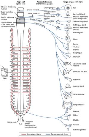
#[[File:Parasympathetic Nervous System.jpg|right|frameless]]Autonomic ganglia belong to the autonomic nervous system, which is divided into the sympathetic and [[Parasympathetic System|parasympathetic]] nervous systems. '''See image 3''' Shows the Autonomic Ganglia (red SNS, blue PNS). The sympathetic chain ganglia constitute a row of ganglia along the vertebral column that receive central input from the lateral horn of the thoracic and upper lumbar spinal cord. At the superior end of the chain ganglia are three paravertebral ganglia in the cervical region. Three other autonomic ganglia that are related to the sympathetic chain are the prevertebral ganglia, which are located outside of the chain but have similar functions. They are referred to as prevertebral because they are anterior to the vertebral column. The neurons of these autonomic ganglia are multipolar in shape, with dendrites radiating out around the cell body where synapses from the spinal cord neurons are made. The neurons of the chain, paravertebral, and prevertebral ganglia then project to organs in the head and neck, thoracic, abdominal, and pelvic cavities to regulate the sympathetic aspect of homeostatic mechanisms<ref name=":1" />. | #[[File:Parasympathetic Nervous System.jpg|right|frameless]]Autonomic ganglia belong to the autonomic nervous system, which is divided into the sympathetic and [[Parasympathetic System|parasympathetic]] nervous systems. '''See image 3''' Shows the Autonomic Ganglia (red SNS, blue PNS). The sympathetic chain ganglia constitute a row of ganglia along the vertebral column that receive central input from the lateral horn of the thoracic and upper lumbar spinal cord. At the superior end of the chain ganglia are three paravertebral ganglia in the cervical region. Three other autonomic ganglia that are related to the sympathetic chain are the prevertebral ganglia, which are located outside of the chain but have similar functions. They are referred to as prevertebral because they are anterior to the vertebral column. The neurons of these autonomic ganglia are multipolar in shape, with dendrites radiating out around the cell body where synapses from the spinal cord neurons are made. The neurons of the chain, paravertebral, and prevertebral ganglia then project to organs in the head and neck, thoracic, abdominal, and pelvic cavities to regulate the sympathetic aspect of homeostatic mechanisms<ref name=":1" />. | ||
== References == | == References == | ||
Revision as of 03:47, 6 February 2021
Original Editor - lucinda hampton
Top Contributors - Lucinda hampton, Uchechukwu Chukwuemeka and Vidya Acharya
Introduction[edit | edit source]

A ganglion is a collection of neuronal bodies found in the somatic and autonomic branches of the peripheral nervous system (PNS).
In addition to the ganglion of the peripheral nervous system, there are also parts of the brain that contains a cluster of interconnected nuceli called the “basal ganglia” or “basal nuclei”[1]
Ganglia can be thought of as synaptic relay stations between neurons. The information enters the ganglia, excites the neuron in the ganglia and then exits[2].
Among vertebrate animals there are three major groups of ganglia. These include:
- Cranial nerve ganglia that contain the neurons of the cranial nerves
- Dorsal root ganglia or spinal ganglia where the cell bodies of sensory or afferent nerves are located See Image 1
- Autonomic ganglia, which contain the cell bodies of the autonomic nervous system. In this system, nerve fibers that run from the central nervous system to the ganglia are called the preganglionic fibers. The nerve fibers originating in the ganglia that connect to effector organs are termed postganglionic fibers.[1]
Image 1: Shows the Autonomic Ganglia (red SNS, blue PNS)
Structure[edit | edit source]

Ganglia are oval in structure and contain
- Neuronal cell bodies (somata)
- Satellite cells, surround neurons in the sensory, sympathetic and parasympathetic ganglia and help regulate the chemical environment. They may contribute to chronic pain.[3]
- A dense connective tissue capsule covers the ganglion, with a single layer of flat shaped satellite cells surrounding each neuronal cell body.
- A basement membrane covers the outer region of the satellite cells.
Three Major Groups Of Ganglia[edit | edit source]
- A spinal ganglion (dorsal root ganglion) is a cluster of nerve bodies positioned along the spinal cord at the dorsal and ventral roots of a spinal nerve. The dorsal root ganglia contain the cell bodies of afferent nerve fibres (those carrying impulses toward the central nervous system); efferent neurons (carrying motor impulses away from the central nervous system) are present in the ventral root ganglia.
- Cranial Nerve Ganglion is analogous to the dorsal root ganglion, except that it is associated with a cranial nerve, instead of a spinal nerve (associated with the spinal cord). The roots of cranial nerves are within the cranium, whereas the ganglia are outside the skull. Eg, The vagus nerve has two sensory ganglia, the superior and the inferior ganglia both outside the skull; the trigeminal ganglion is superficial to the temporal bone . Like the sensory neurons associated with the spinal cord, the sensory neurons of cranial nerve ganglia are unipolar in shape with associated satellite cells[4]. See image 2, shows various CN ganglia (outside of skull).
- Autonomic ganglia belong to the autonomic nervous system, which is divided into the sympathetic and parasympathetic nervous systems. See image 3 Shows the Autonomic Ganglia (red SNS, blue PNS). The sympathetic chain ganglia constitute a row of ganglia along the vertebral column that receive central input from the lateral horn of the thoracic and upper lumbar spinal cord. At the superior end of the chain ganglia are three paravertebral ganglia in the cervical region. Three other autonomic ganglia that are related to the sympathetic chain are the prevertebral ganglia, which are located outside of the chain but have similar functions. They are referred to as prevertebral because they are anterior to the vertebral column. The neurons of these autonomic ganglia are multipolar in shape, with dendrites radiating out around the cell body where synapses from the spinal cord neurons are made. The neurons of the chain, paravertebral, and prevertebral ganglia then project to organs in the head and neck, thoracic, abdominal, and pelvic cavities to regulate the sympathetic aspect of homeostatic mechanisms[4].

References[edit | edit source]
- ↑ 1.0 1.1 New Medical Ganglion Available from:https://www.news-medical.net/health/What-is-a-Ganglion.aspx (accessed 5.2.2021)
- ↑ ken Hub Ganglion Available from:https://www.kenhub.com/en/library/anatomy/nerve-ganglia (accessed 5.2.2021)
- ↑ QBI glial cells Available from:https://qbi.uq.edu.au/brain-basics/brain/brain-physiology/types-glia (accessed 6.2.2021)
- ↑ 4.0 4.1 OSE Ganglia and nerves Available from:https://open.oregonstate.education/aandp/chapter/13-2-ganglia-and-nerves/ (accessed 5.2.2021)

