Femoral Neck Fractures Biomechanics: Difference between revisions
No edit summary |
No edit summary |
||
| Line 2: | Line 2: | ||
== Introduction == | == Introduction == | ||
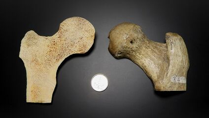
The hip joint contact forces are in excess of 500% body weight (BW) and can be as high as 509 kg of force during during ADL's, but spontaneous fractures typically do not occur in healthy individuals. | [[File:Internal and external architecture.jpg|thumb|Internal and external architecture NOF|alt=|424x424px]] | ||
The [[Hip Anatomy|hip joint]] contact forces are in excess of 500% body weight (BW) and can be as high as 509 kg of force during during ADL's, but spontaneous fractures typically do not occur in healthy individuals. | |||
== Etiology == | |||
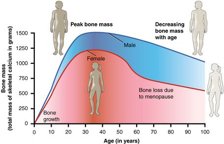
# [[Effects of Ageing on Bone|Ageing,]] [[osteoporosis]], or metastatic lesions, may weaken bone tissue to such an extent that spontaneous [[Hip Fracture/Femoral Neck Fracture|femoral neck fractures (NOF)]] occur. The initiation of [[fracture]] is typically at the superior aspect of the femoral neck, a region with less [[Bone Cortical And Cancellous|cortical]] thickness, and travels along through the trabecular bone.<ref name=":4">Basso T, Klaksvik J, Syversen U, Foss OA. [https://www.sciencedirect.com/science/article/pii/S0020138312001283 Biomechanical femoral neck fracture experiments—a narrative review]. Injury. 2012 Oct 1;43(10):1633-9.</ref> | |||
# [[Bone Stress Injuries|Femoral neck stress fractures]] occur when homeostatic remodelling can not keep up with the strain that is put on the bone. This is often due to repetitive loading of bone with abnormal forces and sudden increase in training volume or activity.<ref name=":7">Robertson GA, Wood AM. [https://www.thieme-connect.com/products/ejournals/html/10.1055/s-0043-103946 Femoral neck stress fractures in sport: a current concepts review]. Sports Medicine International Open. 2017 Feb;1(02):E58-68.</ref> Femoral neck stress fractures are associated with activities like long-distance running, gymnastics, and ballet.<ref name=":7" /> The cyclical loading of the tissue leads to decreased tissue tolerance. The micro-fractures that occur on the bone cannot be repaired fast enough and turn into fractures.<ref name=":6" /> | |||
== Fracture risk == | |||
[[File:Bone Mass.jpeg|thumb|456x456px|Bone mass/Age graph]] | |||
Fracture risk is determined by the bone strength and the applied load. | |||
* Bone strength: [[Bone Density]] at the femoral neck and the cortical bone thickness and bone density. | |||
* Bone porosity: assessed through a bone biopsy, strong predictor of mechanical strength. | |||
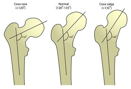
* Bone geometrically: normal femoral neck-shaft angle is between 124-135⁰. Having a femoral neck-shaft angle that is outside of the normal range increases the risk of a femoral neck fracture ([[Coxa Vara / Coxa Valga|coxa vara or coxa valga]]) . | |||
* Increased hip axis length (measured along the femoral neck axis and is defined as the distance from the inferolateral aspect of the greater trochanter to the inner [[Pelvis|pelvic]] brim), increased neck length.<ref name=":2">Augat P, Bliven E, Hackl S. [https://journals.lww.com/jorthotrauma/FullText/2019/01001/Biomechanics_of_Femoral_Neck_Fractures_and.6.aspx Biomechanics of femoral neck fractures and implications for fixation]. Journal of orthopaedic trauma. 2019 Jan 1;33:S27-32.</ref><ref name=":3">Kani KK, Porrino JA, Mulcahy H, Chew FS. [https://link.springer.com/article/10.1007/s00256-018-3008-3 Fragility fractures of the proximal femur: review and update for radiologists]. Skeletal radiology. 2019 Jan;48(1):29-45.</ref> | |||
== | == Biomechanics: Forces == | ||
The | [[File:Femoral neck-shaft angle.jpg|thumb|437x437px|Femoral neck-shaft angle|alt=]] | ||
The NOF experiences the highest stress loads within the femur. <ref name=":6">Neubauer T, Brand J, Lidder S, Krawany M. [https://www.tandfonline.com/doi/abs/10.1080/15438627.2016.1191489 Stress fractures of the femoral neck in runners: a review.] Research in Sports Medicine. 2016 Jul 2;24(3):283-97.</ref> Forces that are acting on the femoral neck are: | |||
# Externally generated: a result of ground reaction forces that translated from the ankle to the NOF as a result of the vertical impact and typically stay below 1.3 times body weight during low-speed walking. <ref name=":2" /> | |||
# Internally generated: a result of the muscles acting on the bone to accomplish the desired movements and maintain balance. Internally generated forces are typically 2-3 times body weight during low-speed walking. <ref name=":4" /> | |||
The internal and external forces produce bending and torsional moments that act on the NOF (reported to be 500-2000 micro-strains for day-to-day activities)<ref name=":2" /> | |||
= | * The bone strain is largest at the inferior aspect of the NOF, resulting in a larger thickness of the inferior aspect of NOF bone cortex compared to the superior aspect.<ref name=":2" /> | ||
* The superior aspect of the femoral neck experiences increased strain during activities like stair climbing where there is an increase in hip flexion and abduction. The increased strain is due to the increase of torsion at the femoral neck.<ref name=":4" /> | |||
=== Categories === | |||
NOF stress fractures are subdivided into two categories: | |||
Fall velocities | # Compressive type: occur at the inferior aspect of the NOF. This type of fracture occurs when the forces applied to the bone are higher than the plastic properties of the bone.<ref name=":7" /> eg Sideway falls (leading cause of [[Hip Fracture|hip fracture]] in the elderly).<ref name=":0">Nasiri Sarvi M, Luo Y. [https://link.springer.com/article/10.1007/s00198-017-4138-5 Sideways fall-induced impact force and its effect on hip fracture risk: a review]. Osteoporosis international. 2017 Oct;28(10):2759-80.</ref> Induce a compressive type of load to the femoral neck.<ref name=":2" /> The factors that are involved in falls that contribute to fractures are femoral strength, fall velocity, and effective mass.<ref name=":0" /> Femoral strength is significantly reduced in a sideways fall scenario. The loading configuration determines the amount of force required to induce a NOF fracture. The force required to induce a fracture during a single leg stance is 7214N vs 3462N during a sideways fall ±1520N. The NOF can withstand a much greater force during vertical loading vs lateral loading on the greater trochanter.<ref name=":0" />; Fall velocities > 3m/s can induce a NOF fracture. Falls due to a slip or stumble have higher fall velocity than falls due to incorrect shifting of weight.<ref name=":0" /> A fall from a standing position is going to have a larger effective mass than a fall from a kneeling position.<ref name=":0" /> | ||
# Tensile type: occur at the superior aspect of the bone. Occurs when the muscles are fatigued, and the tensile forces are translated predominantly by the bone. In normal conditions, the gluteus medius and minimus muscles counteract the high-tension forces acting on the NOF.<ref name=":7" /> | |||
== References == | == References == | ||
<references /> | <references /> | ||
| Line 37: | Line 41: | ||
[[Category:Hip]] | [[Category:Hip]] | ||
[[Category:Hip - Bones]] | [[Category:Hip - Bones]] | ||
[[Category:Biomechanics]] | |||
Revision as of 02:50, 10 December 2022
Introduction[edit | edit source]
The hip joint contact forces are in excess of 500% body weight (BW) and can be as high as 509 kg of force during during ADL's, but spontaneous fractures typically do not occur in healthy individuals.
Etiology[edit | edit source]
- Ageing, osteoporosis, or metastatic lesions, may weaken bone tissue to such an extent that spontaneous femoral neck fractures (NOF) occur. The initiation of fracture is typically at the superior aspect of the femoral neck, a region with less cortical thickness, and travels along through the trabecular bone.[1]
- Femoral neck stress fractures occur when homeostatic remodelling can not keep up with the strain that is put on the bone. This is often due to repetitive loading of bone with abnormal forces and sudden increase in training volume or activity.[2] Femoral neck stress fractures are associated with activities like long-distance running, gymnastics, and ballet.[2] The cyclical loading of the tissue leads to decreased tissue tolerance. The micro-fractures that occur on the bone cannot be repaired fast enough and turn into fractures.[3]
Fracture risk[edit | edit source]
Fracture risk is determined by the bone strength and the applied load.
- Bone strength: Bone Density at the femoral neck and the cortical bone thickness and bone density.
- Bone porosity: assessed through a bone biopsy, strong predictor of mechanical strength.
- Bone geometrically: normal femoral neck-shaft angle is between 124-135⁰. Having a femoral neck-shaft angle that is outside of the normal range increases the risk of a femoral neck fracture (coxa vara or coxa valga) .
- Increased hip axis length (measured along the femoral neck axis and is defined as the distance from the inferolateral aspect of the greater trochanter to the inner pelvic brim), increased neck length.[4][5]
Biomechanics: Forces[edit | edit source]
The NOF experiences the highest stress loads within the femur. [3] Forces that are acting on the femoral neck are:
- Externally generated: a result of ground reaction forces that translated from the ankle to the NOF as a result of the vertical impact and typically stay below 1.3 times body weight during low-speed walking. [4]
- Internally generated: a result of the muscles acting on the bone to accomplish the desired movements and maintain balance. Internally generated forces are typically 2-3 times body weight during low-speed walking. [1]
The internal and external forces produce bending and torsional moments that act on the NOF (reported to be 500-2000 micro-strains for day-to-day activities)[4]
- The bone strain is largest at the inferior aspect of the NOF, resulting in a larger thickness of the inferior aspect of NOF bone cortex compared to the superior aspect.[4]
- The superior aspect of the femoral neck experiences increased strain during activities like stair climbing where there is an increase in hip flexion and abduction. The increased strain is due to the increase of torsion at the femoral neck.[1]
Categories[edit | edit source]
NOF stress fractures are subdivided into two categories:
- Compressive type: occur at the inferior aspect of the NOF. This type of fracture occurs when the forces applied to the bone are higher than the plastic properties of the bone.[2] eg Sideway falls (leading cause of hip fracture in the elderly).[6] Induce a compressive type of load to the femoral neck.[4] The factors that are involved in falls that contribute to fractures are femoral strength, fall velocity, and effective mass.[6] Femoral strength is significantly reduced in a sideways fall scenario. The loading configuration determines the amount of force required to induce a NOF fracture. The force required to induce a fracture during a single leg stance is 7214N vs 3462N during a sideways fall ±1520N. The NOF can withstand a much greater force during vertical loading vs lateral loading on the greater trochanter.[6]; Fall velocities > 3m/s can induce a NOF fracture. Falls due to a slip or stumble have higher fall velocity than falls due to incorrect shifting of weight.[6] A fall from a standing position is going to have a larger effective mass than a fall from a kneeling position.[6]
- Tensile type: occur at the superior aspect of the bone. Occurs when the muscles are fatigued, and the tensile forces are translated predominantly by the bone. In normal conditions, the gluteus medius and minimus muscles counteract the high-tension forces acting on the NOF.[2]
References[edit | edit source]
- ↑ 1.0 1.1 1.2 Basso T, Klaksvik J, Syversen U, Foss OA. Biomechanical femoral neck fracture experiments—a narrative review. Injury. 2012 Oct 1;43(10):1633-9.
- ↑ 2.0 2.1 2.2 2.3 Robertson GA, Wood AM. Femoral neck stress fractures in sport: a current concepts review. Sports Medicine International Open. 2017 Feb;1(02):E58-68.
- ↑ 3.0 3.1 Neubauer T, Brand J, Lidder S, Krawany M. Stress fractures of the femoral neck in runners: a review. Research in Sports Medicine. 2016 Jul 2;24(3):283-97.
- ↑ 4.0 4.1 4.2 4.3 4.4 Augat P, Bliven E, Hackl S. Biomechanics of femoral neck fractures and implications for fixation. Journal of orthopaedic trauma. 2019 Jan 1;33:S27-32.
- ↑ Kani KK, Porrino JA, Mulcahy H, Chew FS. Fragility fractures of the proximal femur: review and update for radiologists. Skeletal radiology. 2019 Jan;48(1):29-45.
- ↑ 6.0 6.1 6.2 6.3 6.4 Nasiri Sarvi M, Luo Y. Sideways fall-induced impact force and its effect on hip fracture risk: a review. Osteoporosis international. 2017 Oct;28(10):2759-80.


