Scaphoid Fracture
Original Editor - Dawn Waugh
Top Contributors - Dawn Waugh, Mats Vandervelde, Abbey Wright, Inoa De Pauw, Admin, Chrysolite Jyothi Kommu, Kim Jackson, Evan Thomas, Rachael Lowe, Amanda Ager, Anas Mohamed, Lucinda hampton, Johnathan Fahrner, WikiSysop, Wanda van Niekerk, 127.0.0.1, Nupur Smit Shah, Vanessa Rhule, Naike De Win and Claire Knott
Clinically Relevant Anatomy
[edit | edit source]
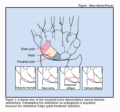
Scaphoid fractures are classified according to their 2-dimensional radiographic appearance, and transverse waist fractures are considered the most common. The scaphoid is the most commonly fractured of the 8 carpal bones of the wrist, with frequent complications that are predisposed by its anatomical location, anatomical configuration (shape and length), and vascular supply.
It is made up of a proximal and distal pole which are joined by a waist. Scapohid fractures make up 50-80% of all carpal fractures. [1] Blood supply is provided by a subdivision of the radial artery and travels distal to proximal.The proximal pole has no direct blood supply and is prone to avascular necrosis (AVN).[2]
The most common mechanism of injury is a fall into an outstretched hand. Proper identification and classification of scaphoid fracture and its complications is necessary for appropriate treatment.
Mechanism of Injury / Pathological Process
[edit | edit source]
Mechanism of injury is usually a fall onto an outstretched arm with wrist hyperextended and radial deviation. This position causes axial loading through the scaphoid. Less common mechanisms of injury are:
- wrist extension with deceleration such as with the hand on a steering wheel
- 'kickback' injuries from machinery
- hyperflexion injuries
- direct impact to the scaphoid
Most injuries are seen in men aged 15-30. 75-80% of fractures occur through the waist of the bone. 15-20% occur at the proximal pole. 10-15% of fractures occur at the distal pole.[2]
Clinical Presentation[edit | edit source]
Clinical presentation includes swelling and pain over the anatomical snuff box. The anatomical snuff box is the area between the extensor pollicis longus and the extensor pollicis brevis. Patient will usually also complain of pain with pressure over the scapoid tubercle. [2]
Diagnostic Procedure
[edit | edit source]
AP and lateral radiographs along with additional scaphoid views (pronated oblique and ulnar-deviated oblique) are used in diagnosing a scaphoid fracture. Up to 15% of scaphoid fractures are not evident on initial radiograph, however. [2] If fracture is suspected,MRI is the next line of imaging to confirm the diagnosis. [3]
Outcome Measures[edit | edit source]
DASH (see Outcome Measures Database)
Management / Interventions
[edit | edit source]
Cast immobilization is the standard treatment for treating a scaphoid fracture. With cast immobilization, chance of non-union is approximately 20%. Therefore, with displaced or unstable fractures, operative treatment is recommended. [4] Though this improves the rate of non-union, the complication rate for ORIF is 30%.[5]
Fractures are usually classified by Herbert and Fisher's system:
A: Acute but stable fractures such as fractures of the tubercle, incomplete or undisplaced fractures of the waist
B: Acute unstable fractures such as distal oblique fractures, complete waist fractures, proximal pole fractures, and
fracture dislocation
C: Fractures with evidence of delayed union
D: Fractures with established non-union[2]
Differential Diagnosis
[edit | edit source]
Differential diagnosis includes Colles' fracture,Salter-Harris fracture, other carpal fractures, scapholunate complex injury.[2]
Key Evidence[edit | edit source]
A systemic review suggests that percutaneous fixation may result in faster union and return to work or sport. There was no difference noted between cast fixation and ORIF. The authors suggest that cast treatment is a good treatment option for most. Surgery should be reserved for high level athletes and manual workers who cannot work in a cast.[5]
Resources
[edit | edit source]
Physical Therapy Management[edit | edit source]
The scaphoid fracture is a troublesome fracture.
Failure of treatment can result in avascular necrosis (up to 40%), non-union (5-21%) and early osteo-arthritis (up to 32%) which may seriously impair wrist function. Impaired consolidation of scaphoid fractures results in longer immobilization, with significant psychosocial and financial consequences.
In dislocated fractures complication rates are even higher. Part of the explanation for this high incidence of avascular necrosis can be found in the special blood supply of the scaphoid bone. Branches of the radial artery enter the os scaphoideus dorsally, thus supplying the bone from distal to proximal. Therefore fractures of the mid or distal third of the scaphoid can result in avascular necrosis of the proximal part of the bone. Non-union is defined as the absence of healing at four to six months after injury. This may be due to delay in treatment, inadequate immobilization, localization of the fracture, instability due to displacement of the fracture fragments or combination with ligamentous injury of the carpus. All these conditions are responsible for severe impairment in wrist function and even permanent disability.
Current treatment strategies are unable to deal with this problem. The number of complications following conventional treatment (immobilization in a cast) is quite high and surgery is generally performed only if complications in healing occur. This is often initiated in a late phase, most often months after the fracture occurred, which again can have severe socio-economical consequences.
Physical forces used in fracture healing are direct current, Pulsed Electromagnetic Fields (PEMF) and ultrasound.
Inductively-coupled electromagnetic fields have been used in medicine since 1974. Physical forces stimulate osteogenesis, in that callus was formed around the cathode. In non-union scaphoid fractures Bora et al reported a 71% reduction in non-union within twelve weeks after initiating the electrical stimulation. Later this rather invasive method was replaced by PEMF, a non-invasive technique.
Treatment of fresh scaphoid fractures by physical forces (ultra sound) accelerates healing by 30%.
Conservative treatment by cast immobilisation below the elbow with thumb metacarpophalangeal joint inclusion is a widely accepted method in the management of stable scaphoid fractures, because the rate of healing is considered to be satisfactory. Conservative treatment appears to have an advantage over surgical treatment due to the possibility of numerous surgical complications. But conservative treatment with long immobilization periods results in nonunion from 1.5 to 37 % of the time. Although cast immobilization is associated with low rates of morbidity and long-term disability, the time until patients can resume work and daily activities may also be prolonged compared to surgery.
The introduction of intramedullary fixation with the Herbert screw represented a revolutionary development in scaphoid surgery. Via minimally invasive screw fixation, even small displaced scaphoid fractures can be treated surgically, with subsequent advantages of early mobilization and faster return to work.
Recent Related Research (from Pubmed)[edit | edit source]
Failed to load RSS feed from http://eutils.ncbi.nlm.nih.gov/entrez/eutils/erss.cgi?rss_guid=1H9AR3ZQQCaD17U2nZhRHO0f8iHnyie26kobpe8zfqRJgmvYyC|charset=UTF-8|short|max=10: Error parsing XML for RSS
References[edit | edit source]
References will automatically be added here, see adding references tutorial.
- ↑ Alshryda SJM, Shah AB, Rhodes S, Odak SS, Murali SR, Ilango B. Interventions for treating acute fractures of the carpal scaphoid bone in adults. Cochrane Database of Systematic Reviews 2007, Issue 2. Art. No.: CD006523. DOI:fckLR10.1002/14651858.CD006523.fckLRA B
- ↑ 2.0 2.1 2.2 2.3 2.4 2.5 Bethel J. Scaphoid Fracture: diagnosis and management. Emergency Nurse. July, 2009. 17(4): 24-29.
- ↑ Henriksen et al. Two-Dimensional Image Fusion of Planar Bone Scintigraphy and Radiographs in Patients with Clinical Scaphoid Fracture: An Imaging Study. Acta Radiologica. February, 2009. 50(1): 71-77.
- ↑ Pfeiffer et al. A prospective multi-center cohort study of acute non-displaced fractures of the scaphoid: operative versus non-operative treatment. BMC Musculoskeletal Disorders. May, 2006. 7:41.
- ↑ 5.0 5.1 Modi et al. Operative versus nonoperative treatment of acute undisplaced and minimally displaced scaphoid waist fractures-A systemic review. Injury, Int. J. Care Injured. 2009. 40: 268-273.


