Adductor Longus: Difference between revisions
Leana Louw (talk | contribs) No edit summary |
Leana Louw (talk | contribs) No edit summary |
||
| Line 10: | Line 10: | ||
== Description == | == Description == | ||
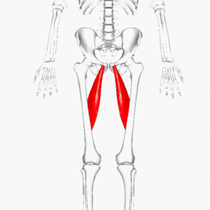
Adductor longus is one of the adductor muscles of the medial thigh.<ref name=":0">Moore KL, Dalley AF, Agur AMR. Clinial oriented anatomy. Philadelphia: Wolters Kluwer, 2010.</ref> Together with [[Adductor Brevis|adductor brevis]], [[Adductor Magnus|adductor magnus]], [[gracilis]] and [[Obturator Externus|obturator externus]], it makes up the adductor compartement.<ref name=":0" /> This large fan-shaped muscle is situated most anteriorly of this group and covers the middle part of [[Adductor Magnus|adductor magnus]] and the anterior part of [[Adductor Brevis|adductor brevis]]. | Adductor longus is one of the adductor muscles of the medial thigh.<ref name=":0">Moore KL, Dalley AF, Agur AMR. Clinial oriented anatomy. Philadelphia: Wolters Kluwer, 2010.</ref> Together with [[Adductor Brevis|adductor brevis]], [[Adductor Magnus|adductor magnus]], [[gracilis]] and [[Obturator Externus|obturator externus]], it makes up the adductor compartement.<ref name=":0" /> This large fan-shaped muscle is situated most anteriorly of this group and covers the middle part of [[Adductor Magnus|adductor magnus]] and the anterior part of [[Adductor Brevis|adductor brevis]].<ref name=":0" /> | ||
[[File:Adductor longus.gif|none|thumb]] | [[File:Adductor longus.gif|none|thumb]] | ||
| Line 30: | Line 30: | ||
== Clinical relevance == | == Clinical relevance == | ||
* [[Adductor Tendinopathy|Adductor tendinopathy]] | |||
* [[Groin Strain|Groin strain]] | |||
== Assessment == | == Assessment == | ||
* [[Manual Muscle Testing: Hip Adduction|Manual muscle testing]]:<ref>Hislop H, Avers D, Brown M. [https://books.google.co.za/books?hl=en&lr=&id=peNOAQAAQBAJ&oi=fnd&pg=PP1&dq=muscle+testing&ots=mxJRzfX8_q&sig=J-rFmGV3dwKthAW-Q0uYki-B8aI#v=onepage&q=muscle%20testing&f=false Daniels and Worthingham's Muscle Testing: Techniques of manual examination.] Elsevier Health Sciences; 2013.</ref> | |||
** Patient positioned in side-lying with side being tested resting at the bottom on the bed. The top leg is supported by the examiner in 25 degrees of abduction. The examiner applies manual resistance to the distal femur proximal to the knee joint in the direction of the bed while the patient actively adducts against the examiner's hand. | |||
** Determine strength by using Oxford grades 0-5. | |||
* [[Adductor Squeeze Test|Adductor squeeze test]] | |||
* Hip adductor length test:<ref>Norris CM. [https://www.sciencedirect.com/science/article/abs/pii/S003194060567068X Spinal stabilisation: 4. Muscle imbalance and the low back.] Physiotherapy 1995;81(3):127-38.</ref> | |||
** Patient is positioned in crook-lying with a posterior pelvic tilt. The patient abducts the hips by moving the knees outwards towards the sides of the bed. | |||
*** Adductor stiffness: Patient not able to maintain position, (+) anterior pelvic tilt | |||
*** Inhibition of [[Gluteus Medius|gluteus medius]] and external rotators of hip can be noted,, causing muscle imbalances and compensatory movement patterns. | |||
** Tests for abdominal weakness as well. | |||
== Treatment == | == Treatment == | ||
== | == References == | ||
<references /> | <references /> | ||
Revision as of 19:34, 12 July 2020
Original Editor - Leana Louw
Top Contributors - Lucinda hampton, Leana Louw, Memoona Awan, Kim Jackson, Vidya Acharya and Ines Musabyemariya
This article or area is currently under construction and may only be partially complete. Please come back soon to see the finished work! (12/07/2020)
Description[edit | edit source]
Adductor longus is one of the adductor muscles of the medial thigh.[1] Together with adductor brevis, adductor magnus, gracilis and obturator externus, it makes up the adductor compartement.[1] This large fan-shaped muscle is situated most anteriorly of this group and covers the middle part of adductor magnus and the anterior part of adductor brevis.[1]
Origin[edit | edit source]
Strong tendon from anterior aspect of pubic body inferior to pubic crest.[1]
Insertion[edit | edit source]
Linea aspera on middle third of femur.[1]
Nerve[edit | edit source]
Anterior devision of obturator nerve (L2, L3, L4).[1]
Artery[edit | edit source]
Function[edit | edit source]
Adduction of femur.[1]
Clinical relevance[edit | edit source]
Assessment[edit | edit source]
- Manual muscle testing:[3]
- Patient positioned in side-lying with side being tested resting at the bottom on the bed. The top leg is supported by the examiner in 25 degrees of abduction. The examiner applies manual resistance to the distal femur proximal to the knee joint in the direction of the bed while the patient actively adducts against the examiner's hand.
- Determine strength by using Oxford grades 0-5.
- Adductor squeeze test
- Hip adductor length test:[4]
- Patient is positioned in crook-lying with a posterior pelvic tilt. The patient abducts the hips by moving the knees outwards towards the sides of the bed.
- Adductor stiffness: Patient not able to maintain position, (+) anterior pelvic tilt
- Inhibition of gluteus medius and external rotators of hip can be noted,, causing muscle imbalances and compensatory movement patterns.
- Tests for abdominal weakness as well.
- Patient is positioned in crook-lying with a posterior pelvic tilt. The patient abducts the hips by moving the knees outwards towards the sides of the bed.
Treatment[edit | edit source]
References[edit | edit source]
- ↑ 1.0 1.1 1.2 1.3 1.4 1.5 1.6 Moore KL, Dalley AF, Agur AMR. Clinial oriented anatomy. Philadelphia: Wolters Kluwer, 2010.
- ↑ 2.0 2.1 University of Washington. Department of Radiology. Adductor Longus. Available from: https://rad.washington.edu/muscle-atlas/adductor-longus/ (accessed 11/07/2020).
- ↑ Hislop H, Avers D, Brown M. Daniels and Worthingham's Muscle Testing: Techniques of manual examination. Elsevier Health Sciences; 2013.
- ↑ Norris CM. Spinal stabilisation: 4. Muscle imbalance and the low back. Physiotherapy 1995;81(3):127-38.


