Brachialis: Difference between revisions
No edit summary |
No edit summary |
||
| Line 2: | Line 2: | ||
'''Top Contributors''' - {{Special:Contributors/{{FULLPAGENAME}}}} | '''Top Contributors''' - {{Special:Contributors/{{FULLPAGENAME}}}} | ||
</div> | </div>[[File:Brachialis muscle1.png|alt=|thumb|386x386px|[[:File:Brachialis muscle1.png]]<ref>By Anatomography (en:Anatomography (setting page of this image)) [CC BY-SA 2.1 jp (<nowiki>https://creativecommons.org/licenses/by-sa/2.1/jp/deed.en</nowiki>)], via Wikimedia Commons</ref> | ||
]] | |||
== Description == | == Description == | ||
The brachialis muscle is the prime flexor of the elbow. This muscle is located in the | The brachialis muscle is the prime flexor of the elbow. This muscle is located in the anterior compartment of the arm along with the [[Biceps brachii]] and coracobrachialis. | ||
=== Origin === | === Origin === | ||
Distal anterior aspect of the humerus, deep to the biceps brachii.<ref name=":0">Brachialis. (n.d.). Retrieved March 12, 2018, from <nowiki>https://rad.washington.edu/muscle-atlas/brachialis/</nowiki></ref> | Distal anterior aspect of the humerus, deep to the biceps brachii.<ref name=":0">Brachialis. (n.d.). Retrieved March 12, 2018, from <nowiki>https://rad.washington.edu/muscle-atlas/brachialis/</nowiki></ref> | ||
=== Insertion === | === Insertion === | ||
Coronoid process | Coronoid process and the ulnar tuberosity.<ref name=":0" /> | ||
=== Nerve === | === Nerve === | ||
The brachialis muscle is innervated by the musculocutaneous nerve and components of the radial nerve. The radial nerve descends in the groove between the brachialis and brachioradialis muscles, above the elbow. Of the muscles in the anterior compartment, the biceps brachii and the brachialis are innervated by C5 and C6 nerve roots.<ref name=":1">TWENTIETH EDITION | |||
THOROUGHLY REVISED AND RE-EDITED BY WARREN H. LEWIS | |||
ILLUSTRATED WITH 1247 ENGRAVINGS | |||
PHILADELPHIA: LEA & FEBIGER, 1918 | |||
[[File:Brachial-plexus-2.png|thumb|336x336px]] | |||
NEW YORK: BARTLEBY.COM, 2000 | |||
</ref>[[File:Brachial-plexus-2.png|thumb|336x336px]] | |||
=== Artery === | === Artery === | ||
| Line 22: | Line 28: | ||
== Function == | == Function == | ||
The brachialis muscle has a large cross sectional area, providing it with more strength than the biceps brachii and the coracobrachialis.<ref name=":2">Marieb, E. N. (2004). ''Human anatomy & physiology'' (6th ed.). San Francisco: Pearson Education.</ref> In order to isolate the brachialis muscle the forearm needs to be in pronation, due to the biceps brachii function as a supinator and flexor. <ref name=":2" /> By pronating the forearm the biceps is put into a mechanical disadvantage. | |||
[[File:Arm cross section.jpg|thumb|<ref name=":1" />]] | |||
== Clinical relevance == | == Clinical relevance == | ||
| Line 34: | Line 41: | ||
== Assessment == | == Assessment == | ||
To assess the strength of the brachialis place the elbow at 90 degrees of flexion with the forearm fully pronated. Then have the patient resist an inferior force placed on the distal forearm.<ref>Muscolino, J. E. (2011). ''Kinesiology: the skeletal system and muscle function'' (2nd ed.). St. Louis, MO: Mosby/Elsevier.</ref> | |||
== Resources == | == Resources == | ||
Revision as of 06:14, 12 March 2018
Top Contributors - Eric Henderson, Matthew Chin, Kirenga Bamurange Liliane, Kim Jackson, Joao Costa and Wendy Snyders
Description[edit | edit source]
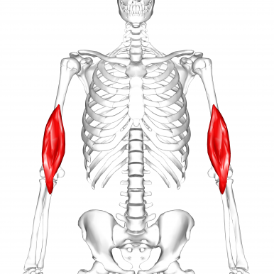
The brachialis muscle is the prime flexor of the elbow. This muscle is located in the anterior compartment of the arm along with the Biceps brachii and coracobrachialis.
Origin[edit | edit source]
Distal anterior aspect of the humerus, deep to the biceps brachii.[2]
Insertion[edit | edit source]
Coronoid process and the ulnar tuberosity.[2]
Nerve[edit | edit source]
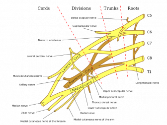
The brachialis muscle is innervated by the musculocutaneous nerve and components of the radial nerve. The radial nerve descends in the groove between the brachialis and brachioradialis muscles, above the elbow. Of the muscles in the anterior compartment, the biceps brachii and the brachialis are innervated by C5 and C6 nerve roots.[3]
Artery[edit | edit source]
Muscular branches of brachial artery, recurrent radial artery.[2]
Function[edit | edit source]
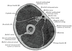
The brachialis muscle has a large cross sectional area, providing it with more strength than the biceps brachii and the coracobrachialis.[4] In order to isolate the brachialis muscle the forearm needs to be in pronation, due to the biceps brachii function as a supinator and flexor. [4] By pronating the forearm the biceps is put into a mechanical disadvantage.
Clinical relevance[edit | edit source]
Cubital Fossa
Posterior interosseous nerve syndrome
Assessment[edit | edit source]
To assess the strength of the brachialis place the elbow at 90 degrees of flexion with the forearm fully pronated. Then have the patient resist an inferior force placed on the distal forearm.[5]
Resources[edit | edit source]
- ↑ By Anatomography (en:Anatomography (setting page of this image)) [CC BY-SA 2.1 jp (https://creativecommons.org/licenses/by-sa/2.1/jp/deed.en)], via Wikimedia Commons
- ↑ 2.0 2.1 2.2 Brachialis. (n.d.). Retrieved March 12, 2018, from https://rad.washington.edu/muscle-atlas/brachialis/
- ↑ 3.0 3.1 TWENTIETH EDITION THOROUGHLY REVISED AND RE-EDITED BY WARREN H. LEWIS ILLUSTRATED WITH 1247 ENGRAVINGS PHILADELPHIA: LEA & FEBIGER, 1918 NEW YORK: BARTLEBY.COM, 2000
- ↑ 4.0 4.1 Marieb, E. N. (2004). Human anatomy & physiology (6th ed.). San Francisco: Pearson Education.
- ↑ Muscolino, J. E. (2011). Kinesiology: the skeletal system and muscle function (2nd ed.). St. Louis, MO: Mosby/Elsevier.


